COMPREHENSIVE ANALYSIS OF DRUGS TO TREAT SARS COV 2 INFECTION: MECHANISTIC INSIGHTS INTO CURRENT COVID 19 THERAPIES (REVIEW)
←
→
Page content transcription
If your browser does not render page correctly, please read the page content below

INTERNATIONAL JOURNAL OF MOlecular medicine 46: 467-488, 2020

 Comprehensive analysis of drugs to treat SARS‑CoV‑2 infection:
 Mechanistic insights into current COVID‑19 therapies (Review)
                 GEORGE MIHAI NITULESCU1, HORIA PAUNESCU2, STERGHIOS A. MOSCHOS3,4,
               DIMITRIOS PETRAKIS5, GEORGIANA NITULESCU1, GEORGE NICOLAE DANIEL ION1,
                  DEMETRIOS A. SPANDIDOS6, TAXIARCHIS KONSTANTINOS NIKOLOUZAKIS5,
                            NIKOLAOS DRAKOULIS7 and ARISTIDiS TSATSAKIS5

     1
      Faculty of Pharmacy, 2Faculty of Medicine, ‘Carol Davila’ University of Medicine and Pharmacy, 020956 Bucharest,
          Romania; 3Department of Applied Sciences, Faculty of Health and Life Sciences, Northumbria University;
        4
         PulmoBioMed Ltd., Newcastle‑Upon‑Tyne NE1 8ST, UK; 5Department of Forensic Sciences and Toxicology,
                Faculty of Medicine; 6Laboratory of Clinical Virology, School of Medicine, University of Crete,
          71003 Heraklion; 7Research Group of Clinical Pharmacology and Pharmacogenomics, Faculty of Pharmacy,
               School of Health Sciences, National and Kapodistrian University of Athens, 15771 Athens, Greece

                                          Received April 20, 2020; Accepted May 18, 2020

                                                     DOI: 10.3892/ijmm.2020.4608

Abstract. The major impact produced by the severe acute                tions, none have yet demonstrated unequivocal clinical utility
respiratory syndrome coronavirus 2 (SARS‑CoV‑2) focused                against coronavirus disease 2019 (COVID‑19). This work
many researchers attention to find treatments that can suppress        represents an exhaustive and critical review of all available
transmission or ameliorate the disease. Despite the very fast          data on potential treatments for COVID‑19, highlighting their
and large flow of scientific data on possible treatment solu-          mechanistic characteristics and the strategy development ratio-
                                                                       nale. Drug repurposing, also known as drug repositioning, and
                                                                       target based methods are the most used strategies to advance
                                                                       therapeutic solutions into clinical practice. Current in silico,
                                                                       in vitro and in vivo evidence regarding proposed treatments
Correspondence to: Professor George Mihai Nitulescu, Faculty           are summarized providing strong support for future research
of Pharmacy, ‘Carol Davila’ University of Medicine and Pharmacy,       efforts.
6 Traian Vuia, 020956 Bucharest, Romania
E‑mail: george.nitulescu@umfcd.ro
Professor Aristidis Tsatsakis, Department of Forensic Sciences         Contents
and Toxicology, Faculty of Medicine, University of Crete,
71003 Heraklion, Greece                                                1. Introduction
E‑mail: tsatsaka@uoc.gr                                                2.	Virological, genomic, and host aspects related to COVID‑19
                                                                           treatment
Abbreviations: AP2, adaptor protein 2; AMPK, AMP‑activated
                                                                       3. Pathogenesis
protein kinase; AT1, Ang1‑8 receptor 1; ACE2, angiotensin‑converting
enzyme 2; AAK1, AP2‑associated protein kinase 1; BECN1, Beclin1;       4. Treatment strategies
COVID‑19, coronavirus disease 2019; EMA, European Medicines            5.	Discussion
Agency; ExoN, exoribonuclease; EC50, half‑maximal effective
concentration; IC50, half‑maximal inhibitory concentration; IFN,
interferon; IL, interleukin; JAK, Janus kinases; MERS‑CoV, Middle      1. Introduction
East respiratory syndrome virus; mAb, monoclonal antibody; MCP1,
monocyte chemoattractant protein 1; nsp, non‑structural proteins;      The severe acute respiratory syndrome coronavirus 2
NAK, numb‑associated kinases; ORFs, open reading frames; RBD,          (SARS‑CoV‑2), provisionally named as 2019‑nCoV and collo-
receptor binding domain; rhACE2, recombinant human ACE2;               quially known as the coronavirus, is the pandemic‑causing
RdRp, RNA‑dependent RNA polymerase; SNPs, single nucleotide
                                                                       pathogen behind coronavirus disease 2019 (COVID‑19), which
polymorphisms; SPK2, S‑phase kinase‑associated protein 2; TLR,
                                                                       originated in Wuhan, Hubei Province, China in the 4th quarter
Toll‑like receptor; TNF‑α, tumour necrosis factor alpha; TMPRSS2,
type II transmembrane protease serine 2; FDA, US Food and Drug         of 2019 (1). Despite the rapid delivery of the SARS‑CoV‑2
Administration                                                         genome by the group originally recognizing this novel viral
                                                                       pneumonia and the subsequent production of a number
Key words: SARS‑CoV‑2, COVID‑19, treatment strategies, drug            of highly specific and sensitive molecular diagnostics for
testing                                                                SARS‑CoV‑2, progress in fighting this disease is balanced by
                                                                       the continued lack of ‘quick win’ treatments that can suppress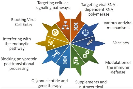
468                                       NITULESCU et al: Current Covid-19 therapies

transmission or ameliorate disease. Of the more than 100             (CoVs) (12) (Fig. 2). The first ORFs are translated into two
clinical trials evaluating a cadre of approved, or clinical stage    large polyproteins (pp1a and pp1ab) cytosolically processed
antivirals and related compounds, none have yet demonstrated         into the non‑structural proteins (nsp) 1‑16. These contain the
unequivocal clinical utility against COVID‑19.                       polyprotein‑processing protease nsp5, a host RNA restriction
    The eventual containment and mitigation of this emerging         factor (nsp1) arresting canonical translation, and collectively
disease will depend on our exponentially increasing knowledge        function as the viral replicase‑transcriptase complex (2) by
around SARS‑CoV‑2 virology, pathology, and cross‑species             rearranging rough endoplasmic reticulum (RER) membranes
transmission. With viral genome organization resolved in             into double‑membrane vesicles (DMVs) wherein replica-
unprecedented speed, molecular pathogenesis and immuno-              tion, transcription and virion assembly take place (13,14).
pathology related to protein products, modes of transmission,        The immunomodulatory protein nsp16 is 2'‑O‑ribose methyl
host cell entry mechanism and kinetics, vesicular trafficking,       transferase that renders the virus a toll‑like receptor (TLR) 7/8
protein and small molecule interaction, biological and replica-      antagonist (15) and is believed to enhance the purported
tion machinery mechanisms of action, are only beginning to           capacity of the virus to evade antiviral interferon‑generating
be understood. Likewise, the propensity for deleterious inflam-      cytosolic pathogen associated molecular pattern recognition
matory responses is episodically studied against SARS‑CoV‑2          receptors, such as the double stranded RNA (dsRNA) sensing
antigen presentation, antigen‑specific antibody and cell medi-       protein kinase R (PKR) and TLR3, on account of the DMV
ated responses contributing to protective adaptive immunity          replication organelle structure (16). An additional protein
and the attenuation of disease severity, as well as pulmonary and    of key pharmacological interest the RNA-dependent RNA
systemic tissue destruction. Comprehensive interrogation of          polymerase (RdRp) nsp12, which is the primary target of the
the viral‑human interactome, and associated chemoinformatic          so‑called directly acting antiviral (DAA) drug family. The
analyses (2,3) nevertheless offer hope for the identification of     remaining ORFs encode the so‑called spike (S or SARS‑2‑S)
nodal intervention points across the continuum of COVID‑19           protein responsible for virus tropism, as well as envelope I,
disease (4,5). As elucidation of these phenomena will undoubt-       nucleocapsid (N) and membrane (M) structural proteins.
edly incur delays in developing appropriate pharmacological          The mechanistic role of many of the remaining accessory
interventions, clinical attention is understandably focused on       proteins that are redundant to viral replication remains unde-
US Food and Drug Administration (FDA) approved drugs                 termined (7).
which present putative treatments aligned to our present under-          The human angiotensin‑converting enzyme 2 (ACE2) cell
standing of SARS‑CoV (the agent of 2002‑2003 Guangdong               surface recaptor has been confirmed as the primary cell attach-
Corinavirus epidemic) and Middle East respiratory syndrome           ment factor for SARS‑CoV‑2 (18). This single transmembrane
virus (MERS‑CoV) infections, followed by compounds in                domain protein is expressed at appreciable protein levels on
clinical, or even late pre‑clinical investigation (6,7).             terminal bronchiole as well as type I and II lung alveolar
                                                                     epithelium cells, which aligns with the primary pathology
2. Virological, genomic, and host aspects related to                 of COVID‑19: loss of O2 saturation due to poor pulmonary
COVID‑19 treatment                                                   gas exchange. Likewise, noteworthy ACE2 levels have been
                                                                     reported among enterocytes of the small intestine, arterial, and
SARS‑CoV‑2 is a pleomorphic, enveloped, positive‑sense               venous endothelial cells, and arterial smooth muscle cells in
single‑stranded RNA virus [(+)ssRNA] which encodes                   most organs, as well as the vagus nerve innervating the lung,
15 or 16 replicate‑related proteins, 4 or 5 structural proteins      heart and digestive system (19,20), which mirror the diversity
and 1‑8 group‑specific or accessory proteins from the                of symptoms and pathologies associated with COVID‑19:
Orthocoronavirinae subfamily, Coronaviridae family, order            diarrhea, myocarditis, fatigue, encephalopathy (headaches,
Nidovirales (8). Coronaviridae are a large family that includes      confusion, anosmia, stroke‑like symptoms, and seizures).
at least four viruses responsible for common colds: four human       Following cellular attachment, pivotal to cellular entry is
coronaviruses (HCoV 229E, NL63, OC43, and HKU1) account              the type II transmembrane protease serine 2 (TMPRSS2)
for up to a third of adult upper respiratory tract infections (9).   which process the SARS‑CoV‑2‑S to expose a cell‑membrane
A phylogenetic tree of alpha‑beta coronavirus is presented in        fusion peptide (21). Once membrane fusion is achieved,
Fig. 1.                                                              viral RNA is released into the infected host cell to initiate
     SARS‑CoV‑2 shows great phylogenetic similarity to several       pathogenic responses and viral replication. In a first step, the
strains of coronavirus isolated from bats in China, sharing          open reading frame 1a/b (ORF1a/b) of the viral genome is
88% identity with bat‑SL‑CoVZC45 and bat‑SL‑CoVZXC21                 translated to produce the replicate proteins, after which the
which were isolated in Zhoushan, eastern China (10), more            replicase‑transcriptase complex DMVs are assembled. In a
than 96% genomic similarity with BatCoV RaTG13 collected             second step, the replication complex reverse‑transcribes the
in Yunnan Province (11), and to a lesser extent to SARS‑CoV          positive RNA genome into full‑length negative‑sense RNAs,
(approximately 79% homology). By contrast, the camelid‑orig-         which in turn template the production of daughter full‑length
inating MERS‑CoV shares approximately 50% homology to                genomes, and a subset of translation‑focused mRNAs. These
SARS‑CoV‑2 (10).                                                     translation‑dedicated transcripts contain a common 5' leader
     Measuring a sizeable 29.8 kb, the SARS‑CoV‑2 genome             sequence cytosolically spliced to downstream genes, which are
is flanked by short 5' and 3' highly structured untranslated         added by discontinuous synthesis of minus sense subgenomic
regions (UTRs) commonplace in RNA viruses and contains               RNAs templating the positive RNA genome (22).
at least 14 open reading frames (ORFs) encoding 27 proteins              The genetic variance of SARS‑CoV‑2 is relatively small
organized in a manner typical of other coronaviruses                 compared to other RNA viruses (23), since CoVs possess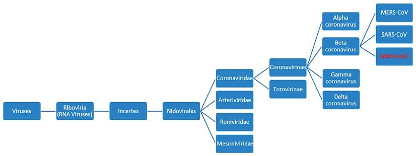
INTERNATIONAL JOURNAL OF MOlecular medicine 46: 467-488, 2020                                            469

                                              Figure 1. Phylogenetic tree of alpha‑beta coronavirus.

                   Figure 2. Structure of RNA translation products of SARS‑CoV‑2 and untranslated regions. Adapted from (17).

mechanisms of transcription error correction through the                   variant, which harbours the noteworthy D614G mutation on
proofreading function of nsp14 (24), a protein common to                   SARS‑CoV‑2‑S (28), has been ascribed to mutation recurrence
MERS‑CoV and SARS‑CoV, which renders them collec-                          and multiple geographical introductions as elucidated through
tively immune to ribavirin (25), but is also reported to                   the analysis of 7,666 published SARS‑CoV‑2 genomes (29).
drive recombinatory promiscuity (24). Consequently, ciral                  However, the impact of these mutations, particularly on protein
quasi‑species are relatively suppressed within patients, and               core to the virus replication cycle such as the cell tropism‑ and
mutation primarily leads to single nucleotide polymorphisms                attachment affinity‑defining SARS‑CoV‑2‑S remains experi-
(SNPs). Nearly 200 such mutations have been reported as of                 mentally undetermined.
mid‑April 2020 (26). Based on the analysis of more than 100                    The current consensus among the scientific community
virus isolates, two main genetic variants of SARS‑CoV‑2 were               remains that the main sites of SARS‑CoV‑2 infection and
initially identified, which are referred to by some members of             replication are both the upper and lower respiratory tracts,
the community as L‑type and S‑type, differing only by 2 SNPs.              despite viral loads being consistenly higher, and more reliable
The L‑type probably evolved from the S‑type and is the more                to detect in lower respiratory tract speciments. However, this
widespread and ‘aggressive’ variant (27). Another study with               position, largely driven by RT‑PCR detection of SARS‑CoV‑2
160 virus isolates from different countries classified the virus           and therefore, by extrapolation replication, does not align with
into A, B and C type, the type A being the oldest virus variant.           the near dearth of ACE2 expression in the upper respiratory
Types A and C are most common in Europe and America,                       tract and oral mucosa (20). The only exceptions to these data
while type B predominates in Asia (26). More recently, contro-             are recent reports of olfactory epithelium ACE2 expression
versy over the transmission propensity of the so‑called L‑type             in mice (30) which is in line with anosmic symptomatology
470 NITULESCU et al: Current Covid-19 therapies
in man, and early evidence of interferon‑induced increase in least appreaciable amounts of virus in the peripheral blood of
ACE2 expression in upper respiratory tract sites, which would patients admitted with COVID‑19. One possible explanation
be secondary to infection and replication in either distal, or of the practically undetectable levels of SARS‑CoV‑2 in blood
physiologically non‑epithelial local cell types (31). Curiously, might be the arrival time of patients to hospitals (week 2), by
endothelial ACE2 expression appears to be ubiquitous even which time seroconversion and neutralizing antibodies are
in the upper respiratory tract (20). These findings postulate being generated and thus viraemia suppressed. Alternatively,
three hypotheses on the infection and replication cascade of endothelial ACE2 expression reduces blood viral load below
SARS‑CoV‑2. The simplest of these assumes direct infection of the limit of detection of RT‑PCR (~15,000 copies/ml whole
upper respiratory tract epithelia from fomites or large droplets blood) by sequestering free viral particles from suspension.
(cough), followed by progressing infection to the lower lung However, such extent of ACE2 expression systemically
by week 2, which would explain deteriorating clinical image across endothelia is not corroborated in public protein
in cases requiring hospitalization. However, this hypothesis expression data repositories (e.g., https://www.proteinatlas.
would rely on transcytotic epithelial mechanisms delivering org/ENSG00000130234‑ACE2/tissue), nor is it physiologi-
SARS‑CoV‑2 to ACE2‑expressing basal upper respiratory/oral cally evidenced by the loss of endothelial barrier function
epithelial cells or endothelia (20). Though transcytotic mecha- as is common in haemorrhagic fever viral infections. Taken
nisms are not implausible (32,33), these cells would in turn together, these findings cast doubts on hypotheses regarding
propagate the virus locally, causing interferon‑induced ACE2 the mechanism of transfer of SARS‑CoV‑2 from the respira-
epithelial overexpression, epithelial cell infection, and luminal tory tract to the intestines (34), which is manifested through
virus shedding, ultimately leading to the observations of faecal detection of SARS‑CoV‑2 RNA (35) and protein in
low‑level detection in nasopharyngeal and oropharyngeal lower GI tract (36). By stark contrast, the main sites of ACE2
swabs by RT‑PCR. However, the immunomodulatory capacity expression, i.e., the lung, GI tract, kidneys, liver and gall-
of SARS‑CoV‑2 manifest through DMVs and nsp16 (13,14,16), bladder, and indeed the testis in men, wherein SARS‑CoV‑2
in addition to the consistently higher viral load in the lower replication has also been documented (37), are all innervated
respiratory tract as opposed to the relatively virus‑free upper by the vagus nerve.
respiratory tract strongly disfavour this hypothesis. The second The median time from symptom onset to acute respiratory
hypothesis would require direct infection of the olfactory distress syndrome, the principal reason for COVID‑19 patients
epithelium again by fomites or droplets, local replication and seeking medical attention, is approximately 8 days; around this
retrograde transport to the lower lung, whereupon the virus timepoint COVID‑19 appears to morph from an acute viral
would establish disease‑causing replication. However, the infection to what might appear to be a second attack, causing
infrequent incidence of anosmia coupled to the opposite direc- the patient's condition to worsen over the second week after
tion of travel for tracheobronchial mucocilliary clearance, symptom onset. Critically, continued deterioration beyond day
and the need for sub‑3 micron aerosol particle dimensions to 14 is associated with high mortality. Thus the disease could be
access the alveoli that would have to be generated in the nose, divided into three major stages after symptom onset: in the first
present a formidable set of physiological barriers to the virus stage symptomatic patients report initially mild‑to‑moderate,
for accessing the lower lung. The last hypothesis would require but quality of life impacting non‑specific symptoms (days 0‑7)
direct infection of the lower respiratory tract through inhala- associated with a progressive drop in viral load; stage II,
tion of SARS‑CoV‑2 containing aerosols, directly infecting wherein severe dyspnoea and associated pneumonia drive
the ACE2‑overexpressing alveoli, sequentially driving upper patients to seek medical attention; and, finally stage III, a severe
respiratory tract infection through mucocilliary clearance respiratory symptomatic stage potentially with high viral load,
and aerosol generation in exhaled breath. Poignantly, this but most commonly characterised by runaway inflammatory
putative infection progression sequence aligns with ACE2 responses associated with multiple organ failure (38,39).
protein expression data, SARS‑CoV‑2 levels reported by Thus, the ensuing innate immune response might be
RT‑PCR across the respiratory tracts of patients, and offers two‑phased; instead of the early innate immunity containing
a mechanistic explanation for the rarity of anosmia and low the virus towards the developement of adaptive immunity,
nasopharyngeal carriage of the virus, as diminishing quanti- in some patients there would appear to be a loss of key
ties of aerosolized virus would need to diffuse into the mucous checkpoint functions driving a secondary, damaging inflam-
found in these tissues upon exhalation. Critically, data docu- matory phase. The second inflammatory phase is driven by
menting viral particle production in upper respiratory tract (or the cytokine release syndrome (the so‑called ‘cytokine storm’)
oral) epithelial or indeed endothelial cells remain elusive from and affects patients with severe conditions. Although genetic
both human and animal studies. differences may underpin innate response amplitude between
individuals, viral amplification appears to ensue (40) causing
3. Pathogenesis direct or, more likely, indirect destruction of tissues affected
by the so-called ‘happy hypoxia’ associated with COVID‑19,
Even though the replication site of the virus is admittedly the the otherwise asymptomatic critical reduction of blood oxygen
lower respiratory system the virus can affect the function of saturation below 95%, sometimes reportedly as low as 50%,
secondary organs that also express ACE2, such as the kidney, i.e., well below the level normally associated with respiratory
GI tract, liver, and gall bladder. Typically, this results in arrest.
symptom severity that drives patients to seek medical atten- The early antibody response against SARS‑CoV‑2
tion, usually by week 2 after symptom onset. However, to date including IgA, IgM, and IgG response was examined by using
no single clincal report has documented measurable, or at an ELISA‑based assay on the recombinant SARS‑CoV‑2INTERNATIONAL JOURNAL OF MOlecular medicine 46: 467-488, 2020 471
protein against the recombinant proteins for the other four three major classifications. The first relies on the repurposing
common cold‑causing human coronaviruses. The median of approved, broad‑spectrum antiviral drugs, through clinical
time to IgM and IgA antibody detection was 5 days with inter- trial and error. The major advantage of this approach consists
quartile range (IQR) 3‑6, while IgG was detected on 14 days of the clear knowledge about the drugs' metabolic profile,
(IQR 10‑18) after symptom onset, with a positive rate of 85.4, dosages needed and supported, potential side effects, and risks.
92.7 and 77.9%, respectively (41). In a study of 23 patients with The disadvantage of these types of drugs is represented by their
COVID‑19, viral load was reported to be declining across the low selectivity and, often, poor utility between virus families.
first week of illness, dropping below detection limits amongst The second strategy is based on high‑throughput screening,
most patients by week 2. With analytical sensitivity lacking in silico, in cellulo or, more recently, by means of artificial
between days 7‑10, IgG and IgM antibodies were detect- intelligence drug discovery, to identify approved, or, at best,
able more reliably at approximately 10 days after symptom late stage drugs with promising therapeutical effects either
onset, with most patients seroconverting within the first through their intended mode of action (e.g., RNA dependent
3 weeks. Importantly, both IgG and IgM were observed to RNA polymerase inhibition), or through their well‑described
neutralize SARS‑CoV‑2‑N and the recaptor binding domain off‑target effects (similar to kinase inhibitor promiscuity). The
of SARS‑CoV‑2‑S (42). third strategy involves identifying the molecular and patho-
Research data on late humoral response and persistence logical characteristics of the disease in question, to develop
of neutralizing antibodies in SARS‑CoV‑2 infection is still entirely novel drugs that may target them. The advantage
limited. A major cause for concern are reports in China, here would be the high selectivity and potentially high effi-
Japan and South Korea of patients testing still positive by cacy (48). Thankfully, the 18 years of head start knowledge
RT‑PCR days, or even weeks after clinical discharge presum- gained against SARS‑CoV‑2 on the back of the SARS‑CoV
ably on the basis of two consecutive negative RT‑PCR results. and MERS‑CoV outbreaks of the early 21st century, have
Some patientsa are even reported to relapse, indicating that a resulted in an unprecedented speed by which the molecular
sustained immune response may be difficult to obtain, or that mechanisms of SARS‑CoV‑2 infection are being elucidated,
at least humoral immunity is inadequate for infection elimina- not least because of the rapid elucidation of the genomic
tion (40). Although the World Health Organization (WHO) has organization of SARS‑CoV‑2. These datasets have informed
recently pronounced these observations as a consequence of several rational, high confidence potential therapeutic targets
RNA shedding from infected apoptotic epithelia in the respi- against SARS‑CoV‑2 (10,49) (Fig. 3).
ratory tract, corroborating data in the literature are lacking. On As the development of new drugs is a long and costly
the other hand, patients who have recovered from SARS‑CoV process, the urgency of the pandemic targeted early efforts
have been reported to harbour potent antibody responses to on identifying candidate treatments among marketed drugs or
SARS‑S protein and its receptor binding domain (RBD) for approved, but later abandoned compounds. Drug repurposing,
at least three years, indicating that RBD‑targeting vaccines also known as drug repositioning, is one of the best solutions to
under development might be adequately protective against identify therapeutic solutions for COVID‑19 in a time‑critical
COVID‑19 (43,44). fashion (50).
Another aspect meriting attention is the progressive
lymphopenia documented during COVID‑19, which is princi- Blocking virus cell entry. Penetration of host cells is an obliga-
pally manifest and most pronounced in severe cases, affecting tory step in the life of any virus rendering it a powerful target
CD4+ and CD8+ T cell levels, as well as B lymphocyte concen- for drug development (51). The discovery of the intrinsic
tration, potentially adversely affecting antibody production. mechanism of SARS‑CoV‑2‑S to mediate cell entry focused
Thus, T cell lymphopenia and associated cytokine concentra- efforts on ACE2 and the whole renin‑angiotensin‑aldosterone
tions have been proposed as biomarkers of poor prognosis, with system (RAAS) (18,51). ACE2 is monocarboxypeptidase that
interleukin (IL)‑6, IL‑10, and D‑dimer continued elevation hydrolyzes the bond between the carboxyl‑terminal phenylala-
marking transition to a severe disease state between days 7‑14 nine and proline in angiotensin II (Ang1‑8) generating Ang1‑7,
after symptom onset, followed by further elevation of D‑dimer, a physiological antagonist of Ang1‑8 with vasodilator and
neutrophilia, blood urea nitrogen, and creatinine marking in the anti‑inflammatory properties (52). Considering these effects,
later, irreversible stages of disease. In contrast, IL‑8 and tumor a soluble recombinant human ACE2 (rhACE2) was devel-
necrosis factor alpha (TNF‑α) elevation appears to be associ- oped under the name APN01 (known also as GSK2586881)
ated and indeed peak early during the recovery phase (45,46). and evaluated in patients with pulmonary arterial hyper-
tension (53) and acute pulmonary injury (54). In healthy
4. Treatment strategies volunteers rhACE2 was administered intravenously in doses
from 100 to 1,200 µg/kg and was generally well‑tolerated,
In the absence of a clinically proven treatment for COVID‑19, with no significant effect on blood pressure and heart rate, and
frontline clinicians are restricted to offering supportive symp- with only mild or moderate adverse events reported (52).
tomatic treatment such as oxygen therapy, steroids on a per SARS‑CoV‑2‑S engages ACE2 as the entry receptor in
case basis, fluid management, and broad‑spectrum antibiotics a similar way as the homologous SARS‑CoV spike protein.
against co‑ or secondary bacterial infections (45). Beyond The expression and distribution of the ACE2 on alveolar
these, as with any viral disease, therapeutic intervention may epithelial cells is consistent with the dry cough and dyspnea
extend to either direct acting antivirals (DAAs) or immuno- experienced by the patients. Consequently, it is anticipated that
modulatory adjuvants and substances (47). Considering the rhACE2 may act as a competitive interceptor of SARS‑CoV‑2
drug discovery process, research efforts can be grouped in thereby reducing cell penetration and offering protection472 NITULESCU et al: Current Covid-19 therapies
Figure 3. Major therapeutical strategies in COVID-19.
against infection (55). A randomized placebo‑controlled study Camostat is an orally active serine protease inhibitor of
(NCT04335136) is scheduled to test rhACE2 in SARS‑CoV‑2 trypsin and various inflammatory proteinases including
patients (56). Furthermore, several peptide derivatives of plasmin, kallikrein, thrombin and urokinase (64). It is
ACE2 were designed using molecular dynamics simulations as approved clinically as mesylate salt in Japan, but not Europe
potential decoys that could be used as future inhaled therapeu- or the USA, for the treatment of chronic pancreatitis and
tics (57). However, drug development history (inhaled insulin, postoperative reflux esophagitis due to its ability to inactivate
Exubera) denotes the brief residence time of soluble proteins trypsin and prevent autodigestion (65). Camostat mesylate has
in the respiratory lumen, necessitating modifications (58) that demonstrated inhibitory activity against TMPRSS2 and other
would sustain rhACE2 decoy potency at the primary infection cell‑surface proteases involved in SARS‑CoV activation (66).
site of interest for SARS‑CoV‑2, the respiratory epithelium. Indeed TMPRSS2 emerged as a promising antiviral drug
Another strategy focused on ACE2 is based on the hypoth- target after it was identified to have a pivotal role for viral
esis that ACE2 activity is reduced by SARS‑CoV‑2‑S, exposing pathogenesis of monobasic H1N1, H3N2 and H7N9 influ-
the organism to higher levels of Ang1‑8 which increases alveolar enza A viruses (67).
permeability and causes lung injury (59). Two compounds, resor- Exposure of human bronchial epithelial Calu‑3 cells to
cinolnaphthalein and 1‑[(2‑dimethylamino)ethylamino]‑4‑(hydro 10 µM camostat mesylate was reported to cause a 10‑fold
xymethyl)‑7‑[(4‑methylphenyl)sulfonyloxy]-9H‑xanthene‑9‑one, decrease in SARS‑CoV viral entry and a 13‑fold decrease
were identified in a large docking study as ACE2 activators and in SARS‑CoV replication (66). Although from a virology
were demonstrated to enhance ACE2 activity in a dose‑depen- perspective such inhibitory effects are considered mediocre,
dent manner, albeit with half‑maximal effective concentration animal experiments in BALB/c mice at 30 mg/k administered
(EC50) values of 19.5±0.4 and 20.1±0.8 µM, respectively (60). orally twice daily for 9 days starting 10 h before SARS‑CoV
Diminazene aceturate is a veterinary‑approved antitrypano- inoculation increased survival rate to 60 vs. 0% for untreated
somal drug that similarly activates ACE2 at comparable in vitro animals (65). Similarly, Calu‑3 cells pre‑incubated with
EC50, rendering protective effects in cardiovascular and type 1 camostat mesylate at very high concentrations (100 µM)
diabetes experimental models (61,62). A recent review study before infection with SARS‑CoV‑2 significantly reduced,
presents the toxic effect of diminazene aceturate on the nervous but failed to abrogate Calu‑3 infection with SARS‑CoV‑2
system of some animals, but provides evidence that it was safe as measured by SARS‑CoV‑2‑specific RT‑PCR (21). Thus,
in thousands of trypanosomiasis patients from Uganda (62). a randomized, placebo‑controlled, phase IIa clinical
These compounds could be a potential therapy in patients with trial (NCT04321096) is evaluating camostat mesylate at
SARS‑CoV‑2, provided topical dosing and pharmacology afford 200 mg x 3/day for 5 days as treatment for SARS‑CoV‑2.
adequate activation of respiratory ACE2. However, it is noteworthy that the study design of this trial
Likewise, use of Ang1‑8 receptor 1 (AT1) antagonists to fails to meet the pre‑treatment or, at least, early treatment
reduce the aggressiveness and mortality of SARS‑CoV‑2 requirement common to most efficacious antivirals and
infection based on two mechanisms, the blockade of the effects indeed the in vitro and in vivo performance requirements
of excess Ang1‑8, and the upregulation of ACE2 (63). Several exhibited previously. Elsewhere, the CLOCC‑2020 clinical
clinical studies are underway to asses various AT1 antagonists: study (NCT04338906) will evaluate the efficacy and safety
losartan 25 mg once daily (NCT04335123, NCT04328012, of camostat mesylate in combination with the controversial
and NCT04311177) or 50 mg daily (NCT04312009), losartan agent hydroxychloroquine in hospitalized patients with
100 mg once daily alone or in combination with aspirin moderate COVID‑19 infection (56).
150 mg once daily or simvastatin 80 mg once daily in an Nafamostat, a chemical analog of camostat, has potent
eight arms factorial trial (NCT04343001), telmisartan 80 mg inhibitory effects on several types of serine proteases
twice daily (NCT04355936), valsartan 80 mg up to 160 mg including trypsin, thrombin and plasmin (68) and was
(NCT04335786) (56). identified to effectively reduce the MERS‑CoV viral entryINTERNATIONAL JOURNAL OF MOlecular medicine 46: 467-488, 2020 473
in Calu‑3 cells at the physiologically relevant concentra- chemically related pyrazole‑pyrolopyrimidine derivatives
tion of 1 nM (69). Nafamostat at 100 µM demonstrated baricitinib and ruxolitinib are clinically approved Janus
promising inhibitory effects against SARS‑CoV‑2 infec- kinase (JAK) inhibitors with important inhibitory effects on
tion of Vero E6 cells (70), but it is anticipated that its very AAK1 (85), likewise predicted to reduce SARS‑CoV‑2 infec-
short half‑life (71) significantly hinders clinical utility in tion (82). A clinical trial (NCT04348071) is programmed to
preventing SARS‑CoV‑2 infection. evaluate ruxolitinib in patients receiving 10 mg twice daily for
Bromhexine, a widely used mucolytic cough medicine, was 14 days and a similar trial (NCT04340232) will test baricitinib
identified as a TMPRSS2 inhibitor in a screening on various administered 2 mg once daily for 14 days (56).
chemical libraries, including over 1,200 FDA approved drugs. The role of cathepsins in coronavirus entry has been estab-
With an IC50 of 0.75 µM, inhibition was reported as selective, lished for SARS‑CoV and MERS‑CoV. In order to investigate
with low effects on the related hepsin and matriptase prote- their role in SARS‑CoV‑2 cell entry a pseudovirus model was
ases (72). However, preliminary data on influenza‑infected tested in ACE2‑expressing 293 cells. While cell treatment with
Calu‑3 cells treated with bromhexine showed no inhibi- the cysteine protease cathepsin B inhibitor CA‑074 (30 µM)
tory effect (73). Despite a lack of in vitro or in vivo data on had no significant effect, cathepsin L inhibitor (SID 26681509,
SARS‑CoV‑2, it has been proposed as a solution for COVID‑19 2 µM) treatment reduced viral entry up to 76%. An inhibitory
patients based on its effect on TMPRSS2 and favorable phar- effect close to 93% was reached after treatment with E64d
macokinetic and safety profile (74). Several clinical trials are (30 µM), an inhibitor of calpain and cathepsin B, H and L (86).
registered to evaluate bromhexine as prophylaxis or therapy Cathepsin L is an attractive target for drug development in
in COVID‑19 patients using various drug combinations several diseases, but despite all the efforts, no cathepsin L
(NCT04355026, NCT04340349, NCT04273763) (56). inhibitor has advanced to clinical trials (87) making future
Aprotinin is a natural 58 amino acid peptide and cathepsin L‑based SARS‑CoV‑2 therapies improbable.
a serine protease inhibitor that blocks fibrinolysis and
reduces bleeding. It demonstrated significant effects against Interfering with the endocytic pathway. Most viruses use
influenza viruses (75). Preliminary research reports have endocytic entry mechanisms and for the majority of them,
demonstrated that in Caco2 cells aprotinin is more potent a reduction of pH serves as a cell penetration trigger (88).
against SARS‑CoV‑2 (EC50 = 22.9 KIU/ml) than against Earlier studies have described that endocytic pH modification
SARS‑CoV (EC50 = 118 KIU/ml) cytopathogenic effects (76) suppresses viral replication and may also have a virucidal
as measured in kallikrein inactivator units (KIU). To this effect. Such a minimal pH value change, with no negative
date, no clinical test has been registered in Clinical Trials impact on the patient's health status, could be achieved by
databases (56). administering a simple small molecule with hydrophobic weak
SARS‑CoV nucleocapsid (protein N) binds to cyclophilin base properties, like ammonium chloride, which would lead to
A, which subsequently interacts with a member of the immu- endocellular alkalosis by accumulating in lysosomes following
noglobulin family of receptors, HAb18G/CD147, on the cell diffusion through plasma and lysosomal membranes. Weak
membrane leading to viral cell invasion (77), in a mechanism bases and ionophores, like ammonium chloride, chloroquine,
similar with that of HIV‑1 virus (78). Based on this model, or monensin are frequently used in experimental studies of
a humanized anti‑CD147 antibody was clinically tested in viral cell penetration mechanisms (89). Ammonium chloride
17 patients with SARS‑CoV‑2 (NCT04275245) and published treatment strongly inhibited the entry of pseudotypes bearing
in a preliminary fashion (79). Whilst the results are interesting, SARS‑2‑S in 293T cells lacking TMPRSS2, but was less effi-
interpretation should be approach with caution as the source cient in Caco‑2 cells (TMPRSS2+) (21).
and characteristics of the antibody, ‘meplazumab’, are missing, Chloroquine, chemically known as 7‑chloro‑4‑((4‑(diethy
and the number of tested patients is very low. lamino)‑1‑methyl butyl)amino)quinoline, is a 9‑aminoquino-
Clathrin‑mediated endocytosis is another mechanism line widely used for the treatment of malaria, amebiasis and
described for SARS‑CoV cell invasion and possibly utilized several auto‑immune diseases (90) recently proposed to be
by SARS‑CoV‑2. Chlorpromazine inhibits the relocation of efficient in inhibiting SARS‑CoV‑2 in infected cells (70,91).
clathrin and the adaptor protein 2 (AP2) from the cell surface, Chloroquine has already demonstrated in the past in vitro
significantly inhibiting SARS‑CoV entry into HepG2 cells at anti‑viral activity against several types of both RNA and
the relatively high concentration of 20 µM (80). Unfortunately, DNA viruses, such as rabies virus, poliovirus, several influ-
it showed no significant effect on the SARS‑CoV‑2 cytopathic enza virus types, hepatitis A, B and C viruses, Dengue virus,
effects in Vero E6 cells when tested at 100 µM (81), yet a Zika virus, and many others (92), as well as SARS‑CoV (93)
clinical trial is scheduled to test the effect of 25 mg chlor- and MERS‑CoV (94). The general anti‑viral mechanism of
promazine administrated intravenously every 6 h for 1 week action relates to the ability of chloroquine, as a weak base, to
(NCT04354805) (56). increase the endosomal pH required for viral/host cell fusion,
An artificial intelligence method identified members of preventing endocytosis, and to interfere with viral particles
the numb‑associated kinases (NAK) family as potential thera- bound to the cell surface membrane (95). Additionally, in
peutic targets against SARS‑CoV‑2 (82). The AP2‑associated the case of SARS‑CoV and SARS‑CoV‑2, chloroquine is
protein kinase 1 (AAK1) is an important member of NAK that proposed to inhibit the glycosylation of ACE2 receptor chains,
binds to clathrin and phosphorylates the medium subunit of thus limiting ligand recognition of these receptors, rendering
AP2, playing an important role in regulating clathrin‑medi- the viral spike protein unable to mediate cell entry (92).
ated endocytosis (83,84) and its inhibition was demonstrated In light of the new pandemic, several clinical trials have
to reduce the infectivity of a wide range of viruses (82). The been conducted in different countries in order to test the safety474 NITULESCU et al: Current Covid-19 therapies
and efficacy of chloroquine phosphate in treating SARS‑CoV‑2 Several inhibitors acting on SARS‑CoV main protease,
patients (96), with results confirming a significant improvement which displays 96% similarity to the SARS‑CoV‑2
when compared to control groups, as in the case of clinical Mpro (106), have recently been synthesized and tested both
trial ChiCTR2000029609 (97). When confronted with the in enzymatic assays and in cell lines, showing promise
recent COVID‑19 crisis in China, chloroquine has even been for developing future drugs. Recently, Zhang et al, after
included in the national treatment guidelines, with Chinese structure‑based drug design and optimization of different
clinicians recommending 500 mg chloroquine twice daily for dipeptide α ‑ketoamides, derived several potential M pro
10 days for the treatment of mild, moderate and severe cases inhibitors, of which (S)‑N‑Benzyl‑3‑((S)‑2‑cinnamamido‑3-
of SARS‑CoV‑2‑related pneumonia with no contraindications cyclohexylpropanamido)-2-oxo‑4‑((S)‑2‑oxopyrrolidin‑3‑yl)
to the drug (98). butanamide (referred to as 11r) displayed physiologically rele-
Hydroxychloroquine is a less‑toxic derivative of chloro- vant (nM) antiviral activity against MERS‑CoV and SARS‑CoV
quine also used as an anti‑malarial and in the management of in hepatic (Huh7) but not routine virology (Vero E6) cell lines,
auto‑immune disease, proposed through in vitro studies to act as well as good inhibition of the SARS‑CoV protease in enzy-
also on SARS‑CoV‑2 infected cells (91). In Vero E6 models matic assays (107). Based on previous research, the same study
hydroxychloroquine presented a better antiviral effect with group obtained a pyridone‑containing α‑ketoamide deriva-
EC50 values of 6.14 µM (after 24 h) and 0.72 µM (after 48 h), tive of 11r, a compound (referred to as 13b) with even lower
compared with chloroquine values of 23.90 µM (24 h) and sub‑micromolar concentrations against SARS‑CoV‑2 Mpro in
5.47 µM (48 h) (91). enzymatic assays, with half‑maximal inhibitory concentra-
Similar to chloroquine in many aspects with the exception tion (IC50) 0.67±0.18 µM, and low‑micromolar EC50 values
of a better pharmacotoxicological profile, hydroxychloroquine (4‑5 µM) in Calu‑3 cells, further obtaining favorable results
is more frequently used instead of chloroquine in COVID‑19 in mouse studies in terms of pharmacokinetic parameters,
clinical trials (99). Several studies confirmed hydroxychloro- reflecting lung tropism after either subcutaneous or topical
quine sulfate administration to be significantly associated with administration (108).
a reduction of the viral load in SARS‑CoV‑2 infected patients, Drug repurposing appears to be another interesting method
as in the case of the Chinese randomized clinical trial reported of identification of Mpro inhibitors. Most notable drugs showing
by Chen et al (ChiCTR2000029559) (100) and Gautret et al, inhibition of the main protease of SARS‑COV‑2 were found to
who also reported after 6 days of treatment apparently signifi- be several anti‑retroviral agents approved for HIV‑1 treatment.
cant benefits compared to negative controls when administering Lopinavir is a selective HIV‑1 protease inhibitor orally
daily 600 mg hydroxychloroquine in association with azithro- administered in combination with ritonavir, a CYP3A4
mycin, where necessary (101). The drug is currently perceived inhibitor used to increase its plasma concentration and clinical
in certain quarters to be more promising than chloroquine as a efficacy (109). Lopinavir was tested against SARS‑CoV and
COVID‑19 treatment due to the reduced probability of causing showed a moderate activity in Vero‑E6 cells (110). In a prelim-
retinopathy, psychiatric disorders, or severe cardiovascular inary, not yet peer‑reviewed, study in Vero E6 cells, lopinavir
adverse effects such as arrhythmia (99); however, self‑medi- showed a mediocre EC50 = 5.73 µM against SARS‑CoV‑2 (111).
cation using both drugs is unadvisable, as 9‑aminoquinolines A recent clinical trial investigating the safety and efficacy of
possess a narrow therapeutic window (102) and are associated lopinavir (400 mg) and ritonavir (100 mg) effect in 99 patients
with QT prologation. Currently, larger studies with a more infected with SARS‑CoV‑2 has already ended, unfortunately
flexible design investigate the effect of hydroxychloroquine, concluding that the administration of these drugs showed no
with the Solidarity study (EudraCT no. 2020‑000982‑18) and significant benefit comparing to current standard care (112).
the Discovery study (NCT04315948) randomizing treatment of The ineffectiveness of this combination could be explained
SARS‑CoV‑2‑infected patients using different antiviral drugs, by the delayed treatment initiation, the median time from
including hydroxychloroquine (103). However, preliminary symptoms identification to therapy onset being 13 days (51).
reports are highlighting a 2.61 adjusted hazard ratio of death Whether the ineffectiveness of the treatment was due to the
among COVID‑19 patients treated with hydroxychloroquine late therapy start or because of low efficient concentrations at
alone in the absence of azithromycin, and no reduction in the the site of action remains to be explored.
risk of mechanical ventilation (104). The clinical use of lopinavir/ritonavir is additionally
linked to nausea and diarrhea (up to 28%) as well as hepato-
Blocking polyprotein posttranslational processing. Another toxic effects (2‑10%). Despite the limited data to support use
promising strategy of treatment appears to be interfering with in the treatment of COVID‑19, there is an important number
viral replication, especially by acting at key points following of clinical studies registered to test the lopinavir (400 mg) and
translation. After synthesis, the two newly‑formed viral poly- ritonavir (100 mg) coformulation alone or in combination with
proteins pp1a and pp1b require for posttranslational processing hydroxychloroquine or interferon (51,56).
two important proteinases encoded in these chains of amino Other HIV‑1 protease inhibitors, such as amprenavir,
acids: papain‑like proteinase, PLpro, and chymotrypsin‑like tipranavir, saquinavir, ritonavir, nelfinavir, indinavir, darunavir,
proteinase, 3CLpro, often referred to as the main protease or and atazanavir that have been predicted to inhibit SARS‑CoV‑2
Mpro (3). The activity of these two cysteine proteases is essen- Mpro (113) were also tested on Vero E6 cells infected to
tial for generating the 16 non‑structural proteins, critical to SARS‑CoV‑2. Among these, nelfinavir was observed to be the
the formation of the replicase complex, and it is known that most efficient, with a mediocre EC50 of 1.13 µM suppressing
inhibiting the activity of either of these two proteases affects viral replication in the post‑entry step (111). In a similar
the replication of SARS‑CoV‑2 (3,105). study on SARS‑CoV, nelfinavir inhibited the viral replicationINTERNATIONAL JOURNAL OF MOlecular medicine 46: 467-488, 2020 475
at non‑toxic doses with a selectivity index (SI) close to 300, active compound, GS‑441524, a 4‑amino‑5‑cyano‑pyrrolotri-
while ritonavir, lopinavir, saquinavir, and indinavir, did not azine analog of adenosine (123). Remdesivir is metabolized
have a significant effect (114). Saquinavir and tipranavir more efficiently than its metabolite GS‑441524 into the active
showed mediocre replication inhibition against SARS‑CoV‑2 nucleoside triphosphate that function as a chain‑terminating
in cell lines (EC50 = 8.83 µM, respectively 13.34), although nucleotide analog blocking RdRp (116). Remdesivir is not
with a lower selectivity index, while amprenavir, darunavir approved by the FDA and not by the European Medicines
and indinavir had low effects on viral replication with EC50 Agency (EMA), but recently received FDA emergency use
values over 30 µM (111). Another preprint publication reports authorization for the treatment of COVID‑19 on the basis of
the SARS‑CoV‑2 viral replication inhibition by atazanavir reduced hospitalisation time (124). However, this outcome
with EC50 values of 2.0±0.12 µM (115). is not without controversy as the trial primary endpoint was
No clinical study is registered in the Clinical Trials altered days before outcome announcement, raising important
database, nor in the Chinese Clinical Trial Registry to test concerns with respect to utility under the purported mecha-
atazanavir, nelfinavir, tipranavir, or saquinavir as COVID‑19 nism of action. Given the structural similarity of remdesivir
therapies. The clinical trials NCT04252274, NTC04303299 to naturally occurring 2'‑O‑methyl nucleoside analogues,
and NCT04366089 will test darunavir in association with antagonists of toll like receptor 7, and the initiation of treat-
other antivirals (56). ment with the pre‑symptomatic/early symptomatic optimal
treatment window, it is postulated remdesivir may actually
Targeting viral RNA‑dependent RNA polymerase. The affect immunosuppression of cytokine storm syndrome, as
RNA‑dependent RNA polymerase (RdRp) is the central opposed to directly acting antiviral effects.
subunit of the RNA synthesizing process for all positive‑strand Remdesivir was developed to treat Ebola virus infection, but
RNA viruses and therefore an attractive target for antiviral demonstrated a broad antiviral spectrum against SARS‑CoV,
inhibitors. In SARS‑CoV‑2 nsp12 functions as RdRp (116) MERS‑CoV, and various other RNA viruses (116). In Vero E6
and plays a central role in the viral replication and transcrip- cells infected with SARS‑CoV‑2, remdesivir potently blocked
tion by catalyzing the synthesis of viral RNA, assisted by virus infection at high nanomolar concentrations with an EC50
nsp7 and nsp8 that serve as co‑factors (117). The polymerase value of 0.77 µM demonstrating a low toxicity as the half‑cyto-
of RNA viruses is prone to replication mistakes, lacking the toxic concentration was reported to be above 100 µM (70).
proofreading capacity of its analog from DNA viruses. This A separate study, however, estimated EC50 at 26.90 µM (81).
high mutation rate (10‑3 to 10‑5 mutations/nucleotide/round of On the other hand, in Caco2 cells remdesivir inhibited
replication) enables RNA viruses to better adapt to environ- SARS‑CoV‑2 cytopathogenic effects with an EC50 value of
mental changes, but also introduces damaging mutations that 0.23 µM (70). The protective effect was increased 10‑fold by
corrupt essential functions, a process called lethal mutagen- the addition of 8 µM of omeprazole. Omeprazole alone has
esis (118,119). also demonstrated anti‑SARS‑CoV‑2 protective effects, but
In CoVs, nsp14 has both 3'‑5' exoribonuclease (ExoN) at physiologically irrelevant concentrations (EC50 = 34 µM),
and guanine‑N7‑methyltransferase (N7‑MTase) functions. substantially above those used in clinical practice. These
ExoN can hydrolyze RNA acting as a proofreading enzyme results are presented in a yet to be peer‑reviewed preprint (76).
that is capable of removing mismatched nucleotides (120). Remdesivir was clinically tested in 175 patients in 2018
Targeting viral RNA‑dependent RNA polymerase has proven during the Ebola outbreak in the Democratic Republic of
a successful strategy in treating various viral infections (121), Congo by intravenous administration of 200 mg on day 1
but is difficult in the case of CoVs because of the ExoN followed by daily maintenance doses of 100 mg from day 2
activity (116,119). Importantly, the catalytic site of RdRp and continuing for 9 to 13 days. In pediatric patients, the
shares structural similarities with the hepatitis C virus (HCV) doses were adjusted based on body weight. The study revealed
ns5b RdRp (117), introducing the prospect of repurposing a favorable safety profile with higher mean levels of creati-
ns5b HCV antivirals as COVID‑19 therapies. The results of nine and aspartate aminotransferase but failed to achieve its
a docking study indicate that tenofovir and sofosbuvir, two primary endpoint (125).
approved HCV antivirals, can bind tightly to SARS‑CoV‑2 A case report of the first COVID‑19 patient identified in
RdRp (122). Still, these results should be view with caution the U.S. presents their treatment with intravenous remdesivir
considering their effect could be theoretically easily reversed on the seventh day of hospitalization and the improvement of
by nsp14 activity. It is thus hypothesized that finding inhibitors their condition afterward (126). A similar case report described
of ExoN activity could be a future direction for COVID‑19 the remdesivir treatment of three hospitalized patients under
therapeutics (119). compassionate use protocol. The patient described as number 6
A number of compounds with proven inhibitory effects was the same as the first case report. All three patients toler-
on other virus RdRps were tested against SARS‑CoV and ated the remdesivir treatment, with transient gastrointestinal
SARS‑CoV‑2, triaging some of them to clinical evaluation. symptoms and a serum aminotransferase elevation (127). The
Ribavirin has the advantage of being clinically approved, but administered doses were not mentioned in the article.
is associated with important side effects and low efficacy. By A randomized, double‑blind, placebo‑controlled clinical
contrast, remdesivir, and to a lesser extent, favipiravir and study (NCT04257656) on confirmed SARS‑CoV‑2 patients
galidesivir, look like the most promising RdRp inhibitors for a found that remdesivir (n=158, vs. 79 placebo) intravenous
future COVID‑19 solution. administration of a 200 mg loading dose on day 1 followed by
Remdesivir, formally known as GS‑5734, is a 2‑ethyl- 100 mg daily as maintenance doses, up to a 10 days, produced
butyl‑L‑alaninate phosphoramidate prodrug that generates the no significant difference in time to clinical improvement.476 NITULESCU et al: Current Covid-19 therapies
The 28‑day mortality was similar between the remdesivir Galidesivir, known also as BCX4430, is an adenosine
group (14%) compared to placebo (13%). However, remdesivir C‑nucleoside analog similar to remdesivir, but has a nitrogen
reduced the time to clinical improvement compared to placebo replacing the oxygen in the ribose ring. It is converted in the
with a median of 18 vs. 23 days in the placebo group. The cell to the corresponding triphosphate nucleotide and after
study reported no significant impact of remdesivir use on the pyrophosphate's cleavage to galidesivir monophosphate
viral load measured on nasopharyngeal and oropharyngeal is incorporated into nascent viral RNA, blocking viral
swabs, implying either no direct acting antiviral effect, or RNA‑dependent RNA polymerase chain extension (135,136).
an unexpectedly long SARS‑CoV‑2 genome half‑life (128). It was reported to be active against a wide range of viruses
This result should be analyzed considering the long average in cell and animal models (137). However, in a HeLa cell
time (10‑11 days) from symptom onset to treatment start. We model of SARS‑CoV infection, galidesivir had an EC50 value
identified a number of other clinical trials that are trying to of 57.7 µM (135). Considering the high similarity of nsp12 in
evaluate the antiviral activity of remdesivir in COVID‑19 SARS‑CoV‑2 and SARS‑CoV (12), it was evaluated in Vero
patients (NCT04252664, NCT04292899, NCT04292730, E6 cells against SARS‑CoV‑2, but the inhibition of viral
NCT04302766, NCT04323761, NCT04280705, NCT04321616, replication was low at 100 µM (81). Nevertheless, a clinical
NCT04315948, NCT04314817, NCT04349410) using the same placebo‑controlled, randomized study (NCT03891420) will
doses as NCT04257656 (56). test galidesivir administered intravenously at 12 h intervals for
Favipiravir (6‑fluoro‑3‑hydroxy‑2‑pyrazinecarboxamide), 7 days in COVID‑19 patients (56).
also known as T‑705, was developed as a structural analog A ribonucleoside derivative, β ‑D‑N4‑hydroxycytidine
of 3‑hydroxy‑2‑pyrazinecarboxamide (T‑1105) compound (known also as NHC or EIDD‑1931), was reported to demon-
identified in an in vitro anti‑influenza virus activity drug strate potent inhibitory effects on MERS‑CoV, SARS‑CoV, and
screening (129). Favipiravir is a prodrug that is activated by SARS‑CoV‑2 in Calu‑3 and human airway epithelial (HAE)
cellular enzyme phosphoribosylation to favipiravir‑ribofura- cells. Encouragingly, NHC inhibited SARS‑CoV‑2 infection
nosyl‑5'‑triphosphate (favipiravir‑RTP) (130). It is reported in Vero E6 cells with EC50 values of 0.3 µM and a value of
to selectively and potently inhibit the RNA‑dependent RNA 0.08 µM in Calu‑3 cells. The isopropyl ester of the prodrug
polymerase of a broad range of RNA viruses: influenza NHC, EIDD‑2801, was evaluated in C57BL/6 mice infected
viruses, arenaviruses, bunyaviruses, noroviruses, flavivi- with a murine adapted SARS‑CoV to reduce significantly the
ruses and filoviruses (131). Favipiravir‑RTP functions as a viral titer and associated infection symptoms when adminis-
guanosine and adenosine analog, its antiviral activity being tered in doses of 500 mg/kg. The antiviral mechanism of action
significantly inhibited by purine nucleotides and nucleosides, proposes the inhibition of RdRp, with the effect of NHC being
but not by pyrimidine nucleotides in an MDCK cell model. only slightly affected by ExoN proofreading activity (138,139).
Yet favipiravir and its ribofuranosyl‑5'‑monophosphate Despite the potent antiviral profile of EIDD‑2801 and its
showed no significant inhibitory effect on influenza virus advantage of oral administration development for COVID, use
RNA polymerase (100 µM). Favipiravir has a similar structure is hindered by the lack of any clinical data.
and also a related antiviral spectrum as ribavirin increasing Ribavirin (1‑ β ‑D‑ribofuranosyl‑1,2,4‑triazole‑3‑carbox
the lethal mutations in the viral genome; this activity raises amide) is a broad‑spectrum antiviral agent clinically used
concerns about teratogen risk potential (131). The phar- primarily for treating hepatitis C. The antiviral mechanism
macokinetic profile was characterized in Japanese healthy is complex, the most important effects being the inhibition of
volunteers to indicate a short half‑life of 2 to 5.5 h and urinary human cells inosine monophosphate dehydrogenase (IMPDH)
excretion in the form of the inactive 5‑hydroxy‑metabolite by the monophosphate derivative of the drug, and the incorpo-
(T‑705M1) (132). Because of its teratogenicity and embryo- ration of ribavirin 5'‑triphosphate in the nascent viral genome
toxicity risks, favipiravir was approved in Japan with strict by RdRp (25). Both ribavirin and favipiravir act as guanine
regulations for use only in epidemics (133). analogs due to their carboxamide group that attaches to cyto-
Similar to remdesivir, favipiravir blocked SARS‑CoV‑2 sine by hydrogen bonds (137).
viral infection in Vero E6 cells, but at almost 80‑fold higher In cell models of SARS‑CoV infection ribavirin had a
concentrations, with a calculated EC50 value of 61.88 µM (70). limited effect on viral replication (110). Similarly high concen-
The results of an open‑label comparative controlled study of trations are needed to block SARS‑CoV‑2 viral infection in
patients with COVID‑19 (ChiCTR2000029600) indicated that Vero E6 cells (EC50 = 109.5 µM) (70) limiting clinical poten-
patients (n=35) receiving favipiravir (1,600 mg x 2, day 1 and tial against COVID‑19. The resistance of SARS‑CoV and
600 mg x 2, days 2‑14) had faster viral clearance and better chest SARS‑CoV‑2 to ribavirin is produced by ExoN's proofreading
imaging changes than those (n=45) treated for 14 days with activity during virus replication (119).
lopinavir (400 mg x 2) and ritonavir (100 mg x 2). Both groups A systematic review of the therapies tested in the SARS‑CoV
also received aerosol inhalation of 5 million U IFN‑α twice outbreak of 2002‑2003 found that among the 20 studies on
daily (134). Several clinical trials are registered to test favipi- ribavirin, results were inconclusive, with no discernible clinical
ravir in COVID‑19 patients (NCT04358549, NCT04346628, differences to other treatments. The review highlights various
NCT04310228, NCT04349241, NCT04336904, NCT04319900, toxic effects presented by several studies, the most frequent
NCT04359615, NCT04333589, NCT04303299, NCT04351295, being hemolytic anemia and liver dysfunction (140). The high
NCT04356495, NCT04345419) administered orally at a doses needed to reduce the viral replication highlight impor-
loading dose of 3,200 up to 3,600 mg (1,600‑1,800 mg at 12 h) tant toxic effects limiting the therapeutic potential of ribavirin
on day‑1, followed by 1,200 to 1,800 mg maintenance dose treatments in COVID‑19 cases. Despite these reservations, an
(600‑800 mg at 12 h) on days 2‑10 (56). open‑label, non‑randomized, clinical trial (NCT04356677) isYou can also read