Idiopathic epilepsy in Finnish Spitz dogs
←
→
Page content transcription
If your browser does not render page correctly, please read the page content below
Department of Equine and Small Animal Medicine
                        Faculty of Veterinary Medicine
                             University of Helsinki
                                    Finland

   Idiopathic epilepsy in Finnish Spitz dogs

        Epidemiological, clinical and diagnostic aspects

                                Ranno Viitmaa

                              Academic dissertation

                                 To be presented,
 with the permission of the Faculty of Veterinary Medicine, University of Helsinki,
for public examination in the Auditorium of Arppeanum, Snellmaninkatu 3, Helsinki,
                        on 15 November 2013, at 12 noon.

                                 HELSINKI 2013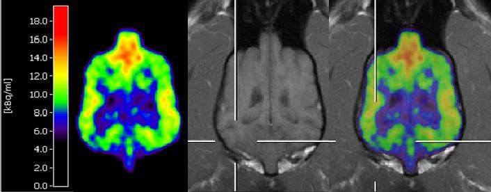
Supervised by:

Professor Emerita Marjatta Snellman, Dipl ECVDI
Department of Equine and Small Animal Medicine
Faculty of Veterinary Medicine
University of Helsinki, Finland

Professor Outi Vapaavuori, Dipl ECVS
Department of Equine and Small Animal Medicine
Faculty of Veterinary Medicine
University of Helsinki, Finland

Reviewed by:

Dr. Kathelijne Peremans, Dipl ECVDI
Department of Medical Imaging and Small Animal Orthopaedics
Faculty of Veterinary Medicine
University of Ghent, Belgium

Dr. Holger A. Volk, Dipl ECVN
Department of Veterinary Clinical Sciences
The Royal Veterinary Collage, UK

Dissertation opponent:

Professor Dr. Andrea Tipold, Dipl ECVN
Department of Small Animal Medicine and Surgery
University of Veterinary Medicine Hannover, Germany

Cover photo by Jouni Keltainen: Suomenpystykorva              Hilla   haukkumassa
metsäkanalintua metsästysretkellä Sodankylässä

ISBN 978-952-10-9274-9 (paperback)
ISBN 978-952-10-9275-6 (PDF, http://ethesis.helsinki.fi/)
Unigrafia
Helsinki 2013
To my parents
CONTENTS
CONTENTS ............................................................................................................ 5
ABSTRACT ............................................................................................................ 7
LIST OF ORIGINAL PUBLICATIONS ................................................................ 9
ABBREVIATIONS ............................................................................................... 10
1.      INTRODUCTION .......................................................................................... 11
2.     REVIEW OF THE LITERATURE ................................................................ 13
     2.1. Epilepsy definitions and classifications .................................................. 13
     2.2. Epidemiology of epilepsy ....................................................................... 14
     2.3. Clinical phenotype of epileptic seizures ................................................. 16
     2.4. Diagnosis of idiopathic epilepsy ............................................................. 18
        2.4.1. Laboratory diagnostics .................................................................... 18
        2.4.2. Magnetic resonance imaging (MRI) ................................................ 19
        2.4.3. Electroencephalography (EEG) ....................................................... 21
        2.4.4. Positron emission tomography (PET) ............................................. 24
AIMS OF THE STUDY ........................................................................................ 28
3.     MATERIALS AND METHODS ................................................................... 29
     3.1. General inclusion criteria ........................................................................ 29
     3.2. Animals and study designs ..................................................................... 29
        3.2.1. Epidemiology, inheritance and phenotype of epilepsy (I) ............... 29
        3.2.2. Structural brain imaging with MRI (II) ........................................... 30
        3.2.3. Cerebral electrical activity measured with EEG (III) ...................... 30
        3.2.4. Imaging cerebral glucose metabolism with FDG-PET (IV) ............ 30
     3.3. Ethical considerations ............................................................................. 30
     3.4. Questionnaire and phone interview (I) ................................................... 31
     3.5. Diagnostic methods ................................................................................. 31
        3.5.1. Laboratory analyses (II-IV) ............................................................. 32
        3.5.2. MRI (II, IV) ..................................................................................... 32
        3.5.3. EEG (II-IV)...................................................................................... 33
        3.5.4. FDG-PET (IV) ................................................................................. 34
     3.6. Statistics .................................................................................................. 35
4.     RESULTS....................................................................................................... 37
     4.1. Epidemiology, inheritance (I) and phenotype (I-IV) of epilepsy ........... 37
     4.2. MRI (II, III, IV) ...................................................................................... 41
     4.3. EEG (III, II, IV) ...................................................................................... 41
     4.4. FDG-PET (IV) ........................................................................................ 45

                                                                 5
5. DISCUSSION ................................................................................................ 47
5.1. Methodological issues ............................................................................. 47
5.1.1. Animals and study design (I-IV) ..................................................... 47
5.1.2. MRI (II) ........................................................................................... 48
5.1.3. EEG (II-IV)...................................................................................... 49
5.1.4. FDG-PET (IV) ................................................................................. 49
5.2. Epidemiology, inheritance and phenotype of epilepsy in FSDs (I) ........ 51
5.3. MRI of epileptic FSDs (II, IV) ............................................................... 55
5.4. EEG of epileptic FSDs (II-IV) ................................................................ 56
5.4.1. Visual evaluation of EEG ................................................................ 56
5.4.2. Quantitative EEG ............................................................................. 58
5.5. FDG-PET of epileptic FSDs (IV) ........................................................... 58
5.5.1. Visual evaluation of FDG-PET ....................................................... 58
5.5.2. Semi-quantitative analysis of FDG-PET ......................................... 59
5.6. Comparisons between MRI, EEG, PET and phenotype (I-IV)............... 60
5.6.1. Comparison of diagnostic modalities .............................................. 60
5.6.2. Comparison of epilepsy phenotype in FSDs and humans ............... 61
6. CONCLUSIONS ............................................................................................ 63
7. ACKNOWLEDGEMENTS ........................................................................... 64
8. REFERENCES ............................................................................................... 67
6ABSTRACT
Epilepsy, a common neurologic disorder in dogs, has also been recognized in the
Finnish Spitz dog (FSD) since the 1980s, but scientifically verified data has been
lacking. In this thesis, epilepsy in FSDs was characterized. Diagnostic investigations,
using tools such as magnetic resonance imaging (MRI) and electroencephalography
(EEG), have not been used consistently in veterinary medicine to diagnose epilepsy in
dogs. The usefulness of these modalities to diagnose different forms of canine epilepsy
needs to be proven. Thus, FSDs with and without focal epilepsy were studied by MRI
and EEG. In addition, the novel functional method to investigate epileptic dogs, 2-
deoxy-2-[18F]fluoro-D-glucose (FDG) positron emission tomography (PET), was
described and results were compared with EEG.
Epidemiological information was based on 2141 FSDs, of which 143 were epileptic,
and prevalence on 2069 living FSDs, of which 111 had epilepsy. The prevalence of
suspected idiopathic epilepsy (IE) in FSDs was found to be 5.3%; males were more
predisposed to epilepsy. The median age at seizure onset was 3 years, seizure frequency
was 3 per year, and duration of seizure episode was 12 min. Focal onset seizures,
characterized by frequent behavioral and autonomic signs were the main phenotype of
epilepsy in FSDs. Although epilepsy in FSDs follows a generally benign course,
generalization of seizure indicate a progressive course of epilepsy. The heritability
estimate of IE in FSDs (0.22) was best explained by polygenic traits.
Although characterized with focal seizures, FSDs have non-lesional epilepsy based on
1.5T MRI examinations. Infrequent reversible brain changes can be found, as a
consequence of seizures.
Visual evaluation of EEG in epileptic FSDs showed interictal epileptiform paroxysmal
activity (20%) less frequently than had been described previously. This activity was
expressed by spikes, polyspikes, and spike slow-wave complexes in the posterior-
occipital derivation. Epileptiform activity, consisting of midline spikes, was recognized
in healthy FSDs. Sleep transients, which were frequently found in FSDs from both
groups, could be easily misinterpreted as epileptiform activity. Quantitative EEG
showed significant difference in various frequency bands related to diseased status or
medication.
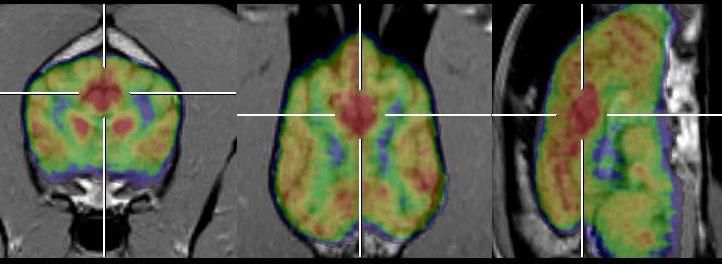
Cerebral glucose utilization was examined by FDG-PET in 11 epileptic and 8 healthy
FSDs. Glucose uptake abnormalities/asymmetries were detected in various brain
regions of 82% of epileptic and in 50% of control FSDs; findings in the occipital cortex
specifically associated with epilepsy. The epileptic dogs had significantly lower
standardized uptake values in numerous cortical regions, cerebellum, and hippocampus
compared to the control dogs. The low cortical glucose uptake values found in the
occipital lobe in both groups of FSDs is an unique finding and may indirectly reflect the
lowered seizure threshold in that region characteristic for this dog breed. Inability to
reveal significant difference of white matter normalized uptake values and left-right
7asymmetry indexes between epileptic and control groups might be related to the method
used to define regions of interest.
Based on these results, epilepsy in FSDs is defined as idiopathic epilepsy, as FSDs lack
changes on the brain MRI and epilepsy is genetically determined. EEG and FDG-PET
suggest involvement of the occipital region, although also wider posterior cortical areas
could be related to epileptogenesis in FSDs.
Visual evaluation of both EEG and FDG-PET can support the diagnosis of IE in dogs.
Although diagnostic yield of EEG to diagnose epilepsy seems to be lower than
suggested for dogs, it is a method of choice for everyday clinical settings. FDG-PET is a
useful research modality for examining epileptic dogs, where visual detection of
hypometabolic areas provides the highest sensitivity. Quantitative assessment methods
of EEG and FDG-PET can be beneficial, but should be used alongside visual evaluation
in epileptic dogs.
8LIST OF ORIGINAL PUBLICATIONS
This thesis is based on the original articles (I-III) and to the unpublished manuscript
(IV). These articles are referred to in the text by their Roman numerals:
I. Viitmaa R, Cizinauskas S, Orro T, Niilo-Rämä M, Gordin E, Lohi H, Seppälä
EH, Bragge H, Snellman M. Phenotype, inheritance characteristics, and risk
factors for idiopathic epilepsy in Finnish Spitz dogs. J Am Vet Med Assoc
2013;243:1001-1009.
II. Viitmaa R, Cizinauskas S, Bergamasco L-A, Kuusela E, Pascoe P, Teppo A-M,
Jokinen TS, Kivisaare L, Snellman M. Magnetic resonance imaging findings in
Finnish Spitz dogs with focal epilepsy. J Vet Intern Med 2006;20:305-310.
III. Jeserevics J, Viitmaa R, Cizinauskas S, Sainio K, Jokinen TS, Snellman M,
Bellino C, Bergamaso L. Electroencephalography findings in healthy and
Finnish Spitz dogs with epilepsy: visual and background quantitative analysis. J
Vet Intern Med 2007;21:1299-1306.
IV. Viitmaa R, Haaparanta-Solin M, Snellman M, Cizinauskas S, Orro T, Kuusela
E, Johansson J, Pääkkönen T, Bergamasco L.A, Metsahonkala L. Cerebral
glucose utilization measured with high resolution positron tomography in
normal and epileptic Finnish Spitz dogs. Submitted to Vet Radiol Ultrasound
The original publications are published in the printed version of this thesis with the kind
permission of the copyright holders.
9ABBREVIATIONS
AI Left-right asymmetry index
ANOVA Ordinary analysis of variance
CBC Complete blood count
CF Complex focal
CFS Complex focal seizures
CI Confidence Interval
CSF Cerebrospinal fluid
CT Computed tomography
EEG Electroencephalography
FDG 2-deoxy-2-[18F]fluoro-D-glucose
FLAIR Fluid-attenuated inversion recovery
FSBC Finnish Spitz Breeder Club
FSD Finnish Spitz dog
HRRT High resolution research tomography
IE Idiopathic epilepsy
ILAE International League Against Epilepsy
im Intra muscular
iv Intra venous
MPR Multiplanar reconstruction
MRI Magnetic resonance imaging
OR Odds ratio
PET Positron emission tomography
ROI Region of interest
RR Reference range
SD Standard deviation
SE Standard error
SF Simple focal
SFS Simple focal seizures
SPECT Single-photon emission computed tomography
SUV Standardized uptake value
T1W T1-weighted
T2W T2-weighted
VOI Volume of interest
10Introduction
1. INTRODUCTION
Idiopathic epilepsy (IE) is a common canine neurological problem with an estimated
prevalence of 0.6% in the general dog population (Kearsley-Fleet et al. 2013). There is
remarkable variability in published prevalence figures for different dog breeds (1 to
33%) and study populations (Falco et al. 1974, Famula & Oberbauer 1998, Knowles
1998, Berendt et al. 2002, Casal et al. 2006, Berendt et al. 2009, Gulløv et al. 2011).
Breeding studies and pedigree analyses have supported a genetic basis for IE in a wide
variety of dog breeds (Srenk et al. 1994, Famula et al. 1997, Jaggy et al. 1998,
Kathmann et al. 1999, Patterson et al. 2005, Licht et al. 2007). It is most likely,
however, that a number of genetic mutations can determine the IE in different dog
breeds, but possibly also for distinct bloodlines within the same breed (Licht et al.
2002). Increasing numbers of genetic studies of canine epilepsy have reported first loci
and candidate genes for IE in dogs (Ekenstedt et al. 2011, Seppälä et al. 2011, 2012).
Therefore a break-through in our understanding of the pathophysiological mechanisms
underlying canine epilepsies may occur in the near future.
Diagnosis of IE in dogs is generally made per exclusionem and based on a history of
more than two seizures in the absence of other medical problems, normal physical and
neurological examination, as well as clinical evaluations (Schwarz-Porsche 1984, Jaggy
& Bernardini 1998, Licht et al. 2002). The Neuroimaging Commission of the
International League Against Epilepsy (ILAE) suggested that magnetic resonance
imaging (MRI) examination should be performed on every human patient with epilepsy
(Kuzniecky et al. 2002). The situation in veterinary epileptology differs however, as a
limited number of studies documenting MRI findings in dogs with seizures have been
available (Kärkkäinen et al. 1991, Kärkkäinen 1995, Mellema et al. 1999, Mariani et al.
2001, Bush et al. 2002). Nonetheless, there is a growing collection of publications
exploring that field. In canine IE, similar to humans, structural brain imaging is
considered to be normal. (Chandler 2006, Thomas 2010).
Modern electroencephalography (EEG) has unfortunately not become a routine
diagnostic procedure when diagnosing epilepsy in dogs (Pakozdy et al. 2012). It is
surprising, as interictal EEG changes can be the only positive finding in dogs with IE. In
addition to confirmation of epileptic cerebral activity, EEG can supply localization of
the epileptogenic foci and play an important role in seizure classification (Commission
on Classification and Terminology of the ILAE 1981, 1989, Flink et al. 2002).
Diagnostic yield of EEG when examining dogs with epilepsy has been reported to be
highly variable however (0% till 86%) (Jaggy & Bernardini 1998, Pakozdy et al. 2012).
Thus, multiple methodological questions need to be answered regarding the use of EEG
to diagnose epilepsy in dogs.
Positron emission tomography (PET) using different radioligands have been used in
epileptic human patients to confirm the epileptic region during presurgical evaluation.
The most widely used tracer used with PET is 2-deoxy-2-[18F]fluoro-D-glucose (FDG)
which reflects cerebral glucose utilization (Juhasz et al. 2005). Glucose metabolism in
11Introduction
focal epilepsy is usually reduced interictally in the region of seizure onset, and increased
during the ictal event. The usefulness of FDG-PET when examining epileptic veterinary
patients, however, remains unknown.
The Finnish Spitz dog (FSD) is a relatively rare breed used traditionally in the Northern
parts of Scandinavia as a “barking hunting dog” for game birds and also as a guard dog.
Since the 1980s there was information available about the incidence of epilepsy in
FSDs, but scientifically proved information was missing. This thesis characterizes
epilepsy in FSDs and explores the usefulness of diagnostic modalities (MRI, EEG, and
FDG-PET) and methods of analysis to investigate epileptic dogs.
12Review of the literature
2. REVIEW OF THE LITERATURE
2.1. Epilepsy definitions and classifications
Epileptic seizure is defined in human medicine as a transient occurrence of signs and/or
symptoms due to abnormal excessive or synchronous neuronal activity in the brain
(Fisher et al. 2005). From the pathophysiological point of view, it is important to
appreciate that normal brain can also have an acute seizure as a natural response to
transient insult or loss of homeostasis (provoked seizure) (Engel 2006). Epilepsy is
defined for practical purposes as two or more unprovoked seizures occurring at least
24 h apart (Thurman et al. 2011). The conceptual definition of epilepsy is rather
complex and also includes the neurobiologic, cognitive, psychological, and social
consequences of this condition, and is characterized by enduring predisposition of the
brain to generate epileptic seizures (requires occurrence of at least 1 seizure) (Fisher et
al. 2005). Some parts of the previous definition might be difficult to apply directly to
animals, however, the definitions and terminology used in veterinary medicine follow
the ILAE 1981–1989 recommendations for the most part (Commission of Classification
and Terminology of the ILAE 1981, 1989), and are more recently modified after the
2001–2006 update (Engel 2001, 2006). Epilepsies are classified according to etiology as
idiopathic (primary), symptomatic (secondary), and probably symptomatic
(cryptogenic) epilepsy (Berendt & Gram 1999). Some authors exclude cryptogenic from
the classification and include reactive epilepsy (Bush et al. 2002, Lorenz et al. 2011a).
Idiopathic epilepsy is defined as recurrent, unprovoked seizures for which no
underlying brain abnormalities can be identified and a familial or genetic predisposition
may be suspected (Knowles 1998). Symptomatic epilepsy refers to a seizure disorder
where seizures are a consequence of a structural brain disorder (March 1998). Probably
symptomatic epilepsy is suspected to be symptomatic, but where etiology is not
determined (Berendt & Gram, 1999). Extracranial metabolic or toxic insults are causing
reactive epilepsy and therefore not considered as true epilepsy (Chandler 2006).
One recent study from the veterinary field has applied the term “genetic epilepsy”
(Gulløv et al. 2012), adapted from the latest ILAE proposals (Berg et al. 2010). A
modified concept of genetic, structural/metabolic, and unknown cause has been
suggested to replace idiopathic, symptomatic, and cryptogenic. Accordingly, genetic
epilepsy was defined as a direct result of a known or presumed genetic defect(s) in
which seizures are core symptom of the disorder; structural/metabolic as a distinct other
structural or metabolic condition or disease that has been demonstrated to be associated
with a substantially increased risk of developing epilepsy and unknown cause with
neutral meaning that the nature of the underlying cause is as yet unknown. (Berg et al.
2010)
Seizure type classification is based on the clinical signs of seizure onset.
Symptomatology of a focal (partial) seizure is consistent with initial activation of only a
part of one cerebral hemisphere (Blume et al. 2001) whereas involvement of both
cerebral hemispheres from the beginning of seizure onset indicates generalized seizure
13Review of the literature
(Blume et al. 2001). Although a descriptive approach of consciousness and seizures is
recommended by the newest ILAE guidelines for humans (Berg et al. 2010), dividing
focal seizures based on the consciousness level as simple focal seizures (SFS) and
complex focal seizures (CFS) (CFS impaired and SFS preserved consciousness) is still
common in veterinary literature. Seizure propagation can occur when SFS progresses to
a CFS or focal onset seizure to generalize (Berendt & Gram 1999).
Epileptic seizures can be self limiting or continuous. When the duration of a seizure
episode is longer than 30 min or a cluster of epileptic seizures lasts the same time
without regaining a normal baseline of function between, it is defined as status
epilepticus (Bateman et al. 1999, Walker 2005). Also a 5 min (Saito et al. 2001, Brophy
et al. 2012) and a 10 min (Monteiro et al. 2012) duration have been used to define SE in
the latest literature. The occurrence of more than one seizure episode during 24 h is
named a cluster seizure (Monteiro et al. 2012).
A seizure episode itself is named ictus and the period between the seizure episodes as an
interictal period. An event shortly prior to the ictus characterized by sensory signs is
referred as an aura (Berendt & Gram 1999). Behavioral signs without impairment of
consciousness and without motor signs which precede hours or days (more than 1h)
before the seizure are defined as prodrome. A period of behavioral changes, blindness
etc., following the seizure episode (lasting minutes to hours) is called the post-ictal
period. Prodrome and post-ictal signs are not counted as part of ictus, but aura (Berendt
& Gram 1999) or short lasting behavioral, autonomic signs preceding ictus (< 1 h)
(Licht et al. 2002) are considered to be part of the focal seizure onset.
2.2. Epidemiology of epilepsy
A broader spectrum of clinical epidemiological studies of epilepsy deals with classical
concepts of incidence, prevalence, basic risk factors, and etiology in addition to a wider
scope of socioeconomic questions, co-morbidities, and factors affecting outcome
(Thurman et al. 2011).
Epilepsy is the most common chronic neurological disorder in humans; approximately
65 million people suffer from the disease worldwide. The annual incidence of epilepsy
is 50 per 100,000 population in industrial countries (100-190/100,000/year in resource-
poor countries) and the prevalence of active epilepsy generally 5-10 per 1000 (Sander
2003, Thurman et al. 2011). Lifetime prevalence rates in humans are much higher, as up
to 5% of the population may experience seizures at some point in life (Sander 2003).
Naturally occurring epilepsy is reported in many animal species including rodents, dogs,
cats, horses, cattle, goats, and non-human primates (Chandler 2006). Seizures are also
the most frequent neurological problem in dogs, with an estimated prevalence from 0.5
to 5.7% (Licht et al. 2002), comprising 2-3% of canine patients treated at veterinary
teaching hospitals (Podell et al. 1995) and involving 10% of neurological problems
(Jaggy & Bernardini 1998). IE has been diagnosed in 25% (Berendt & Gram 1999) to
80% of dogs with seizures (Schwarz-Porshe 1994). A number of dog breeds have been
14Review of the literature
described to have an increased risk of IE with the highest reported prevalence of 33% in
the Belgian Shepherd family (Berendt et al. 2009).
The genetic mechanisms of IE are likely to vary not only between breeds, but possibly
even within the same breed (Licht et al. 2002). Recently described first loci and
candidate genes for IE in dogs (Ekenstedt et al. 2011, Seppälä et al. 2012) represent the
first steps to defining the epilepsy genes. LGI2 truncation has been demonstrated to
cause benign focal onset juvenile epilepsy in Lagotto Romagnolos (Seppälä et al. 2011).
In clinical medicine, approximately 70% of all epilepsy patients lack an obvious
extraneous cause and are presumed to have a predominantly genetic basis and 40% are
thought to have a complex genetic basis with an unknown number of susceptibility
genes (Heron et al. 2007, Dibbens et al. 2007). Some dominant and autosomal recessive
epilepsy genes are already known (Heron et al. 2007). Until now, the suggested modes
of inheritance for IE in dogs include: autosomal recessive in Keeshonds (Hall &
Wallace 1996), Vizsla (Patterson et al. 2003), Standard Poodles (Licht et al. 2007),
Belgian Shepherds (Berendt et al. 2009), and Border Collies (Hülsmeyer et al. 2010);
partially penetrant autosomal recessive in Irish Wolfhounds (Casal et al. 2006); and
polygenic recessive in Golden Retrievers (Srenk & Jaggy 1996), Labrador Retrievers
(Jaggy et al. 1998), and Bernese Mountain dogs (Kathmann et al. 1999). In Beagles,
autosomal recessive and sex-linked suppressors have been reported (Bielfelt et al.
1971). The male predisposition for IE has been reported for Golden retrievers (Srenk et
al. 1994), Bernese Mountain dogs (Kathmann et al. 1999), and Irish Wolfhounds (Casal
et al. 2006), when female prevalence was found in Belgian Shepherds (Berendt et al.
2008). Overall 1.5 times higher predisposition to epilepsy in male dogs has been found
in one study from the UK which included 539 dogs involving multiple dog breeds
encompassing patients collected from veterinary practices (Kearsley-Fleet et al. 2013).
Epidemiological studies of human epilepsy indicate a slightly higher incidence of
epilepsy in males overall (Kotsopoulos et al. 2002), but gender susceptibility varying
for specific epilepsy subtypes (Christensen et al. 2005).
Many predisposing factors for epileptic seizures have been investigated in dogs,
including feeding habits, time of day or year, the weather, lunar cycle, and sex cycle,
but no significant associations have been made (Berendt et al. 2002, Hülsmeyer et al.
2010, Browand-Stainback et al. 2011). Some authors have found that seizures usually
occur at the time of rest or sleep (Langweiler & Jaggy 1998, Morita et al. 2002, Weissl
et al. 2012). On the other hand, various nonspecific stress situations have also been
reported to trigger seizures in a high proportion of dogs (Heynold et al. 1997, Berendt et
al. 2008, Hülsmeyer et al. 2010). High stress levels and significant life events acting as
precipitating factors for seizure occurrence have also been documented in both humans
(Temkin & Davis 1984, Koutsogiannopoulos et al. 2009) and some animal epilepsy
models (Joels 2009).
Numerous predictors of seizure outcome have been suggested in human epilepsy, such
as sex, seizures in relatives, prior neonatal seizures, prior febrile convulsions, age at
15Review of the literature
seizure onset, abnormal neurological status, seizure frequency at onset of seizures,
seizure etiology, type and number of seizure types, type of epilepsy syndrome, time
prior to the onset of drug therapy, number of seizures prior to the onset of drug therapy,
age at the onset of drug therapy, the early effect of drug therapy, and the number
seizures during early drug therapy (Sillanpää 2000). In contrast, only a few factors have
been found to be predictors of epilepsy outcome in dogs, such as early initiation of
treatment, an advanced age at seizure onset, and a high body weight (Heynold et al.
1997, Saito et al. 2001, Berendt et al. 2007, Hülsmeyer et al. 2010). A significantly
shorter life span has been reported for dogs in which euthanasia or death was directly
caused by epilepsy, and epileptic females live longer than males (Berendt et al. 2007).
Some breeds have a shorter life span with IE, including Australian Shepherd dogs
(Weissl et al. 2012), Border Collies (Monteiro et al. 2012, Hülsmeyer et al. 2010), and
Irish Wolfhounds (Casal et al. 2006). Other breeds, like German Shepherds and Boxers,
have been found to have a high occurrence of cluster episodes as a criterion for poor
prognosis (Monteiro et al. 2012). On the contrary, no significantly shortened life span
has been found in the family of Belgian Shepherds with genetic epilepsy, despite a
majority of deaths related to epilepsy (Gulløv et al. 2012). Expected life span in dogs
with IE has also been reported in another study, but status epilepticus was found as a
factor shortening survival time (Saito et al. 2001). In dogs with juvenile IE, survival
time was shortened with a history of status epilepticus. Diagnosis of symptomatic
epilepsy and number of antiepileptic drugs used before investigation also shortened
survival in dogs with juvenile epilepsy (Arrol et al. 2012). One study found a negative
influence of neutering on the occurrence of cluster episodes (Monteiro et al. 2012),
whilst others found that neutering may influence seizure frequency and seizure severity
in female Belgian Shepherds (Berendt et al. 2008). Genetic studies have demonstrated
polymorphism in the ABCB1 gene to be associated with Phenobarbital treatment and
seizure outcome in border collies (Alves et al. 2011) and collies (Muñana et al. 2012),
but not in Australian Shepherd dogs (Weissl et al. 2012).
Epilepsy remission rates have been reported to vary from 13.7% in Belgian Shepherds
(Gulløv et al. 2012) to 24% in Danish Labrador retrievers (Berendt et al. 2002), but
nearly all Lagotto Romagnolos with benign juvenile epilepsy have had remission
(Jokinen et al. 2007). However, as the remission has not been determined in a constant
way (seizure freedom minimally 1 to 3 years), direct comparison between the studies
and breeds may be complicated (Berendt et al. 2002, Berendt et al. 2007, Hülsmeyer et
al. 2010). In humans over 70% of epileptic patients will be suspected to enter remission
(Sander 2003).
2.3. Clinical phenotype of epileptic seizures
An epileptic seizure is a clinical event and can affect motor, sensory, and autonomic
function, consciousness, emotional state, memory, cognition, or behavior, depending on
location of onset in the brain (Fisher et al. 2005). Some signs from this wide range of
epileptic phenomena recognized in humans are complicated or nearly impossible to
16Review of the literature
assess in animals. Therefore a simplified approach to classify seizures in animals should
be more appropriate.
Canine ictus may be evident from motor, behavioral or autonomic signs as a single
nominator or in different combinations. Motor signs are isolated to a body part or are
unilateral when focal and presented as tremors, tonic or clonic movements, difficulties
to walk or stand, or abnormal body deviations. They have been recognized from 45%
(Hülsmeyer et al. 2010) to 100% (Licht et al. 2002) of dogs. Psychic or behavioral signs
have been occurring from 10% (Licht et al. 2002) to 92% (Gulløv et al. 2011) of dogs
and are defined as paroxysmal signs of anxiety, restlessness, unprovoked aggression,
and/or out of context behavior. Compulsive tail chasing, fears, hyperactivity, and
phobias in Bull terriers might be related to focal seizure activity (Dodman et al. 1996).
Epigastric sensations, papillary dilatation, lacrimal secretion, and urinary or fecal
incontinence are described autonomic signs and occur in between 23% (Berendt et al.
2004) and 67% (Licht et al. 2002) of dogs. Automatism has been defined in clinical
medicine as more or less coordinated, repetitive, motor activity usually occurring with
impaired cognition (Blume et al. 2001). Seizure related behaviors such as chewing or
swallowing movements, lip smacking, body scratching, as well as changing position or
circling when performed repeatedly have been interpreted in veterinary literature as
automatisms and therefore indicative to impaired consciousness (Licht et al. 2002). For
assessment of consciousness in dogs, responsiveness when owner tries to get their
attention and ability to keep attention, in addition to whether the animal is able to
navigate in any way (to walk, run or jump) have been applied (Licht et al. 2002).
Focal seizures, with or without secondary generalization, have been reported to be the
predominant seizure type of IE for multiple dog breeds, including Dalmatians and
Standard Poodles (Licht et al. 2002), Labradors retrievers (Berendt et al. 2002), Vizslas
(Patterson et al. 2003), Belgian Shepherds (Berendt et al. 2008), Border Collies
(Hülsmeyer et al. 2010), and Petit Basset Griffon Vendeen (Gulløv et al. 2011). Focal
seizures with generalization, however, are the most frequent seizure type. A high
incidence of generalized seizures (75-90%) has been previously reported in dogs with
IE (Schwarz-Porshe 1994, Jaggy & Bernardini 1998). Since more recent veterinary
studies have started to pay attention to the initial clinical signs preceding the generalized
phase of seizure, seizures have been classified as generalized less frequently in dogs
(Licht et al. 2002). Generalized seizures have been reported to be the predominant
seizure type in Irish Wolfhounds (Casal et al. 2006), and also involving almost half of
the seizures in English Springer spaniels with IE (Patterson et al. 2005).
During generalized seizure, animals may fall to the side and lose consciousness at some
point. Motor signs are bilateral and largely symmetrical, most frequently tonic-clonic
movements are observed during generalized seizure phase, but tonic, clonic, myoclonic,
or atonic signs are also described in dogs. Canine absence seizures are complicated to
define, although described when accompanied with myoclonic features (Poma et al.
2010). Automatisms as coordinated paddling of four limbs can be seen. In generalized
17Review of the literature
seizures, autonomic signs can be observed nearly as frequently as motor signs (Licht et
al. 2002). The post-ictal phase in dogs is characterized by restlessness, thirst, hunger,
aggression, or brief post-ictal blindness (Hülsmeyer et al. 2010). The duration of the
post-ictal phase has been shown to increase with disease progression (Weissl et al.
2012).
A number of characteristics have been shown to be significantly different when
comparing seizures in dogs with IE and symptomatic epilepsy, including: age, body
weight, presence of partial seizures, cluster seizures, status epilepticus, ictal vocalization
and neurological deficits (Pakozdy et al. 2008). IE has been reported to be more
probable in dogs that are between 1 and 5 years at the time of seizure onset, if the
seizure occurred between 8 am and midnight, or in dogs over 15 kg or if the interval
between the first and the second seizure exceeded 4 weeks (Podell et al. 1995). Median
seizure onset has been reported to be between 2 and 3 years for numerous dog breeds,
but the seizure onset ranged from 2 months to 10 years (Jaggy & Bernardini 1998,
Kathmann et al. 1999, Patterson et al. 2003, Patterson et al. 2005, Casal et al. 2006,
Hülsmeyer et al. 2010). Dogs with juvenile forms of IE might start to have seizures as
early as 5 weeks of age (Jokinen et al. 2007, Arrol et al. 2012).
2.4. Diagnosis of idiopathic epilepsy
Diagnosis of epilepsy is primarily based on the history of recurrent seizures, but general
physical and detailed neurological examinations are basic assessments that should be
performed on every animal with the history of seizures in order to detect changes
indicative of symptomatic epilepsy or of general disease conditions (Lorenz et al.
2011a). Dogs diagnosed with IE are supposed to have no neurological deficits when
examined interictally. Some neurological deficits (i.e. disorientation, blindness) may
persist when the animal is examined during the post-ictal period even longer than 24 h
(Hülsmeyer et al. 2012).
2.4.1. Laboratory diagnostics
Laboratory diagnostics, including complete blood count, serum chemistry profile, and
urine analysis, are often considered to be the minimal required examinations for the
diagnosis of IE in order to exclude extracranial causes for seizures (Thomas 2010). A
serum chemistry profile includes glucose, total protein, albumin, globulin, blood urea
nitrogen, creatinine, total bilirubin, alanine aminotransferase, aspartate
aminotransferase, alkaline phosphatase, creatine kinase, cholesterol, and electrolytes
(Na, K, Ca, P, Mg). Paired serum bile acids, collected after 12 h fasting and
postprandial, have been used in young animals to rule out a portosystemic shunt
(Webster 2005). Specific gravity, chemistry, and sediment examinations should be
performed from urine samples. An increasing number of diseases that cause seizures in
dogs can also be confirmed from various tissue samples using genetic tests (Lohi et al.
2005, Awano et al. 2006).
18Review of the literature
Cerebrospinal fluid (CSF) is collected by a cerebellomedullary cisternal puncture under
general anesthesia and used to rule out intracranial inflammatory, infectious, or
neoplastic diseases. Total cell counts can be measured using a hemocytometer or mirror
counting chamber. Differential white blood cell counts and cytological examinations
can be performed on samples prepared with a cytocentrifuge or after sedimentation
methods to concentrate cells (Lorenz et al. 2011b). For measurements of protein
concentration in the CSF, qualitative methods such as the Pandy test, or quantitative
analysis can be performed. Increased protein level in CSF is a non-specific indicator of
central nervous system pathology. In the dogs with IE, CSF should remain within
reference range regarding total nucleated cell count (< 5 cells/ul) and protein level (<
250 mg/l). A mild increase in total cell count of the CSF early after ictus may occur
(Goncalves et al. 2010). Electrophoresis is used to quantify the levels of protein
fractions or immunoglobulins (Lorentz et al. 2011b) and measurements of C-reactive
proteins in CSF in order to exclude inflammatory processes within central nervous
tissue (Martinez-Subiela et al. 2011). Levels of neurotransmitters, such as γ-
aminobutyric acid, glutamate, and aspartate, may be lower in dogs with IE, but not
equally in all dog breeds (Ellenberger et al. 2004). The ability to identify a definitive
diagnosis from the CSF can be improved with microbiologic testing, measurement of
antibody titers, polymerase chain reaction (PCR) testing, and immunocytochemistry
(Lorenz et al. 2011b). CSF analysis has been demonstrated to be of value as it is an
indicator of central nervous system pathology; MRI shows changes in only 6% of dogs
with seizures and with normal interictal neurological examination and CSF results while
MRI abnormalities were present in 43% of dogs with abnormal CSF and normal
neurologic examination results and in 97% of dogs with abnormalities detected in both,
in CSF and neurologic examination (Bush et al. 2002).
2.4.2. Magnetic resonance imaging (MRI)
The physical nuclear magnetic resonance phenomenon was discovered 1946, but the
first applications of medical imaging appeared much later – in the 1980s (Jackson et al.
2005a). MRI is based on the visualization of the hydrogen atoms nucleus using strong
magnetic field and radiofrequency pulses (Jackson et al. 2005b, Gavin 2009). For the
routine imaging of the neurocranium, T1-weighted (T1W) and T2-weighted (T2W)
images in 3 standard planes (i.e. sagittal, transverse, dorsal) are used. Multiplanar
reconstruction (MPR) which is 3D of T1W sequence could be used instead of T1W
images and reconstructed later in all standard planes. Inversion recovery sequences as
fluid-attenuated inversion recovery (FLAIR) can be used to improve delineation of
primary lesions from the oedematous changes and distinction between the gray and
white matter (Blümcke 2011, Bagley et al. 2009). Diffusion-weighted imaging could be
applied when brain infarction is suspected (Major et al. 2012). Repeating T1W images
or MPR after injection of paramagnetic contrast agent (i.e. gadolinium - dimeglumine
gadopentate) is indicated when inflammatory or neoplastic changes, altering the blood-
brain-barrier of the cerebral vasculature are suspected (Bagley et al. 2009). Contrast
agents generally cause an increase in T1W signal which is recognized as increased
hyperintensity in T1W images (Gavin 2009).
19Review of the literature
MRI is considered the most valuable diagnostic tool for investigating the etiology of
epilepsy in vivo because the detection of lesions is an important factor in planning
epilepsy management and in predicting the prognosis of individual human patients.
Over 80% of humans with epilepsy have a focal epilepsy, and only 43% have a cerebral
lesion (Lee et al. 2002). MRI identifies lesions more frequently in patients with
temporal lobe epilepsy (76%) than with extratemporal epilepsy (47%) (Casse et al.
2002). Hippocampal sclerosis, the main cause of temporal lobe epilepsy in humans, is
detected by MRI in approximately 55% of the patients (Lehericy et al. 1997). The
detection of lesions has improved since the application of higher strength magnetic
fields and computer-based postprocessing analysis (Kuzniecky et al. 2002, Knake et al.
2005). In addition to visual evaluation of the images, different quantitative
measurements can be applied. Hippocampal MRI volumetry is an established technique
and performed in epileptic humans with high confidence (Jackson & van Paesschen
2002, Jeukens et al. 2009).
The majority of veterinary MRI studies are performed using low magnetic field
equipment (Konar & Lang 2011); although, some recent publications indicate that
animals also might benefit from the use of 3 or 7 T MRI machines (Kang et al. 2009,
Martin-Vaquero et al. 2011). MRI examinations are used increasingly for epileptic
canine patients, but are more frequently performed to examine patients with suspected
symptomatic epilepsy. MRI findings from the dogs having seizures as a result of brain
tumors (Thomas et al. 1996, Kraft et al. 1997, Bush et al. 2002, Cherubini et al. 2005,
Schwartz et al. 2011), inflammation (Sawashima et al. 1996, Kuwabara et al. 1998,
Mariani et al. 2001), vascular problems (Kitagawa et al. 2008, Timm et al. 2008),
malformations (Kitagawa et al. 2003, Jeffery et al. 2003, Jeffery 2005, Davies et al.
2013), and metabolic diseases (Garosi et al. 2003, Moon et al. 2012) have been
described. In dogs, MRI has been approved to be both sensitive and specific for
identifying brain lesions and classifying disease as inflammatory or neoplastic, with the
exception of cerebrovascular disease in general or specific inflammatory or neoplastic
disease, which can frequently be misclassified (Wolf et al. 2012). A low-field MRI
study on canine seizures, associated with normal interictal neurological examination and
no identifiable metabolic cause, has reported brain changes in 2.2% of dogs younger
than 6 years and 26.7% of dogs older than 6 years (Smith et al. 2006). Structural
imaging with MRI is suspected to be within normal limits in dogs with IE (Chandler
2006, Thomas 2010). MRI results might also be invariably normal (Bagley et al. 2009)
or reveal only mild nonspecific changes (Koie et al. 2004) in dogs with degenerative
and metabolic diseases.
Seizure activity may cause structural brain changes that are most likely to occur when
MRI is performed within 24 h from the seizure episode. These changes are seen as focal
hyperintensive regions on T2W sequences and may be reversible when adequate seizure
control is reached. Changes are suspected to resolve between 10 to 16 weeks following
seizures.(Mellema et al. 1999) Hippocampal sclerosis is known to be a secondary
pathology in humans and dogs, because hippocampal neurons are vulnerable to
20Review of the literature
excitotoxic damage by intense and prolonged seizure activity (Jackson & van Paesschen
2002, Buckmaster et al. 2002, Fatzer et al. 2000). Secondary hippocampal changes in
the brains of dogs with suspected IE have been reported (Hasegawa et al. 2002, Morita
et al. 2002). Pseudolesions in the hippocampus region have been described in MRI of
seizuring dogs which appear to be artifacts caused by the fat containing petrous
temporal bone (Cooper et al. 2010).
An automatic voxel-based morphometry technique is described for the canine model of
aging to assess regional gray and white matter brain atrophy and provide higher
diagnostic yield compared to the manual ROI drawing method (Tapp et al. 2006).
Lateralized hippocampal atrophy in epileptic dogs was demonstrated using MRI in one
exceptional study. When hippocampal atrophy was observed in 12% of epileptic dogs
using visual evaluation of images, volumetry then described an asymmetry ratio of more
than 6% observed in 48% of epileptic dogs. (Kuwabara et al. 2010) Another recent
study pointed out difficulties in using hippocampal volumetry in dogs, namely large
variation in canine skull size, poor interobserver agreement, and amount of time
required to perform volumetry (Milne et al. 2013).
2.4.3. Electroencephalography (EEG)
The EEG is a record of the spontaneous electrical activity of the cerebral cortex. This
method has been used in human patients to diagnose and manage epilepsy since 1935
when Gibbs and colleges described spike and wave discharges (Pillai & Sperling 2006).
The EEG became commonly used in the 1950s, but the importance of clinical EEG has
been diminished during the past 30 years, except for epilepsy (Niedermeier & Schomer
2011). In addition to confirmation of epileptic cerebral activity, EEG can supply
detailed localization of epileptogenic foci and thereby play an important role in seizure
classification (Commission of Classification and Terminology of the ILAE 1981, 1989,
Flink et al. 2002). According to guidelines for EEG methodology in the diagnosis of
epilepsy (Flink et al. 2002) the use of “modified combined nomenclature” derived from
10-20 system for electrode location is advised (Klem et al. 1999). Routine montages
should include bipolar montages with longitudinal and transverse chains and referential
montages. The duration of recording time should include a minimum of 30 min of
artifact free recordings. (Flink et al. 2002)
EEG recordings are interpreted on the basis of the frequency and amplitude of
background rhythms, the presence or absence and distribution of abnormal events and
precise characteristics of abnormal events (Pillai & Sperling 2006).
Background rhythms are distributed to the spectral frequency bands namely defined in
humans as delta (0.5–4 Hz), theta (4–8 Hz), alpha (8–13 Hz), and beta (14–40.0 Hz)
(Noachtar et al. 1999). The gamma frequency band also described in humans (>40Hz) is
not considered in veterinary studies. Epileptiform activity can be found in interictal
EEG of epileptic patients and consists of transients clearly distinguishable from
background activity, with characteristic spike morphology (Noachtar et al. 1999). These
21Review of the literature
paroxysms of epileptiform activity represent the summation of excitatory and inhibitory
postsynaptic potentials associated with hypersynchronous neuronal firing with
paroxysmal depolarization shift and following hyperpolarization (Pillai & Sperling
2006).
In human patients with a history of seizures, focal interictal discharges suggest
diagnosis of localization related epilepsy, and the character and location of changes may
suggest etiology of the epilepsy syndrome and location of the epileptogenic region
(Pillai & Sperling 2006). Generalized epileptic discharges are a hallmark of one of the
generalized epilepsy syndromes, including idiopathic and symptomatic generalized
epilepsies. (Stern 2005)
Interictal epileptic discharges have been detected by EEG in 29% to 55% of human
patients (Pillai & Sperling 2006). When outpatient EEG is repeated up to four times,
detection of interictal epileptic discharges could rise up to 90%. EEG sensitivity is
highest in children and lowest in elderly patients. (Stern 2005) Epilepsy medication with
benzodiazepines and barbiturates decrease the occurrence of interictal epileptic
discharges, but only when given acutely. Importantly, not all interictal spikes and sharp
waves are associated with epilepsy (i.e. benign epileptiform transients of sleep, wicket
spikes, rhythmic midtemporal theta discharges, vertex sharp waves, and midline theta
rhythm) and are considered as benign findings unrelated to seizure disorders. (Pillai &
Sperling 2006)
Sleep has been known to increase detection of interictal epileptiform discharges and
ideally EEG recording should include 30 min of sleep. Sleep recording has been
reported to detect epileptic discharges in 40% of humans on whom no epileptic
discharges where seen at wakefulness. Sleep deprivation for 24 h increases the detection
of epileptic discharges by 20% or more. (Pillai & Sperling 2006)
The effect of hyperventilation is most impressive with absence seizures when it
increases generalized spike–wave activity in 50-80% of patients. The hyperventilation
may activate focal epileptic discharges in 6% of patients with complex focal seizures
and clinical seizures in over 4% of patients. (Pillai & Sperling 2006)
Photic stimulation induces epileptic discharges in many epileptic patients. It usually
signifies a variety of generalized epilepsy syndromes. According to some authors,
photic stimulation may trigger epileptiform discharges in patients with focal seizures
when they arise from the occipital lobe. (Pillai & Sperling 2006)
Interictal epileptic discharges may rarely occur in children (1.9–3.5%) and adults (0.2–
0.55) without epilepsy. Interictal epileptic discharges in some locations are less likely to
indicate epilepsy, as only 40% of patients with central-mid-temporal spikes, or 50%
with occipital spikes, have seizures (Stern 2005, Pillai & Sperling 2006). Therefore
EEG findings should always be interpreted in the context of clinical history, physical
22Review of the literature examination, and neuroimaging findings, otherwise misdiagnosis is possible. (Pillai & Sperling 2006) Long-term video-EEG may be the only method to distinguish epileptic from non- epileptic seizures and is performed on hospitalized patients. When suspecting ictal signs, patient responsiveness and muscle tone, etc. could be tested. (Flink et al. 2002) Video-EEG recording could also potentially be employed in the future for animal patients. However, multiple methodological issues need to be resolved before good quality long-term recordings on awake animals could be applied to everyday epileptic canine patients. In the backwash of human EEG research, veterinary investigators started to publish about the EEG in dogs and cats. Methods of interpreting animal recordings were adapted from human EEG. Despite extensive work done in the 1960s until the mid-80s (Croft 1962, Redding 1964, Klemm 1968a, 1968b, Redman et al. 1973, Knecht et al. 1984), EEG never reached a similar importance in diagnosing epilepsy in dogs, relative to human patients. Early studies were made on conscious animals (Croft 1964). Later numerous restraints were employed when examining dogs with the EEG, associated with a variety of needle placements and montage protocols, but no consensual agreements about standard EEG protocols have been reached (Itamoto et al. 2002, Bergamasco et al. 2003, Pellegrino & Sica 2004, Davis et al. 2011, James et al. 2011). In sedated dogs, there is a predominance of high frequency and high amplitude background rhythms (δ and θ) of the EEG (Moore et al. 1991, Itamoto et al. 2002, Bergamasco et al. 2003) whilst in conscious animals, low amplitude (
Review of the literature

suggested a much lower diagnostic yield of EEG (0-25%) when examining dogs with IE
interictally (Brauer et al. 2012, Pakozdy et al. 2012).

In canine EEG, the effects of selected activation techniques like photic stimulation and
hyperventilation have been shown to be of little value (Holliday et al. 1970, Brauer et
al. 2011, 2012). Continuous EEG has been used for monitoring the status epilepticus in
dogs and cats (Raith et al. 2010). There is some evidence that interictal EEG would
show a decrease of paroxysmal discharge detection rate when following efficacy of
antiepileptic medication in epileptic dogs (Wrzosek & Nicpon 2012). EEG has been
used in evaluating the efficacy of gold wired implants inserted to acupuncture points in
dogs with uncontrolled seizures (with IE); no changes were noted in quantitative EEG
despite clinical seizure reduction by at least 50% (Goiz-Marquez et al. 2009). In
addition, EEG has been used to distinguish paroxysmal dyskinesia in Chinook dogs
from epileptic seizures (Packer et al. 2010). The radio-telemetric method to record EEG
in awake dogs to evaluate proconvulsant risk of the medication in safety pharmacology
has been also described (Dürmüller et al. 2007).

2.4.4. Positron emission tomography (PET)
PET was developed in the 1970s and has been increasingly used during last couple of
decades as a research tool, but also for many clinical fields, e.g. in neurology, oncology,
and cardiology (Casse et al. 2002, Juhasz al. 2005, Alavi et al. 2011). PET scanning
allows noninvasive measurements of physiological and biochemical processes in vivo
using compounds of interest labelled with short-living positron emitting isotopes such
as 15O (half-life 2 min), 11C (20 min), and 18F (110 min) (Brooks 1991). These positron-
emitting radionuclides are produced with accelerators, typically cyclotrons (Saha et al.
1992). The PET scanners are based on detection of the annihilation photons in
coincidence. The positron is the antiparticle of the electron and it has the same resting
mass, but has a positive charge. Positrons are emitted when a proton in the proton-rich
nucleus of an atom becomes a neutron. The positron combines with the electron after it
has been traveling a few millimeters in the tissue. After the formation of the extremely
short living positronium, the two particles annihilate, converting their mass to energy
which is in the form of two 511-keV annihilation photons (gamma ray). (Fahey 2001)
These photons move away from each other at the angle of 180 degrees, allowing
external detection of them (Phelps & Mazziotta 1985). Circumferentially located
radiation detectors of a PET scanner detect the number of emitted photons, but counting
only coincidence photons (detected within 10 ns) (Fahey 2001). Spatial resolution of
PET is limited by the factor that the scanner detects not the place of positron emission
which is question of interest, but the point of annihilation (Phelps & Mazziotta 1985).
Accuracy of the PET scanner largely depends on the size and cross sectional geometry
of the detectors (Fahey 2001, Juhasz et al. 2005) Therefore spatial resolution of even
modern very high resolution small animal PET scanners is close to a millimeter (Park et
al. 2007), which is about the distance the positron travels (Fahey 2001).

                                            24
Review of the literature
Data collected with PET is reconstructed to a cross-sectional image of the distribution
of the PET tracer in the subject (Phelps & Mazziotta 1985). In static imaging, the tracer
uptake is averaged during a determined time in steady-state, whereas in dynamic
scanning, series of images are produced showing changes across time.
PET examinations are usually available only from special institutions as they require
expensive specialized equipment, generally including a cyclotron on site because of
short isotope half-lives (Saha et al. 1992). Importantly, radioactivity exposure may limit
the possibility of several PET investigations on the same subject or to control
individuals.
The most widely used PET tracer in epilepsy in humans with PET is FDG which
reflects glucose utilization. FDG is transported to the tissue and phosphorylated like
glucose by hexokinase. Unlike glucose, 2-deoxyglucose-2-fluoro-6-phosphate cannot be
further phosporylated and gets trapped in the tissue in proportion to the local rate of
glucose utilization. In the brain, this reflects synaptic density and functional activity of
the brain tissue (Juhasz et al. 2005). A 3-compartment tissue model was developed by
Sokolof to describe the uptake and metabolism of 2-deoxyglucose (Sokolof 1981). This
model has been accepted for PET studies using the FDG, which is found to retain the
same chemical properties with 2-deoxyglucose. Importantly, this model is applicable
only for the steady-state condition.
Cerebral metabolism and blood flow are markedly increased in the epileptic focus
during the ictal period, but interictally cerebral metabolic activity declines below normal
levels (Alavi et al. 2009). Suggested mechanisms for interictal cerebral glucose
hypometabolism include neuronal loss, reduction of synaptic density in pathways
involved in seizure onset and spread, and interictal inhibitory processes. In IE, the
hypometabolism is most likely an indication of interictal inhibitory processes. (Casse et
al. 2002)
In clinical epileptology FDG-PET is mainly indicated for human patients with
medically refractory seizures for presurgical evaluation when brain MRI is normal or
shows only nonspecific abnormalities (Commission of Classification and Terminology
of the ILAE 2000). Interictal FDG-PET has the highest diagnostic yield in patients with
temporal lobe epilepsy (up to 90%) (Juhasz et al. 2005). Importantly, FDG-PET has
shown correctly lateralized hypometabolism in 80% of patients with temporal lobe
epilepsy and with normal MRI (la Fougere et al. 2009) and in more than 50% of patients
with non-lateralizing surface ictal EEG (Juhasz et al. 2005). Therefore in many patients,
FDG-PET could replace invasive intracranial EEG recordings in presurgical evaluation.
However, it has been demonstrated, using ictal intracranial EEG recordings, that the
hypometabolic regions seen in FDG-PET are not strictly confined to the seizure onset
zone, but commonly extend beyond that and some hypometabolic cortical regions are
not involved in seizure onset or early seizure propagation (Juhasz et al. 2005, Alkonyi et
al. 2009). Hypometabolic cortical areas in interictal FDG-PET which are distant from
25You can also read