MOLECULAR APPROACHES FOR CHARACTERIZATION, DIAGNOSIS AND THERAPY OF ALLERGY TO PETS - From THE DEPARTMENT OF CLINICAL NEUROSCIENCE Karolinska ...
←
→
Page content transcription
If your browser does not render page correctly, please read the page content below
From THE DEPARTMENT OF CLINICAL NEUROSCIENCE

       Karolinska Institutet, Stockholm, Sweden

  MOLECULAR APPROACHES FOR
CHARACTERIZATION, DIAGNOSIS AND
  THERAPY OF ALLERGY TO PETS

                   Anna Wintersand

                    Stockholm 2020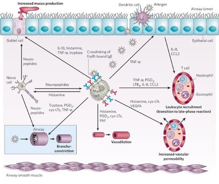
All previously published papers and figures were reproduced with permission from the
publisher.
Published by Karolinska Institutet.
Printed by Eprint AB 2020.
© Anna Wintersand, 2020
ISBN 978-91-7831-746-2
Molecular approaches for characterization, diagnosis
and therapy of allergy to pets

THESIS FOR DOCTORAL DEGREE (Ph.D.)
Public defense in CMM Lecture Hall, Visiongatan 18, ground floor, L8:00, Karolinska
University Hospital, Solna

Friday the 27th of March 2020 at 9.00 am

By

Anna Wintersand

Principal Supervisor:                      Opponent:
Associate Professor Guro Gafvelin          Professor Peter Schmid-Grendelmeier
Karolinska Institutet                      University of Zürich
Department of Clinical Neuroscience        Department of Dermatology

Co-supervisors:                            Examination Board:
Associate Professor Hans Grönlund          Professor Susanne Gabrielsson
Karolinska Institutet                      Karolinska Institutet
Department of Clinical Neuroscience        Department of Medicine, Solna

Professor Thomas Kündig                    Adjunct Professor Lennart Nilsson
University of Zürich                       Linköping University
Department of Dermatology                  Department of Biomedical and Clinical Sciences

                                           Professor Catarina Almqvist Malmros
                                           Karolinska Institutet
                                           Department of Medical Epidemiology and
                                           Biostatistics
To all my fur babies, past, present and future
The most exciting phrase to hear in science, the one that heralds new discoveries,
                                               is not 'Eureka!' but 'That's funny...'

                                                                      Isaac Asimov
ABSTRACT Allergy to furred animals is a common affliction, where up to 20% of the population in affluent countries are sensitized to one or more pet allergens. Several causative allergens from an array of species have been identified, but there are still gaps in our knowledge. This complicates diagnosis and treatment of allergy to pets. Conventional methods rely on naturally derived allergen extracts, with poor characterization and lack of broadly accepted standards. Due to the nature of the extracts, some allergens could be missing or present in clinically non-significant concentrations. The aim of the thesis is to improve diagnostics by characterizing extracts and evaluating component based microarrays to conventional methods. Further, to identify new allergens as well as describe previously only partially characterized allergens, their distribution and properties. Paper I compares two multiplex microarray assays in allergy diagnostics to the standard methods ImmunoCAP and skin prick test with doctor’s diagnosis in a cohort of children with persistent asthma (n=71). The results showed that 75% of all children were sensitized to at least one allergen and that nearly half was multisensitized to three or more allergens. The accuracy of the methods was comparable, they all had a pair-wise concordance of 90% and above. However, the microarray assays contributed with positive IgE responses to new allergens not described in the diagnosis in almost half the patients (47%). The added value of microarray analysis in multisensitized patients is not to belittle. In paper II, an analysis of the content and composition of dog dander extracts for skin prick test is conducted. Results from five commercial manufacturers display a large variation, not only between the different suppliers, but also between batches albeit to a lesser extent. Anatomical location of the source material also differs, hair, dander and epithelia was analyzed. Two of the allergens were detected only at low amounts and patient sera sensitized to these failed to induce an allergic response to the extracts in a basophil activation test. Moreover, a population of dogs was investigated in regard to their allergen profile, also resulting in a substantial divergence between individuals. The heterogeneity of the extracts calls for better content characterization and standardization. Substandard quality could render flawed diagnosis and jeopardize patient safety. Paper III characterizes sensitization to horse allergens, where several allergens are only described partially, or analysis of IgE binding performed in a small cohort. We present sensitization data for four horse allergens including a novel protein, designated Equ c 7, with a prevalence of 38%. Equ c 7 is a homologue to the cat allergen Fel d 1, suggesting possible cross-reactivity. Further, we describe two forms of full-length Equ c 2 and the potential roles of isoforms. Here we contribute to the panel of horse allergens. Allergy to horse is less investigated than to other pets and likely more allergens await discovery. Aerodynamic properties of allergens are investigated in paper IV, where three dog allergens were sampled, however, only two could be detected. Particles associated with airborne allergen differed in size. Can f 1 was only detected on particles larger than 2.8 µm, diversely from Can f 4 which was found on particles of all sampled sizes >8.3-
LIST OF SCIENTIFIC PAPERS
 I. Annica Önell*, Anna Whiteman*, Björn Nordlund, Francesca Baldracchini,
    Giorgio Mazzoleni, Gunilla Hedlin, Hans Grönlund & Jon R. Konradsen
    Allergy testing in children with persistent asthma: comparison of four
    diagnostic methods
    Allergy, 2017; 72: 590–597
    *Shared first authorship
    Note: Anna Wintersand changed name from Whiteman

 II. Anna Wintersand, Klara Asplund, Jonas Binnmyr, Erik Holmgren, Ola B.
     Nilsson, Guro Gafvelin och Hans Grönlund
     Allergens in dog extracts: Implication for diagnosis and treatment
     Allergy, 2019;74:1472–1479

III. Jonas Binnmyr*, Anna Wintersand*, Erik Holmgren, Ola B. Nilsson, Mattias
     Bronge, Guro Gafvelin och Hans Grönlund
     IgE profiling using a panel of horse allergens, including the novel
     allergen Equ c 7 and full length Equ c 2
     Manuscript
     *Shared first authorship

IV. Anna Wintersand, Malin Alsved, Jonas Jakobsson, Sasan Sadrizadeh, Hans
    Grönlund, Jakob Löndahl och Guro Gafvelin
    Airborne allergens from dogs - quantitation and particle size
    Manuscript
CONTENTS
1 INTRODUCTION........................................................................................................... 1
1.1 The immune system............................................................................................... 1
1.1.1 The innate immune system ....................................................................... 1
1.1.2 The adaptive immune system ................................................................... 1
1.1.3 T cells ........................................................................................................ 2
1.1.4 B cells ........................................................................................................ 3
1.1.5 Antibodies ................................................................................................. 3
1.1.6 Immune dysregulation............................................................................... 4
1.2 Allergy ................................................................................................................... 5
1.2.1 Hypersensitivity ........................................................................................ 5
1.2.2 IgE-mediated allergy ................................................................................. 6
1.2.3 Sensitization .............................................................................................. 7
1.2.4 Effector response ....................................................................................... 8
1.2.5 The mucosa in allergy ............................................................................... 9
1.2.6 Mast cells and basophils .........................................................................10
1.3 Allergens ..............................................................................................................11
1.3.1 Structure ..................................................................................................11
1.3.2 Sensitization to pets.................................................................................12
1.3.3 Dog allergens ...........................................................................................12
1.3.4 Horse allergens ........................................................................................13
1.3.5 Cross-reactivity .......................................................................................14
1.3.6 Distribution ..............................................................................................14
1.4 Allergy diagnostics ..............................................................................................15
1.4.1 Skin prick test ..........................................................................................15
1.4.2 Serology...................................................................................................15
1.4.3 Extracts and components ........................................................................16
1.5 Treatment of allergy ............................................................................................16
1.5.1 Allergen specific immunotherapy...........................................................16
1.5.2 Immunomodulation .................................................................................17
2 THESIS AIM .................................................................................................................19
3 MATERIAL AND METHODS....................................................................................21
3.1 Material ................................................................................................................21
3.1.1 Patient material ........................................................................................21
3.1.2 Animal material .......................................................................................21
3.2 Methods ...............................................................................................................21
3.3 Ethical statement..................................................................................................24
4 RESULTS AND DISCUSSION ...................................................................................254.1 Allergy testing in children with persistent asthma: comparison of four
diagnostic methods [I] ......................................................................................... 25
4.2 Allergens in dog extracts: Implication for diagnosis and treatment [II] ............ 27
4.3 IgE profiling of horse sensitized subjects using a panel of horse allergens,
including the novel allergen Equ c 7 and full length Equ c 2 [III] .................... 30
4.4 Airborne allergens from dogs - quantitation and particle size [IV] ................... 32
5 CONCLUSIONS ........................................................................................................... 35
6 FUTURE PERSPECTIVES.......................................................................................... 37
7 POPULÄRVETENSKAPLIG SAMMANFATTNING .............................................. 41
8 ACKNOWLEDGEMENTS.......................................................................................... 45
9 REFERENCES .............................................................................................................. 49LIST OF ABBREVIATIONS AIT Allergen specific immunotherapy ADCC Antibody-dependent cellular cytotoxicity APC Antigen presenting cell BCR B cell receptor CD Cluster of differentiation DAMPs Damage-associated molecular patterns ELISA Enzyme-linked immunosorbent assay Fab Fragment antigen binding Fc Fragment crystallizable HDE Horse dander extract HDM House dust mite HLA Human leukocyte antigen IEC Ion exchange chromatography IFNγ Interferon gamma Ig Immunoglobulin IL Interleukin ILIT Intralymphatic immunotherapy ILC2 Innate lymphoid cells type 2 IMAC Metal chelate affinity chromatography MHC Major histocompatibility complex NK cell Natural killer cell PAMPs Pathogen-associated molecular patterns PBMC Peripheral Blood Mononuclear Cells PSA Prostate-specific antigen SDS-PAGE Sodium dodecyl sulfate polyacrylamide gel electrophoresis SEC Size exclusion chromatography SLIT Sublingual immunotherapy SCIT Subcutaneous immunotherapy TCR T cell receptor TH T helper cell
1 INTRODUCTION
As living beings, we are constantly under attack from pathogens, which require our
physiology to reproduce and thrive. The downside for us is that our bodies are damaged in
that process, sometimes unrepairable or worse even fatally. Thus a war is constantly being
waged between us and invasive microorganisms. To that end, the immune system has
evolved in an arms race, to help us gain the upper hand in survival. Unfortunately, sometimes
the immune system mistakes a harmless object for a pathogen and responds accordingly. This
may result in causing harm instead of protecting the body, manifesting diseases like
autoimmunity and allergy. This thesis contributes with new insights regarding the allergens
causing allergic disease to pets, their variability, properties and distribution, to improve
diagnosis and treatment of allergy.

1.1   THE IMMUNE SYSTEM

1.1.1 The innate immune system

The immune system constitutes two major parts, the innate and the adaptive immune system.
The innate immune system, which is present at birth, includes the first line of defense with
physical outer barriers such as the skin, mucosa and epithelial layers. Their role is to prevent
entry into the body, but if damaged or compromised pathogens might penetrate. When that
occurs, antimicrobial proteins, the complement system and innate immune cells are
circulating to identify and neutralize the threat. The innate immune system is non-specific but
responds quickly to pathogens or cell damage using pathogen-associated molecular patterns
respectively damage-associated molecular patterns (PAMPs/DAMPs) (1). These receptors
have a limited diversity, about 100 receptors have the ability to recognize around 1000
antigens (2). But the antigens identified represent commonly distributed preserved essential
features of pathogens, such as lipopolysaccharide of bacterial cell walls or double stranded
RNA, frequent in viruses but foreign in humans. The PAMPs and DAMPs are found on cells
associated with the innate immune response such as neutrophils, macrophages or NK-cells,
but also on epithelial cells in the barriers. When a pathogen is identified, these cells try to
eliminate the microorganism by phagocytosis, opsonization or cytotoxicity also causing
inflammation (3). Secreting inflammatory mediators as chemokines and cytokines also helps
signal to attract more immune cells to the site of infection and to appropriately activate them.
Specialized antigen presenting cells (APCs) also express PAMP/DAMP receptors. In this
way they constitute a bridge between the innate and the adaptive arm of the immune system,
and dendritic cells for instance, have the ability to activate the adaptive immune system to get
a tailored, more persistent response to the pathogen.

1.1.2 The adaptive immune system

The adaptive immunity takes longer to engage, but carries substantially more specificity. It
also harbors a memory which increases the efficiency towards the same antigen following

                                                                                              1
repeating encounters (4). However, more recent research has shown an advanced interplay between the innate and the adaptive systems, where some cells perform functions or share characteristics attributed to both systems (5-8). The adaptive immune system requires multiple activation signals, including co-receptors and cytokines to upregulate and mature into effector cells. Innate immune cells engage and activate the adaptive cells, namely lymphocytes during an infection. Lymphocytes comprises of T and B cells, these cells have a much broader repertoire of antigen recognition through the T or B cell receptors (TCR/BCR), compared to the innate cells. However, each individual cell carries only one specificity, determined through hypervariable regions and somatic gene rearrangement. These features allow a vast array of possible combinations and specificities (9). 1.1.3 T cells T cells are lymphocytes that are specialized for specific antigen recognition through the TCR and responsible for cell mediated immunity. All T cells display the surface marker CD3 (cluster of differentiation), a co-receptor in conjunction with the TCR and ζ-chain forming the TCR complex. T cells are classified in two major groups depending on the presence of a second surface co-receptor, CD4 or CD8, giving them different capabilities and function. The CD8+ cells, or cytotoxic T cells, recognize major histocompatibility complex (MHC) class I molecules presented on all of the own body’s cells when infected with an intracellular pathogen. The MHC complexes are encoded in the human leukocyte antigen (HLA) genes, and are like the TCR/BCR, highly diverse. Together they represent most polymorphic genes in the genome, to provide a broad repertoire of specificity. When activated the CD8+ T cell kills off the infected cell, mainly through cell lysing mechanisms by releasing their granules containing perforin and granzymes. CD4+ T cells are called T helper cells due to their ability to activate and facilitate differentiation of other immune cells. The CD4+ T cell recognizes antigens presented on the MHC class II molecule of antigen presenting cells, which are extracellular foreign antigens digested and displayed as peptides on the surface of the APC. The activated CD4+ cell differentiate into an effector cell which produces cytokines to recruit and activate different target cells depending on the present microbe (2). To that end different subpopulations of CD4+ cells have evolved. TH1 type cells are characteristic of combating viruses and intracellular pathogens, macrophages have been shown to induce this pathway (10) as well as dendritic cells (11) through interleukin (IL)-12 and interferon gamma (IFNγ). The TH1 cells in turn produce more IFNγ and express the CD-40 ligand to enhance macrophages’ ability to kill ingested microorganisms. The TH2 pathway is initiated by IL-4 from mast cells or epithelial cells, in response to extracellular parasites (12). The typical cytokines produced by TH2 are IL-4 to continue the TH2 differentiation, IL-5 to recruit eosinophils for killing parasites and IL-13 to increase mucus secretion and contractions of smooth muscle (13). These cytokines also promote the alternative pathway of macrophages, to produce growth factors and stimulate fibrosis (14). TH17 cells differentiation occurs in response to bacterial or fungal infections, where dendritic cells secrete IL-1, IL-6, IL-23 and TGF-β (15). These cells 2
produce IL-17 and IL-22 (16) recruiting neutrophils and driving inflammation as well as
repairing epithelia. Recent research has also linked them with the defense against intracellular
bacteria (17). Further, additional subsets have been identified, e.g. TH22, which is a pro-
inflammatory lineage of T cells infiltrating the epidermis, producing IL-22 and TNF-α (18)
and shown to assist in cutaneous immunity (19). TH9 is another subset of T helper cells,
which have been implicated in contributing to allergic responses by secretion of IL-9. The
cytokine, together with IL-4 stimulate B cells to produce IgE (20) and are necessary for mast
cell amassment during inflammation (21).

1.1.4 B cells

B cells belong to the second class of lymphocytes, responsible for the humoral branch of the
adaptive immune system, the major effector function being antibody production. In contrast
to T cells, the antigen receptor immunoglobulin (Ig) can exist either as a membrane receptor
(BCR) or in soluble form as an antibody. Both types share the same specificity on an
individual cell. Another distinction between T and B cell receptors is that while the TCR
recognizes peptides and linear epitopes, the immunoglobulin bind to the whole antigen and
thus conformational epitopes (9). Two membrane bound Ig classes are expressed on naïve B-
cells, IgM and IgD. These receptors recognize and bind antigen, but need a second stimulus
to cause activation. The mode of activation can either be T cell dependent if the antigen in
question is a protein, or T cell independent during polysaccharide or lipid encounters where
the repetitive antigens have the ability to cross-link adjacent Ig molecules or bind innate toll-
like receptors on the B-cell. The activated B cell undergoes clonal expansion and
differentiation into either antibody producing plasma cells or memory B cells. The T cell
dependent pathway also have the ability to induce class switching and affinity maturation to
further tailor the response (22). The remaining three Ig classes are a result of isotype
switching depending on the type of T cell help and the milieu.

1.1.5 Antibodies

Antibodies, the secreted form of immunoglobulin, consist of two variable regions, (fragment
antigen binding, Fab) and a constant one (fragment crystallizable, Fc). The Fab portion of the
antibody is highly diverse and can bind a vast array of antigens. Due to the rearrangement of
the V(D)J domains of the genome this polymorphism is possible. Further specialization
occurs by class-switch recombination and somatic hypermutation, where AID plays a vital
role. AID is a single stranded DNA that causes mismatched DNA by deamination. When
DNA repair systems removes the mismatch, mutations arises causing further diversity (23).
The Fc region determines the isotype of the antibody and thus the function. The region bind
receptors on cell surfaces eliciting a cellular response causing either up- or downregulation.
Hence, Fc receptor signaling determines the immune response from the affected cells (24).

The Ig isotype IgM is either expressed as a surface bound monomer or secreted as a pentamer
during infections where it opsonizes pathogens (25). IgD can also be secreted as a soluble

                                                                                               3
form in blood serum. It’s role in immune responses is still unclear, but it is suggested to be involved in maintaining homeostasis (26). IgG can trigger up-regulation and activation of myeloid cells and some endothelial cells, facilitating phagocytosis, endocytosis and antibody- dependent cellular cytotoxicity (ADCC). IgG can also opsonize pathogens to be recognized by NK-cells and destroyed. However, IgG can also bind to the inhibitory receptor FcγRIIb expressed on mast cells, basophils and monocytes to initiate a down-regulation of the cell (27). IgE is engaged in the defense against helminth infections and are pathologically associated with allergic diseases and asthma (28). IgE binds to the high affinity receptor FcεRI, present on mast cells, basophils, eosinophils and DC. Mast cells, the main expresser of FcεRI, become coated with antibodies when sensitized. Upon antigen interaction the cell degranulates releasing mediators toxic to parasites, causing inflammation. The antibody is also involved in IgE dependent phagocytosis as well as B-cell maturation and survival (29). Lastly, IgA is found in serum, but predominantly on mucosal surfaces and in secretions. IgA can act both pro-inflammatory, when binding an antigen forming an immune complex causing cytokine release, cytotoxicity and phagocytosis, and anti-inflammatory (30). The IgA immune complex can also pass though membranes via the polymeric transmembrane receptor pIgR, eliminating the pathogen from circulation and function as a way of clearing infections (31, 32). 1.1.6 Immune dysregulation The immune system is highly effective in eradicating pathogens and keeping us healthy. The aptitude however comes at a price. Sometimes the immune system recognizes harmless molecules as threats or worse, the own self as foreign. Hereditary defects in the genome or mutations can also cause immune deficiencies. For example, defective T cell development can cause severe combined immunodeficiency (SCID), the individuals lacking functional T cells and generally having a low lymphocyte population. The condition causes critical infections and predisposes to early death (33). Loss of CD40 or CD40L due to mutations resulting in an inability of B-cells to undergo class-switch, causes hyper-IgM syndrome as well as a shortage of the other antibody isotypes. This manifests as recurrent bacterial infections, autoimmunity and malignancy (34). Hyper-IgE syndrome (HIES) patients have a defect in the transcription factor STAT3, critical for differentiation of T cells into TH17 cells and activation of innate lymphoid cells type 3 (ILC3). Absence of these cells causes a multitude of bacterial and fungal skin and pulmonary infections including eczema. The reason for the elevated IgE levels is not fully understood, however these patients lack a pronounced TH2 phenotype and the IgE is not allergen-specific, resulting in less allergic disease compared to atopic patients (35). Immune deficiencies can also be acquired from pathogen exposure. The retrovirus HIV causes acquired immune deficiency syndrome (AIDS), characterized by a depletion of CD4+ cells, eventually causing recurrent infections and malignancies, resulting in death if untreated (36). During lymphocyte development in the thymus and bone marrow, the cells undergo several selection steps to ensure that the naïve lymphocytes have the ability to identify foreign 4
antigens meanwhile discriminating self-antigens, thus inducing self-tolerance. When the
immune system fails to distinguish one’s own antigens from those foreign, the result can lead
to severe tissue damage manifesting as autoimmunity (37). T cell mediated autoimmunity
includes for example diabetes type I where TH1 cells react towards pancreatic islet antigens
on β-cells, destroying these resulting in lack of insulin production. Multiple sclerosis is
another severe form of autoimmunity, where the autologous T cells attack the myelin sheaths
of the central nervous system, creating plaques causing the myelin to dissolve and the nerve
signals to diminish. Common symptoms are muscle weakness, ataxia, blindness and
ultimately paralysis. Several autoantigens have been suggested as targets, myelin
oligodendrocyte glycoprotein (MOG) being one of the first identified, but questioned more
recently. However, advances are being made in detecting novel autoantigens and more
sensitive methods are under development (38).

Allergic disease is also a consequence of a dysfunctional immune response, in this instance
hypersensitivity. The immune system mistakes a harmless molecule, for instance pollen or
food, for a pathogenic one and responds accordingly causing harm to the body in the process.

1.2   ALLERGY

1.2.1 Hypersensitivity

Clemens von Pirquet first defined allergy as the odd disposition of some people to respond
with hypersensitive symptoms to specific substances in 1906 (39). Gell and Coombs, two
British immunologists suggested in 1963 that there were four types of allergic reactions that
was the result of hypersensitivity (40). They proposed a classification, despite lack of
knowledge on immune mechanisms like T cell functions and cytokine repertoire at the time
drafted, which was adopted for decades. The classical classification was defined as:

Type I – Acute type, IgE mediated. When the antigen is recognized and bound by IgE
antibodies on mast cells, IgE crosslinks causing degranulation and release of mediators
instantaneously.

Type II – Semi-delayed, antibody mediated. Autoreactive IgG recognizing host cell and
opsonizes it. Cell is lysed by complement or antibody-dependent cellular cytotoxicity by NK
cells.

Type III – CIC mediated. Immune complexes are formed and deposited in tissues from
abundant antigen bound to antibodies and complement. Causing inflammation and tissue
damage.

Type IV – Delayed-type, cell mediated. The antigen is presented by an APC to a T cell,
activation occurs and cytokines are released.

However, the validity for all types of hypersensitivity reactions have been debated as well as
the lack of a more complex accurate description (41). A new revised classification was

                                                                                            5
proposed by Johansson et al, accepted by EEACI in 2001 (42) and by the World Allergy
Organization in 2003 (43). The consensus of the current hypersensitivity classification is as
follows:
Non-allergic hypersensitivity
Allergic hypersensitivity
IgE-mediated
Atopic
Non-atopic, e.g. insect sting, helminth infection, pharmaceutical,
Non IgE-mediated
T cell-mediated, e.g. celiac disease
Eosinophilic, e.g. eosinophilic esophagitis
IgG-mediated, e.g. allergic alveolitis
Other
1.2.2 IgE-mediated allergy
We are constantly exposed to potential allergens from the environment. People with IgE-
mediated allergy respond to these allergens by producing IgE antibodies, referred to as
sensitization. However, some people develop allergic disease and other do not. Hereditary
predisposition to develop allergies is described as atopy (from Greek atopos, meaning out of
place) (44). Having atopic parents increases the likelihood of the child to develop allergic
disease (45, 46). Further, the gender of the child may influence the parental contribution to
atopy. A higher risk of girls developing allergy has been shown to depend of maternal allergy
and the same has been shown for boys and paternal allergy (47). Certain alleles, like a
polymorphism of the 17q21 locus, have also been linked with high risk of developing asthma,
which could be an explanation for the diverging results of the hygiene hypothesis and pet
keeping (48). Mutations in filaggrin functionality also have an effect on risk of sensitization,
but the outcome differs depending on the allergen and age of exposure (49). However, several
factors influence the risk of developing allergy, besides the genes, and the cause is likely
multifactorial (50).
The hygiene hypothesis has long been a favored explanation for increased risk of
sensitization. It was coined by Strachan in 1989, who observed that children growing up in
larger families had a lower incidence of hay fever and eczema compared to children in
smaller families (51). He postulated that the unhygienic contact with siblings or infected
mother would explain the difference. Thus early exposure to microorganisms would provide
protection against development of allergies and overly clean environments would be
disadvantageous. Some later studies have supported the hypothesis (52), while others have
shown more conflicting results depending on the pathogen exposure (53), where e.g. RSV
infection in infancy seems to increase the risk for allergy (54, 55). However, growing up on a
farm decreases the risk of developing allergies according to several studies (56-59). Living
with cats and dogs during the first year of life also provide a protective effect against allergic
6disease (60-62). The composition and diversity of the gut flora during infancy has proven to
impact the risk of sensitization and asthma (63, 64), where T-regulatory cells are important
for keeping homeostasis (65). Further, even later in life altered microbiota have been linked
with inflammatory disease e.g. rheumatoid arthritis (66). The same is true for the airways,
where an abundance of certain strains of Proteobacteria is linked with incidence of asthma
(67) and Moraxella or Haemophilus infiltration associated with neutrophilia (68).
1.2.3 Sensitization
The first time an allergen is encountered in an atopic individual, sensitization occurs without
symptoms. The allergen breaches the mucosal layers in the airways, gastrointestinal tract or
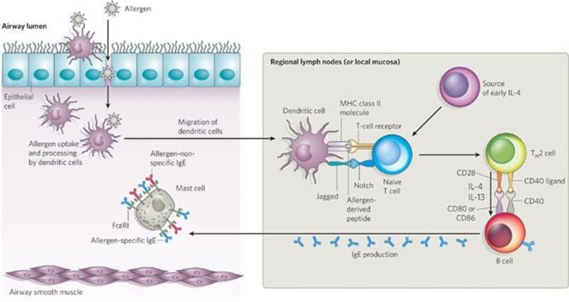
penetrates disrupted epithelia in the skin, see figure 1. House dust mite (HDM) allergen even
have the ability to disrupt tight junctions using protease activity (69). Dendritic cells in the
tissue capture and process the allergen, causing the DC to mature and migrate to the regional
lymph node. In the lymph node the DC presents the processed allergen peptides to naïve T
cells using the MHC class II molecule. If recognized, depending on the milieu and presence
of IL-4 from mast cells or epithelial cells, the naïve T cell will differentiate into an effector
TH2 cell. The TH2 cells start to produce more IL-4 and IL-13, and activate B cells and
phagocytes by engaging their CD40 receptor using the CD40 ligand and CD80/86 with CD28
(70). The B cell undergoes class switch and starts producing IgE, and with help of resident T
follicular helper cells increases the affinity of the antibodies, making them more potent (71).
The T follicular helper cells are also thought to drive inflammation by producing IL-4 (72).
The secreted IgE is spread by the blood systemically, and further into tissues where it
encounters mast cells, to which it binds using the high affinity receptor FcɛRI. The mast cells
are now sensitized and ready to combat the allergen during subsequent exposure.
Figure 1: Sensitization to allergens in the airway. Galli, S., Tsai, M. & Piliponsky, A. Nature
454, 445–454 (2008).
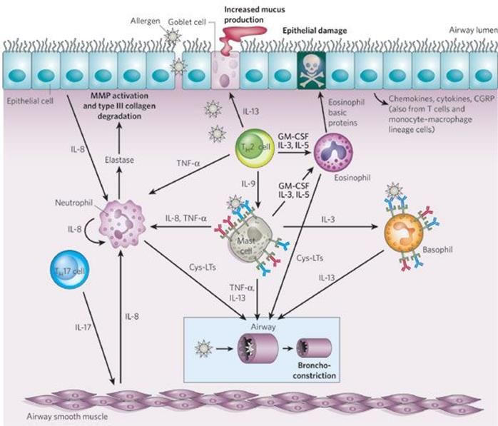
71.2.4 Effector response The following time the allergen is encountered an allergic response initiates. The effector phase can be described as three stages, early, late and chronic phase. A few minutes after the allergen exposure, the early phase occurs when IgE-coated mast cells degranulate upon recognition of the allergen, cross-linking of FcεRI and releasing mediators causing inflammation, see figure 2. Symptoms occur such as asthma due to bronchoconstriction, swelling and redness because of vasodilation and rhinitis from mucus production (73). Figure 2: Early phase of allergen-induced airway inflammation. Galli, S., Tsai, M. & Piliponsky, A. Nature 454, 445–454 (2008). The late phase response occurs hours after the allergen exposure, and is a result of the innate and adaptive immune cells recruited by the chemokines, cytokines and mediators present at the site of inflammation. The innate cells cause damage to the epithelia, neutrophils release elastase causing collagen degradation (74) and eosinophils their major basic protein (MBP) destroying cells (75), see figure 3. If the inflammation is persistent, long-term damage to the airways through remodeling can occur. The smooth muscle cells in the lungs increase both in numbers and size, decreasing the lumen size, while mucus producing goblet cells become more abundant (76). With constant infiltration of immune cells, cytokines are further injuring the tissue (77). In non-allergic individuals, T regulatory cells contribute to keeping the balance of the immune system and produce IL-10 to inhibit activation of the TH2 pathway (78). 8
Figure 3: Late phase of allergen-induced airway inflammation. Galli, S., Tsai, M. &
Piliponsky, A. Nature 454, 445–454 (2008).
1.2.5 The mucosa in allergy
The mucosal layers on the inside of the body are the surfaces that are directly exposed to
pathogens and if breached, the normal entry point for microbes. These layers consist of a
mucus secreting epithelium that covers the entire gastrointestinal tract, the respiratory tract,
the urogenital tract and the middle ear, forming the largest part of the immune tissues (9).
However, these tissues are permeable to allow substances such as nutrients or oxygen to pass,
making defense an important feature. In an allergy context, when the body tries to fight off a
perceived threat in the form of an allergen, the cells driving inflammation also causes damage
in the bronchial tract. Lymphocytes in patients susceptible to allergic disease differentiate
into TH2 cells, that together with innate lymphoid cells type 2 (ILC2) activate and start
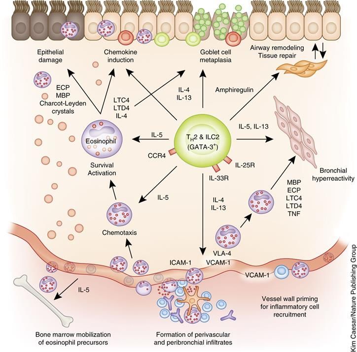
producing cytokines (79), figure 4. Secretion of IL-13 causes airway remodeling and airway
obstruction, features of allergic asthma (80). Recruitment of eosinophils via the cytokine IL-5
further drives inflammation and degranulation causing epithelial damage (81). Production of
IL-4 induces naïve T cells to differentiate into TH2 cells creating a positive feedback loop and
more recently has been shown to synergize with IL-3 to further enhance TH2 promotion (82).
In addition, IL-4 has been shown to polarize macrophages into the alternatively activated M2
macrophages through the inflammasome NLRP3 (83), potentially causing fibroblast
accumulation and tissue remodeling through TGF-β secretion (84).
9Figure 4. Overview of functions of TH2 cells and ILC2 cells in asthma. (Lambrecht B.N. & Hammad H., Nature Immunology 16, 45–56(2015)) The gut also plays a major role in the development of allergies, studies have shown that diversity of microbiota during infancy affects allergy and asthma outcome (85, 86). Further, impaired IgA ability to bind fecal bacteria predisposes children to develop allergies (87) and consuming breast milk with lower diversity of microbiota could facilitate this development (88). Richness of bacterial flora as well as composition in the saliva also impacts the probability to acquire allergic disease and discussions are ongoing regarding efficacy of probiotic prophylaxis (89). 1.2.6 Mast cells and basophils Mast cells are found in all tissue vascularized in the body, but more abundantly close to epithelia, airways and the gastrointestinal tract. Diversely, basophils are encountered in circulation and only migrate into tissue during an immunological event (90). However, both cells express the high affinity receptor for IgE, FcεRI. When sensitized, allergen-specific IgE is bound to the receptor coating the cell. Upon allergen encounter, two or more IgE molecules bind the allergen, crosslinking the receptors leading to an aggregation of FcεRI (91). The aggregation activates a downstream process where the protein tyrosine kinases LYN and 10
FYN initiate a cascade resulting in degranulation, synthesis of pro-inflammatory lipid
mediators and activation of transcription factors encoding cytokines, chemokines and growth
factors (92). When degranulation occurs, histamine and lipid mediators, among others
prostaglandins and leukotrienes, which cause vasodilation and swelling, are released. Further
chemokines which induce migration of leukocytes to the site of inflammation, as well as
TNF-α causing bronchoconstriction and pro-inflammatory cytokines such as IL-4, IL-5 and
IL-13 are secreted (93). FcεRII or CD23 is the low affinity equivalent and can exist both
membrane bound or soluble. It has the ability to inhibit IgE production when engaged on B
cells (94) and when in soluble form has cytokine-like activity, promoting proliferation and
sustaining cell growth (95). FcεRII is expressed on among others T and B cells (96),
granulocytes (97) and monocytes (98). A new study has found a cross-talk between mast cells
and basophils, where IL-33 produced by mast cells induce basophils to drive allergic
inflammation (99).

1.3   ALLERGENS

1.3.1 Structure

Allergens are generally small hydrophilic proteins or glycoproteins derived from the animal,
plant or fungi kingdoms. Allergens contain conformational epitopes or in rare cases
carbohydrate residues recognized by immunoglobulins (100). Galactose‐α‐1,3‐galactose (α‐
Gal), a carbohydrate residue with the capacity to induce IgE antibodies are associated to red
meat allergy, but sensitization has been linked to tick bites (101). However, the mechanism is
still not fully determined. Further, an allergen must have the ability to cross-link IgE on mast
cell and basophil surfaces, which requires at least two IgE epitopes. Consequently, many
allergens naturally exist as dimers or oligomers to facilitate the immune cell activation
capacity (102).

Another inherent ability of allergens is the stability of the protein, especially in regard to food
allergens that have to withstand the gastrointestinal tract and proteases (103). The major
peanut protein Ara h 1 and the hazelnut allergen Cor a 14 for instance, are able to survive
digestion and to elicit severe allergic reactions (104, 105). The capacity to maintain
conformation could also serve to slow down digestion by lysosomes after phagocytosis and
contribute to a prolonged MHC class II presentation (106, 107). Lately, focus has been
shifted to the adherent adjuvant properties of some allergens, such as their function as
proteases and thus the ability to affect epithelial cells directly and drive TH2 inflammation
(108).

A large portion of the pet allergens belong to the lipocalin protein family (109), and the
similarities within the group could explain the common pattern of multi-sensitization across
species (110). The other three major protein families connected with pet sensitization are
kallikrein, serum albumins and secretoglobin (111). Most animal allergens studied belong to
pet mammals like cat and dog, or those in our proximity e.g. house dust mite. Less is known

                                                                                                11
about uncommon pets like reptiles or birds, however a toxin has been described for spiders (112). Allergen nomenclature is derived from the host species name. The Latin name of the Linnean system for dog is Canis familiaris and the allergens are thus named Can f and allocated a number in sequence, for instance Can f 1, Can f 2 and so forth. Equus caballus is Latin for domestic horse and the nomenclature for these allergens Equ c. The World Health Organization and the International Union of Immunological Societies Allergen Nomenclature Sub-Committee are responsible for recording and classifying characterized allergens (113). 1.3.2 Sensitization to pets Sensitization to pet allergens increases the risk of developing allergy and asthma (114) and over 50% of studied children admitted to hospital for asthma exacerbations were shown to be sensitized to pet allergens, signifying the connection (115). The prevalence of allergy however differs between geographic locations and populations, where the westernized countries are more prone to disease development (116-118). Further, exposure to urban environments during childhood was connected with a higher risk of sensitization as an adult compared to rural settlement (119). During the last decades the sensitization rate in childhood to inhalant allergens has increased dramatically (120), although in older children in high prevalence areas, asthma development increase seems to come to a halt (121). Studies suggest a sensitization rate of 20% to dogs and 18% to cat in Sweden, but only 6% and 10% respectively in Palermo (116). In the US, a prevalence of 12% has been recorded for both dogs and cats (122). Sensitization to horse has been reported to 5% in one study in Italy (123) and 3% in another (124). 1.3.3 Dog allergens Regarding dogs, eight allergens have been described to date, Can f 1-8 (Table I). Can f 1 is a major lipocalin allergen 23-25 kDa, found in dander and saliva (125). Approximately 70% of dog allergic individuals are sensitized to Can f 1 (126). Recently, a study revealed that the N and C-terminus of Can f 1 both contained a conformational IgE epitiope, possibly a target for future therapeutics (127). Can f 2 is a minor lipocalin, detectable in saliva, with an IgE reactivity of 23% and a size of 19 kDa (128). Can f 3, is serum albumin, abundant in blood, but can also be found in saliva and skin, possibly leaking through from damaged epithelia or gums. It is a larger protein, 69 kDa and cross-reactive with serum albumins from other mammals (129). Due to this, Can f 3 is less immunogenic with a prevalence of 35% sensitization (130). Can f 4, an 18 kD lipcocalin, was first thought to have a sensitization rate of 35% (131). It was in a later study displaying positive titers of up to 81%, suggesting the classification as a major allergen (132), possibly due conformational stability. Can f 4 also has the ability to form dimers, possibly affecting the allergenicity (133). Can f 5, prostatic kallikrein of 28 k Da, is also a major allergen with prevalence up to 76% among dog sensitized patients (134). Being a homologue to human prostate-specific antigen (PSA), it is 12
only expressed in male dogs, making avoidance easier (135). Case studies have also
confirmed the link between Can f 5 sensitization and allergy to human seminal fluid (136,
137). Another cross-reactive allergen of 27-29 kDa denoted Can f 6, has also been described
in both fur and saliva with a sensitization of 38% (138). Can f 7 or Niemann Pick type C2
protein, has been characterized as a 16 kDa protein with an IgE positivity of 10-20% (139).
Recently, a novel allergen has been added to the IUIS Allergen database, a 14 kDa cystatin
with a prevalence of 13% named Can f 8 (113).

Mentions of hypoallergenic dogs have been frequent in media and popularity of cross-breeds
with these claimed traits has risen. However, no scientific data supports the statement
regarding hypoallergenic dog breeds. On the contrary, one study describes higher
concentrations of Can f 1 in the acclaimed hypoallergenic breeds compared to other breeds
(140). Others report no difference in allergen concentration between homes of so-called
hypoallergenic dogs compared to control dogs (141). Further, a higher incidence of allergy in
children growing up with these claimed hypoallergenic breeds was found. Conversely, a
reduced risk of developing allergy was instead correlated with female dogs and keeping two
dogs or more (142).

Table I. List of currently known dog and horse allergens, their molecular weight, prevalence
of sensitization and biochemical name.

Dog allergens     Size         Prevalence      Biochemical name
Can f 1           23-25 kDa    70%             Lipocalin
Can f 2           19 kDa       23%             Lipocalin
Can f 3           69 kDa       35%             Serum albumin
Can f 4           18 kDa       35-81%          Lipocalin
Can f 5           28 kDa       76%             Kallikrein
Can f 6           27-29 kDa    38%             Lipocalin
Can f 7           16 kDa       10-20%          NPC2
Can f 8           14 kDa       13%             Cystatin
Horse allergens   Size         Prevalence      Biochemical name
Equ c 1           25 kDa       51-70%          Lipocalin
Equ c 2           17 kDa       50%             Lipocalin
Equ c 3           67 kDa       40%             Serum albumin
Equ c 4           17 kDa       77%             Latherin
Equ c 5           "            "               "
Equ c 6           15 kDa       Not reported    Lysozyme

1.3.4 Horse allergens

Six horse allergens have been registered in the IUIS Allergen database (Table I). Equ c 1, the
major horse allergen, is a 25 kDa lipocalin protein (143, 144) with a reported IgE prevalence
of 51-76% (145, 146). Equ c 2 has previously only been described as two incomplete
isoforms of a 17 kDa lipocalin protein. IgE recognition in immunoblot was detected in about
50% of patient samples, however no distinction was made between the different isoforms and
only 23 patients were analyzed (147). Equ c 3 is serum albumin and holds a molecular weight
                                                                                           13
close to other albumins, 67 kDa (148). Sensitization rate reported is approximately 40% (149). Equ c 4, diversely is a latherin of 17k Da, with surfactant properties found in sweat and saliva (150). Equ c 5 was later described as an independent allergen, but found to be identical to Equ c 4 and removed from the IUIS database. Prevalence was only reported in the Equ c 5 paper, 77% of 22 patients (151). Lastly Equ c 6, a lysozyme, was described in a case study and classified both as a contact allergen causing urticaria and a food allergen after ingestion (152). Similarly to dogs, hypoallergenic horses has been a hot topic. However, in concordance with the results for dogs, no evidence for the hypothesis has been found. Curly horses are classified as a hypoallergenic breed, but had significantly higher levels of horse dander antigens, Equ c 1 and Equ c 4, compared to several other breeds (153). Equ c 4 has a high variation both within and between ten different breeds, where only stallions had significant consistently higher concentration compared to mares and geldings (154). 1.3.5 Cross-reactivity Due to pet allergens generally belonging to a select few protein families, cross-reactivity is common. Clinically, pet allergic patients often are multi-sensitized, making diagnosis more complicated, with mono-sensitization being as low as 5% (149). To elucidate the primary allergen source in poly-sensitized patients, molecular based allergy diagnostics is a useful tool (155). Two super-families of pet allergens are similar in structure and reported as cross- reactive, lipocalins and serum albumin (156, 157). Lipocalin Can f 6, shares similar tertiary structure and sequence with the cat allergen Fel d 4 (158). Fel d 4 together with horse allergen Equ c1, also inhibit IgE binding to Can f 6 in vitro, implying cross-reactivity between the three allergens (138). However, in spite of a low sequence identity, Fel d 4 also displayed cross-reactivity with Can f 2 (159). Dog allergen Can f 4 also cross-reacts with an odorant binding cow allergen (131). Further, Fel d 7, a cat allergen that 46% of cat sensitized individuals react to, was shown to moderately correlate to Can f 1 and inhibit IgE-binding in dog sensitized patients (160). Three peptides from horse serum inhibit IgE and IgG from horse allergic patients to not only horse, but also dog and cat albumin (161). One third of patients sensitized to dog, cat and horse displayed cross-reactivity to all the serum albumins (162). Taken together, patients sensitized to above mentioned protein families should be further investigated, because the assumed primary sensitizing allergen might only be a cross- reactivity, and another allergen actually the principal cause of allergic symptoms. 1.3.6 Distribution Pet allergens are abundant in homes with resident pets, but can also be found in public places like schools and hospitals, making avoidance measures difficult for allergic individuals (163, 164). A recent study even detected a higher concentration of allergen in day care centers compared to homes (165). Allergen reduction tools have proven some decrease of allergen 14
concentration and allergic symptoms, including air cleaners and recurrent washing of the
animal (166-169), but avoidance if sensitized is recommended.

The aerodynamic properties of allergens varies, where house dust mite allergens group I are
carried mainly on particles averaging 20 µm (170). Cat and dog allergen associated particles
however, belong to a broader range, where 20% of particles were smaller than 5µm (171).
Smaller particles have the capacity to reach further down the airways, increasing the
probability of uptake and encountering of mast cells, and sensitization to these have been
correlated to inflammation and asthma (172). Comparison of allergens in settled dust in
relation to the airborne compartment, showed correlation for homes with dogs, but not in cat
households, suggesting these particles have further unknown properties (173). Airborne
levels of horse allergen has been shown to disperse quickly, even in the vicinity of stables
(174), where concentration was 500-fold lower just outside compared to inside the stable
increasing to a 3000-fold difference 12 meters away (175).

1.4   ALLERGY DIAGNOSTICS

1.4.1 Skin prick test

The golden standard of allergy diagnostics is still skin prick test (SPT) and the most common
practiced (176). The method is usually performed using naturally derived aqueous extracts
acquired from the allergen source (177). The standard procedure is conducted as described
(178) by placing a grid on the patient’s forearm and adding a small drop of each allergen to
be analyzed, as well as a negative (diluent) and a positive control (histamine solution). The
skin is then pierced using a lancet. The grid is removed and excess solutions wiped off. After
15 minutes the results is recorded by measuring the diameter of the wheal formed by the local
immune reaction. A wheal diameter ≥3 is considered positive. The advantage of using the
skin prick test is the cheap cost, it is quick to perform and well proven (179). To consider
when using the skin prick test is that the positive control can influence the negative control if
in close proximity (180). The physicians interpreting the result differently can also impact
diagnosis (181). Further, because the nature of the extracts and lack of a consensus
standardization (182), false negative responses can occur if an allergen is lacking from the
analytic preparation or not present in adequate amounts to produce a reaction (183).

1.4.2 Serology

Detection of specific IgE antibodies in serum is another well-established method of
diagnosing allergy (184). It was introduced in 1967 as a radioallergosorbent test (RAST) by
Johansson and Wide (185). Several different methods are in practice today, where the single-
plex assays ImmunoCAP and Immulite 2000 are most commonly used (186, 187). The
analyzed allergen can either be in an extract or a component, coupled to a solid phase. Sera is
added and if allergen-specific IgE is present, it binds to the allergen. Detection is done by a
fluorescent anti-human IgE, in proportion to the patient sIgE (188). The advantage of these

                                                                                              15
assays is that they quantify the sIgE in units per liter (kU/L), the sensitivity and precision is high, it is reproducible and IgG does not interfere with the measurement (189). The downside of the method is again the extracts and their content, or lack thereof. Especially in regard to food allergy where different molecules of the allergen source are connected with severity of symptoms (190). Lack of allergens in the extracts can, as in the skin prick test produce false negative results (191). Recently, multiplex analyses have been developed. These assays test for a large panel of common allergens simultaneously and use lesser sera per allergen tested (192). Another advantage is that they are often component resolved, which improves the accuracy (193). Further, in multi-sensitized individuals, deducing cross-reactivities and detecting the primary sensitization profile is less complicated (194). 1.4.3 Extracts and components As mentioned, the extract based assays struggle with sensitivity, due to the varying composition of extracts (195). Preparation of the raw material into aqueous extracts also causes inconsistencies. Some methods destroy conformational epitopes and, additionally, selection of the raw material can impact the composition of allergens (177). Even different batches from the same manufacturer differ in patient SPT responses (196). The varying protein content and composition of SPT solutions have been demonstrated for dog dander and the possible impact on the accuracy of diagnosing allergy to dogs (197). Further, due to the allergen variability of dogs and lack of low molecular weight allergens in extracts, inconsistencies cause difficulties in diagnosis (198). The same phenomenon has been shown for fish extracts, where different manufacturers varied 10-fold in content, (199) and in extracts for cockroach allergy diagnostics (200). Molecular components provides a solution to the inconsistencies of extracts, by having a consistent allergen content, thus making standardization possible (201). Further, the issue with unknown molecules and their properties are non-existent. In cockroach extracts for example, glycinin is not unlikely present, due to contamination from their food source. Patients sensitized to soybean can respond positively although they are negative for cockroach, providing a false diagnosis (202). 1.5 TREATMENT OF ALLERGY 1.5.1 Allergen specific immunotherapy The only curative treatment currently available for allergies is allergen specific immunotherapy (AIT), where desensitization is achieved by administrating the relevant allergen in low doses over a long period of time gradually increasing the dose (203). However, no standardized protocol exist, physicians worldwide practices different doses and frequency of treatment, complicating understanding of efficacy (204). As with the skin prick test, AIT relies on allergen extracts. Due to the absence of a common standardization and wide range of administered doses, knowledge on effective doses are lacking (205). The mechanism of AIT encompasses shifting the TH2 immune response into a TH1 pathway 16
including blocking IgG antibodies and induction of IL-10 secreting T regulatory cells (206).
IgG1 and IgG4 in response to AIT, can compete with IgE-allergen complexes for binding the
low affinity FcγRIIb on B cells, thus inhibit the antigen presentation to T cells promoting
tolerance (207).

Two methods are in practice in the clinics today, subcutaneous immunotherapy (SCIT) and
sublingual immunotherapy (SLIT). SLIT is less invasive, where tablets are orally
administered compared to SCIT, where the allergen is injected under the skin, but suggested
as less effective (208). However, from a safety perspective SLIT is advantageous, less
adverse systemic reactions are reported but local responses are more frequent. Although
uncommon, SCIT can cause potentially lethal anaphylaxis, which should be taken into
account when initiating treatment (209). Studies on treatment of allergy to pets display
divergent results. For dog AIT, a review of clinical trials show inadequate results after
therapy attributed to inferior extracts and the numerous dog allergens in relation to patients’
divergent sensitization profiles (210). A case study report states that AIT to dog can decrease
allergic responses to other furry animals, likely due to cross-reactivity (211). Not many
studies have been conducted on AIT on horse allergic patients, but one study found that
rhinitis lessened for 93% of the patients and asthma symptoms reduced for 90% (212).

1.5.2 Immunomodulation

A large concern regarding immunotherapy is safety, anaphylaxis and severe systemic
reactions poses danger to patients undergoing treatment (213). To combat the issue, as well as
increasing the efficacy of the treatment, different strategies has been applied to modify the
therapeutic allergens. For example, allergoids are chemically modified allergens, by
polymerization with aldehydes conformational IgE-epitopes can be destroyed while
sequential T cell epitopes are retained, thereby reducing allergenicity while maintaining
immunogenicity (214). A cat dander allergoid induced a TH1 and T regulatory response in
vitro and lesser IgE interaction (215). Further, a HDE allergoid has proven well-tolerated in
several clinical trials (216). Another strategy involves DNA containing unmethylated CpG
oligodeoxynucleotide (ODN), a feature of bacterial and viral genomes. It is recognized by
toll-like receptor 9, which has the capability of switching a TH2 response into a TH1 milieu
(217). CpG ODN has been utilized as an adjuvant in treatment of peanut allergy (218, 219).
Traditionally, adjuvants has been added to vaccines for boosting the efficacy, where
aluminum hydroxide has been frequent choice. However, the mechanisms are still not fully
understood, but several advances have been made (220).

                                                                                            17
You can also read