Congenital gallbladder agenesis in a 9-month-old Bull Terrier
←
→
Page content transcription
If your browser does not render page correctly, please read the page content below
Case Report                                                                       Veterinarni Medicina, 66, 2021

                                                                    https://doi.org/10.17221/135/2020-VETMED

Congenital gallbladder agenesis in a 9-month-old
Bull Terrier
Olga Gojska-Zygner1,2,3, Marek Galanty4, Beata Degorska4,
Jan Frymus4, Wojciech Zygner5*
1
  Veterinary Clinic Teodor, Warsaw, Poland
2
  Veterinary Clinic Morskie Oko, Warsaw, Poland
3
  24h Veterinary Clinic Elwet, Warsaw, Poland
4
  Department of Small Animal Diseases with Clinic, Institute of Veterinary Medicine,
  Warsaw University of Life Sciences, Warsaw, Poland
5
  Department of Preclinical Sciences, Institute of Veterinary Medicine, Warsaw University
  of Life Sciences, Warsaw, Poland
*Corresponding author: wojciechzygner@yahoo.pl

Citation: Gojska-Zygner O, Galanty M, Degorska B, Frymus J, Zygner W (2021): Congenital gallbladder agenesis
in a 9-month-old Bull Terrier. Vet Med-Czech 66.

Abstract: Congenital gallbladder agenesis is an extremely rare disorder, which has, to the best of our knowledge, only
been reported in seventeen dogs (mainly in Japan). In almost all of these cases, gallbladder agenesis or hypoplasia
was detected in small dogs. In this report, we present a case of gallbladder agenesis in a 9-month-old intact female
Bull Terrier. The clinical signs included diarrhoea, sporadic vomiting, apathy and decreased appetite. The serum
biochemistry revealed an increased liver enzyme activity, an increased concentration of serum bile acids and mild
hyperbilirubinaemia. A diagnostic laparotomy demonstrated the lack of a gallbladder and dilation of the common
bile duct, which was misinterpreted as the gallbladder in the ultrasonographic examination. The histological exami-
nation of the liver revealed degenerative changes in the hepatocytes with glycogen accumulation and some necrotic
hepatocytes. The therapy included a low protein diet, fluids, silymarin and ursodeoxycholic acid. After nine weeks
of therapy, the dog was in good condition, the diarrhoea and vomiting ceased, and the liver function parameters,
such as the AST and GLDH activities, and the concentration of bile acids had decreased to reference intervals.

Keywords: cholestasis; developmental disorder; liver; therapy; ursodeoxycholic acid

  Canine congenital gallbladder agenesis is a rare de-      (2019) diagnosed the disorder in another dog using
velopmental disorder. To the best of our knowledge,         computed tomography. Gallbladder agenesis is also
only three cases had been described up to 2010, two         rarely detected in humans, and clinical signs are
cases in Maltese dogs and one case in a Chihuahua           observed only in about 25% of the cases (Serour
(Liptak et al. 2000; Austin et al.2006; Kamishina           et al. 1993).
et al. 2010). In 2018, Sato et al. (2018) described           The aetiology of this anomaly is still unknown;
twelve cases of gallbladder agenesis and five cases         however, it is considered to be embryological and
of gallbladder hypoplasia in dogs from Japan. In that       probably a hereditary disorder associated with
study most cases were detected in Chihuahuas.               a failure of the liver and gallbladder primordia
In 2019, Bugyiova et al. (2019) detected this anom-         development or a failure of the vacuolation of the
aly in a Pug using ultrasonography and Kelly et al.         gallbladder (Tang et al. 2015; Thornton et al. 2016).

1
Case Report                                                                         Veterinarni Medicina, 66, 2021

                                                                    https://doi.org/10.17221/135/2020-VETMED

Case description                                             serum biochemical examination one month prior
                                                             to referral did not reveal any abnormalities.
  A 9-month-old, 25 kg, intact female Bull Terrier             On presentation to the clinic, the dog was in good
presented to the veterinary clinic with a three-             condition and, according to the dog’s owner, it had
month history of chronic diarrhoea, flatulence and           a good appetite. A clinical examination did not re-
sporadic vomiting. The owner of the dog described            veal any abnormalities. An abdominal palpation did
the stools as grey-green, abundant, unformed and             not reveal any pain or tension, with no abnormali-
pasty. At another clinic, during this 3-month period,        ties detected during the palpation examination. The
the dog had received four therapies with Tylosin             mucosal membranes were pink without any yellow
which resulted in an improvement for a few days.             or yellowish colouration. The body temperature,
The referring veterinarian reported that an abdomi-          hydration status and superficial lymph nodes were
nal ultrasonography, a complete blood count and              normal. The diagnostic tests included: an abdomi-

Table 1. Serum liver parameters in the 9-month-old Bull Terrier with gallbladder agenesis obtained between the
5th of April and the 1st of July, 2017

                     Reference    Examination     Examination       Examination        Examination     Examination
Parameter
                     intervals    No. 1 (5 Apr)   No. 2 (6 May)    No. 3 (20 May)      No. 4 (3 Jun)   No. 5 (1 Jul)
ALT
                     0.02–1.33        2.98            6.98              2.4                3.6              –
(μkat/l)
AST
                     0.02–1.26        1.32            4.62              1.28               1.55            0.78
(μkat/l)
ALP
                     0.02–2.35        0.55            0.75              0.9                1.2              –
(μkat/l)
GLDH
                     0.01–0.18        0.45            3.87              0.22                   –           0.04
(μkat/l)
GGTP
                     0.01–0.12        0.28             –                 –                     –            –
(μkat/l)
Cholesterol
                      3.1–8.6         9.7             9.6               6.53                   –            –
(mmol/l)
Total bilirubin
                     0.01–3.4         5.4             6.31              8.5                6.8              –
(μmol/l)
Fasting bile acids
                       0–18          30.7            65.4              99.4               22               1.79
(μmol/l)
Albumin
                      25–44          37.9            37.2                –                     –            –
(g/l)
Total protein
                      54–75          70.1            66.3                –                     –            –
(g/l)
Urea
                      3.3–8.3         6.74             –                 –                     –            –
(mmol/l)
Glucose
                     3.05–6.1         4.85             –                 –                     –            –
(mmol/l)
TLI
                       5 – 35        19                –                 –                     –            –
(μg/l)
α-amylase
                     0.17–27.51       8.74             –                 –                     –            –
(μkat/l)
Lipase
                     0.02–2           0.6              –                 –                     –            –
(μkat/l)

ALT = alanine transaminase; ALP = alkaline phosphatase; AST = aspartate transaminase; GGTP = gamma-glutamyltrans-
peptidase; GLDH = glutamate dehydrogenase; TLI = trypsin-like immunoreactivity

2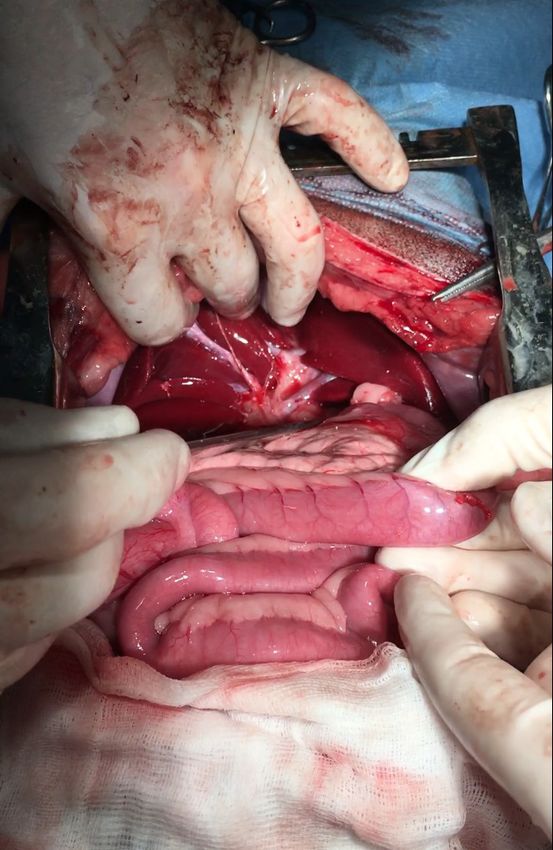
Case Report Veterinarni Medicina, 66, 2021
https://doi.org/10.17221/135/2020-VETMED
nal ultrasonography, faecal examinations (using concentrations of the liver parameters (Table 1: ex-
flotation techniques with oversaturated NaCl and amination No. 1). When repeated the next day, the
33% ZnSO4 solutions), a urine examination, a com- concentration of the fasting serum bile acids was
plete blood count and determination of the serum within the reference interval (6.1 μmol/l). The rea-
biochemical parameters (Tables 1 and 2). son for the increased serum bile acid concentration
The abdominal ultrasonography revealed the in- during the initial examination was considered to be
homogeneous hyperechoic structure of the liver. because the blood sample was probably collected af-
According to the ultrasonographic description, the ter a meal. The hepatoprotective drug Zentonil 400
lumen of the gallbladder was subtly filled with bile (Vetoquinol), a commercial diet supporting liver
without any calculi or changes in its wall. The results function (Hepatic Royal Canine) and a probiotic sup-
of the faecal examinations were negative; however, plement FortiFlora (Purina PRO PLAN Veterinary
a microscopic examination revealed the presence Diets) were prescribed for 4 weeks.
of fat in the stool. The urine examination did not After 3 weeks, the dog presented to the clinic
reveal any abnormalities. The complete blood count again owing to a decreased appetite and apathy. The
did not reveal any pathological changes; however, the diarrhoea had ceased; however, the stools were pale
serum biochemical examination revealed increased and yellow. A clinical examination did not reveal
any abnormalities except apathy. Cholestasis was
Table 2. Results of the complete blood count performed considered as a probable cause of the current state
on the 9-month-old Bull Terrier with gallbladder agen- of the animal. However, an abdominal ultrasonog-
esis on the 5th of April, 2017 raphy showed no obstruction in the bile ducts, and,
according to the ultrasonographic description, the
Reference Result of exa- gallbladder was unchanged and subtly filled with
Parameter
interval mination
bile. An ultrasound examination of the liver did
RBC count not show a portosystemic shunt. Fluid therapy [al-
5.5–8.5 6.32
(T/l)
ternately Solutio Ringeri and 0.9% NaCl; 500 ml,
Haemoglobin concentration subcutaneously (s.c.) twice daily], ursodeoxycho-
9.31–11.79 9.99
(mmol/l) lic acid [5 mg/kg, perorally (p.o.) twice daily] and
Haematocrit Zentonil 400 were administered.
44–55 47.6
(l/l) After 10 days of therapy, the state of the dog was
MCV unchanged, and the stools were still pale and yellow.
60–77 75.3
(fl) Increased liver function indicator values were still
MCHC observed (Table 1: examination No. 2). The owner
19.86–22.34 20.98
(mmol/l) of the dog agreed to a diagnostic laparotomy and the
WBC count collection of material for liver histological exami-
6–12 11.2
(g/l) nations. The surgery revealed a lack of a gallblad-
Neutrophils der on the visceral surface of the liver and dilation
55–75 70
(%) of the common bile duct (Figure 1). A histological
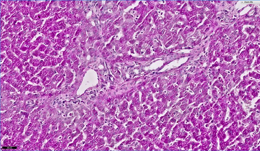
Lymphocytes examination revealed diffuse, moderate hepatocel-
13–30 22 lular cytoplasmic vacuolar degeneration (Figure 2),
(%)
Monocytes and a periodic acid-Schiff staining showed glycogen
1–10 5 accumulation in the hepatocytes (Figure 3).
(%)
After surgery, the dog was treated with a low pro-
Eosinophils
0–6 3 tein diet, ursodeoxycholic acid (5 mg/kg, p.o. twice
(%)
daily) and fluids (alternately Solutio Ringeri and
Basophils
0–1 0 0.9 % NaCl; 250 ml, s.c. twice daily). Maropitant
(%)
at a dosage of 1 mg/kg s.c. once a day for 5 days
Platelet count
150–500 320 was also administered owing to the vomiting. Ten
(g/l)
days after surgery, the dog presented to the clinic
MCHC = mean corpuscular haemoglobin concentration; for a control visit. The owner of the dog described
MCV = mean corpuscular volume; RBC = red blood cell it as having increased vitality. The vomiting had
count; WBC = white blood cell count ceased, the dog had a good appetite, and stools
3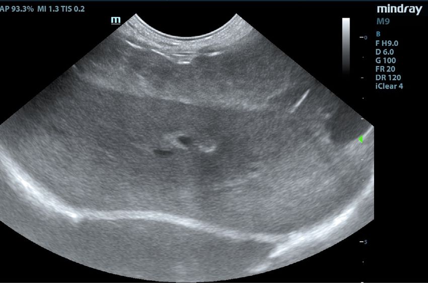
Case Report                                                                      Veterinarni Medicina, 66, 2021

                                                                   https://doi.org/10.17221/135/2020-VETMED

    Figure 1                                                    Figure 2

                                                            Figure 2. Cytoplasmic vacuolar degeneration of the hepa-
                                                            tocytes in the 9-month-old Bull Terrier with gallbladder
                                                            agenesis; magnification × 20, haematoxylin-eosin staining

                                                                 Figure 3

                                                            Figure 3. Glycogen accumulation in the hepatocytes
                                                            in the 9-month-old Bull Terrier with gallbladder agen-
                                                            esis; magnification × 20, periodic acid-Schiff staining
Figure 1. View of the porta hepatis during the diagnostic
laparotomy in the 9-month-old Bull Terrier. Visible lack    the fasting bile acids had decreased in comparison
of a gallbladder and dilation of the common bile duct       to the previous tests (Table 1: examination No. 4).
                                                            The dog was in good condition, had a good appe-
were light brown and formed. A clinical exami-              tite, and the stools were brown and formed. A clini-
nation revealed a lack of fever, abdominal pain,            cal examination did not reveal any abnormalities.
or yellow or pale mucosal membranes. A serum                The therapy (ursodeoxycholic acid, silymarin,
biochemical examination showed increased con-               low protein diet and fluids) was continued for the
centrations of the liver parameters; however, they          next 4 weeks. After this time, a serum biochemi-
were lower than in the previous examinations,               cal examination revealed the AST, GLDH and bile
with the exception of the total bilirubin and bile          acids were within reference intervals (Table 1: ex-
acid concentrations. The glutamate dehydrogenase            amination No. 5). The dog was in good condition;
(GLDH) activity was mildly increased, while the             however, urticaria appeared on both hind legs; this
cholesterol concentration was within the reference          symptom disappeared after a single intramuscular
interval (Table 1: examination No. 3). An increased         injection with 0.2 mg/kg of dexamethasone.
dosage of ursodeoxycholic acid to 7.5 mg/kg twice              A further therapy was based on a low protein diet
daily was recommended, and, additionally, silyma-           and ursodeoxycholic acid. Control examinations
rin at 70 mg in toto orally 3 times a day after a meal      of the serum liver parameters, urine examination
was administered. A fluid therapy, the same as pre-         and liver ultrasonography were recommended ev-
viously, was continued for the next 14 days.                ery 2 months.
  After 2 weeks of therapy, a serum biochemical                Over the next 17 months, the complete blood
examination revealed an increased alanine trans-            counts and serum biochemical examinations were
aminase (ALT) and aspartate transaminase (AST)              performed every 2 months; no abnormalities were re-
activity, but the concentrations of bilirubin and           vealed, except sporadic, mildly increased ALT and

4
Case Report Veterinarni Medicina, 66, 2021
https://doi.org/10.17221/135/2020-VETMED
AST activities. The concentrations of bilirubin, bile activities were within reference intervals (Table 3).
acids, and the alkaline phosphatase (ALP) and GLDH The ultrasonographic examinations of the liver every
Table 3. Monitored serum liver parameters and complete blood count from the 9-month-old Bull Terrier with gall-
bladder agenesis over a period of 17 months between the 30th of August 2017 and the 28th of November 2018
Reference
Parameter 30/08/17 25/10/17 03/01/18 07/03/18 09/05/18 11/07/18 19/09/18 28/11/18
interval
RBC
5.5–8.5 6.68 6.44 6.36 7.29 6.31 6.40 6.12 6.97
(T/l)
Hb
9.31–11.79 9.74 9.87 10.18 10.67 9.99 10.12 9.68 10.61
(mmol/l)
Ht
44–55 45.9 45 47.1 52 46.7 46.9 44.7 48.2
(l/l)
MCV
60–77 69 69.1 74.1 71.3 74 73.3 73 69.2
(fl)
MCHC
19.86–22.34 21.29 21.97 21.6 20.54 21.41 21.53 21.72 22.09
(mmol/l)
WBC
6–12 12.7 11.5 10.8 12.3 10.7 11.2 12.2 11.9
(g/l)
Segmented neutrophils
55–75 77 67 71 73 69 74 73 70
(%)
Band neutrophils
0–4 3 0 2 1 0 2 2 3
(%)
Lymphocytes
13–30 17 28 21 24 25 20 21 23
(%)
Monocytes
1–10 2 2 2 1 2 1 2 1
(%)
Eosinophils
0–6 1 3 4 1 4 3 1 3
(%)
Basophils
0–1 0 0 0 0 0 0 1 0
(%)
Platelet count
150–500 270 277 267 239 242 287 291 236
(g/l)
ALT
0.02–1.33 0.52 1.57 1.43 0.63 0.77 1.02 1.72 1.2
(μkat/l)
AST
0.02–1.26 0.55 1.12 1.2 0.6 0.65 0.83 1.4 1.1
(μkat/l)
ALP
0.02–2.35 0.65 0.73 1.02 0.88 0.78 0.9 1.02 0.87
(μkat/l)
GLDH
0.01–0.18 0.16 0.15 – 0.14 – 0.15 0.16 0.15
(μkat/l)
Tot. bil.
0.01–3.4 3.2 3.34 3.23 3.12 – 3.04 3.4 3.1
(μmol/l)
Fast. BA
0–18 12.9 9.1 11.8 14.3 10.8 13.7 16.4 15
(μmol/l)
ALP = alkaline phosphatase; ALT = alanine transaminase; AST = aspartate transaminase; Fast. BA - concentration of fast-
ing bile acids; GLDH = glutamate dehydrogenase; Hb = haemoglobin concentration; Ht = haematocrit; MCHC = mean
corpuscular haemoglobin concentration; MCV = mean corpuscular volume; RBC = red blood cell count; Tot. bil. = total
bilirubin concentration; WBC = white blood cell count
5Case Report Veterinarni Medicina, 66, 2021
https://doi.org/10.17221/135/2020-VETMED
Figure 4 other cases, appeared at a young age. However,
as opposed to most of the other cases, diarrhoea,
but not vomiting, was the main clinical sign.
Ascites, anorexia and seizures were also reported
in the other dogs with congenital gallbladder agen-
esis. However, in eight out of the seventeen dogs
with agenesis or hypoplasia of the gallbladder, like
in many humans with this anomaly, no clinical signs
were observed (Austin et al. 2006; Kamishina et al.
2010; Scobie and Bramhall 2016; Sato et al. 2018).
In all the cases of the dogs with gallbladder agen-
esis, including this report, the laboratory liver pa-
Figure 4. Ultrasound picture of the liver of the 9-month- rameters were elevated. In these cases, the ALT
old Bull Terrier with gallbladder agenesis showing its and AST activities were increased, probably result-
non-homogeneous and hyperechoic structure ing from the action of the hydrophobic bile acids
(Liptak et al. 2000; Austin et al. 2006; Kamishina
two months showed the same non-homogeneous and et al. 2010; Sato et al. 2018). In few cases, the ALP
hyperechoic structure of the organ (Figure 4). Yet, activity was also increased; however, it was not ob-
the ultrasonography did not reveal a hepatomegaly. served in the present case. Interestingly, a mild in-
During this time, the dog was in good condition, no crease in the bilirubin concentration was observed
vomiting was observed, and the stools were brown in this case, but not in the other cases of the dogs
and formed. with this anomaly (Sato et al. 2018). However, hy-
Currently, over two years after surgery, the dog perbilirubinaemia has also been observed in humans
is still under the care of the first author of this case with this defect (Tjaden et al. 2015). In the present
report. The dog is still treated with ursodeoxycholic report, the increased concentrations of the total
acid and fed a low protein diet. Occasionally, aller- bilirubin and bile acids might be associated with
gic skin reactions are observed; these are probably cholestasis (Berk and Javitt 1978). This may result
caused by the pharmaceutical products contain- from the compensatory dilation of the common bile
ing ursodeoxycholic acid. The dog was castrated duct, with the role of the gallbladder probably taken
17 months after the laparotomy that was compli- over by the common bile duct (Liptak et al. 2000).
cated by severe bleeding, which may have been Moreover, it should be mentioned that a dilated
caused by the improper functioning of the liver. common bile duct may be misinterpreted as a gall-
bladder by an ultrasonographic examination, this is
also observed in humans with this anomaly (Serour
DISCUSSION et al. 1993; Liptak et al. 2000; Sato et al. 2018). Thus,
it is possible that cases like the one described here
Congenital gallbladder agenesis occurs, or is de- are not so extremely rare; the under-reporting may
tected, extremely rarely in dogs. This anomaly is result from the subclinical course of the disease
observed in small and miniature breeds of dogs. in some cases and the low sensitivity of an ultraso-
So far, the lack of a gallbladder has been detected nographic examination for the diagnosis. Currently,
only once in a larger dog. In that case, the gall- in human medicine, computed tomography or mag-
bladder agenesis was recognised in a German netic resonance imaging are recommended for the
Shepherd from Japan (Liptak et al. 2000; Austin diagnosis of this anomaly (Kelly et al. 2019).
et al. 2006; Kamishina et al. 2010; Sato et al. 2018). In this case, the recognition of cholestasis may
To our knowledge, the presented case report is the also be confirmed through a microscopic fae-
first description of congenital gallbladder agenesis cal examination via the detection of fatty drops
in a Bull Terrier, and, so far, the second case of this in the stool before treatment, and improvement
anomaly in a dog other than a small or miniature of the condition of the dog after treatment with
dog. In this case, the clinical signs, such as vomit- ursodeoxycholic acid which increases the bile flow.
ing, diarrhoea, decreased appetite and apathy, were The hepatoprotective action of this hydrophilic
similar to other reported cases, and, like most of the bile acid also results from its anti-inflammatory
6Case Report Veterinarni Medicina, 66, 2021
https://doi.org/10.17221/135/2020-VETMED
properties and inhibition of cellular apoptosis and activity and the pharmaceutical product with urso-
necrosis. Moreover, the competitive displacement deoxycholic acid should be changed to another with
of endogenous hepatotoxic hydrophobic bile acids the same active substance (Plumb 2008). However,
by the ursodeoxycholic acid in the enterohepatic it should be emphasised that glucocorticosteroids
circulation prevents the liver fibrosis and cirrhosis should be used carefully in dogs with cholestasis
induced by the hydrophobic bile acids (Lazaridis and increased bile acids concentrations. Out et al.
et al. 2001; Roma et al. 2011). (2014) showed that the use of prednisolone in mice
In the presented report, the degenerative hepa- caused an increase in the serum bile acid concentra-
tocellular changes were probably caused by the tion. On the other hand, two weeks of using dexa-
increased concentrations of hydrophobic endog- methasone in rats limited the liver injury caused
enous bile acids, such as taurochenodeoxycholic by cholestasis (Eken et al. 2006). Similar conclu-
and taurolithocholic acids, as a result of the cho- sions were drawn by Tiao et al. (2011). Those au-
lestasis. The pro-inflammatory action of these thors observed a decrease in the serum ALT, AST
acids results from the activation of Kupffer cells, and ALP activity, and the bilirubin concentration
stimulating secretion of pro-inflammatory cyto- in rats with cholestasis after a single dose of dexa-
kines such as TNF-α and IL-1β. Moreover, the lipid methasone. Moreover, Gabbia et al. (2018) showed
peroxidation of the hepatocellular membranes, in- that dexamethasone counteracts a hepatic inflam-
duced by the hydrophobic bile acids, leads to injury mation and the oxidative stress in cholestatic rats.
of the hepatocytes and further cellular apoptosis Thus, it is possible that in the presented case report,
and necrosis, and the long-term exposure of hepa- the single dose of dexamethasone had no negative
tocytes to these acids may lead to liver fibrosis and influence on the course of the liver disease or even
cirrhosis (Lazaridis et al. 2001; Copple et al. 2010; contributed to limiting its progress.
Chiang 2013). The excessive glycogen accumula- Gallbladder agenesis is recognised extremely rare-
tion in hepatocytes observed in this case might also ly in veterinary practice. To the best of our knowl-
result from the prolonged exposure of hepatocytes edge, the presented report is the first description
to bile acids which activate the liver enzyme glyco- of this anomaly in a Bull Terrier, and the second
gen synthase (Fang et al. 2007; Chiang 2013). report from Europe. It cannot be excluded that such
Silymarin, a mixture of flavonoids and polyphe- cases may occur more frequently, yet remain undi-
nols with antioxidative and hepatoprotective prop- agnosed, due to the subclinical course of the disease
erties, was used in the therapy for 6 weeks, owing in some cases and the low sensitivity of the ultra-
to the observation of the hepatocellular changes sonographic examinations in the diagnosis of this
in the dog under the histological examination (Feher defect.
and Lengyel 2012). We propose that ursodeoxycho-
lic acid must be administered to the dog for the re-
minder of its life. The purpose of such a therapy is Acknowledgement
to limit the progress of liver disease. The liver in this
dog will probably never function properly. This sup- Photomicrograph kindly provided by Dr. Diego
position may be confirmed by the severe bleeding Caliari Dipl. ECVP from the IDEXX Laboratory,
observed after the dog’s castration. Although the Ludwigsburg, Germany.
dog has allergies, and pharmaceutical products with
ursodeoxycholic acid may contribute to the develop-
ment of urticaria and other skin allergic reactions, Conflict of interest
the authors concluded that ursodeoxycholic acid
should be used permanently in this dog (Hempfling The authors declare no conflict of interest.
et al. 2003). In case of allergic reactions stemming
from products containing ursodeoxycholic acid,
glucocorticosteroids should be used if antihista- REFERENCES
mine drugs are not available despite their influence
on the stimulation of glycogen and lipid accumula- Austin B, Tillson DM, Kuhnt LA. Gallbladder agenesis
tion in the hepatocytes and increasing the serum in a Maltese dog. J Am Anim Hosp Assoc. 2006 Jul-Aug;
ALT and gamma-glutamyltranspeptidase (GGTP) 42(4):308-11.
7Case Report Veterinarni Medicina, 66, 2021
https://doi.org/10.17221/135/2020-VETMED
Berk PD, Javitt NB. Hyperbilirubinemia and cholestasis. Liptak JM, Swinney GR, Rothwell TL, Hunt GB. Aplasia
Am J Med. 1978 Feb;64(2):311-26. of the gallbladder in a dog. J Small Anim Pract. 2000 Apr;
Bugyiova K, Kubovcikova A, Podhorska D. Agenezia zlcoveho 41(4):175-7.
mechura – Prvy publikovany pripad u mopsa [Gallbladder Out C, Dikkers A, Laskewitz A, Boverhof R, van der Ley C,
agenesis in pugs – First published report]. Vetžurnál. Kema IP, Wolters H, Havinga R, Verkade HJ, Kuipers F,
2019 Mar;17(1):10-1. Slovak. Tietge UJ, Groen AK. Prednisolone increases enterohe-
Chiang JY. Bile acid metabolism and signaling. Compr patic cycling of bile acids by induction of Asbt and pro-
Physiol. 2013 Jul;3(3):1191-212. motes reverse cholesterol transport. J Hepatol. 2014 Aug;
Copple BL, Jaeschke H, Klaassen CD. Oxidative stress and 61(2):351-7.
the pathogenesis of cholestasis. Semin Liver Dis. 2010 Plumb DC. Plumb’s veterinary drug handbook. 6th ed. Ames,
May;30(2):195-204. IA, USA: Blackwell Publishing; 2008. 265 p.
Eken H, Ozturk H, Ozturk H, Buyukbayram H. Dose-related Roma MG, Toledo FD, Boaglio AC, Basiglio CL, Crocenzi
effects of dexamethasone on liver damage due to bile duct FA, Sanchez Pozzi EJ. Ursodeoxycholic acid in cholesta-
ligation in rats. World J Gastroenterol. 2006 Sep 7;12(33): sis: Linking action mechanisms to therapeutic applica-
5379-83. tions. Clin Sci (Lond). 2011 Dec;121(12):523-44.
Fang Y, Studer E, Mitchell C, Grant S, Pandak WM, Hy- Sato K, Sakai M, Hayakawa S, Sakamoto Y, Kagawa Y,
lemon PB, Dent P. Conjugated bile acids regulate hepa- Kutara K, Teshima K, Asano K, Watari T. Gallbladder agen-
tocyte glycogen synthase activity in vitro and in vivo via esis in 17 dogs: 2006–2016. J Vet Intern Med. 2018 Jan;
Gαi signaling. Mol Pharmacol. 2007 Apr;71(4):1122-8. 32(1):188-94.
Feher J, Lengyel G. Silymarin in the prevention and treat- Scobie JL, Bramhall SR. Congenital agenesis of the gall-
ment of liver diseases and primary liver cancer. Curr bladder: A UK case report. Oxf Med Case Reports. 2016
Pharm Biotechnol. 2012 Jan;13(1):210-7. Aug 29;2016(8):176-8.
Gabbia D, Pozzo L, Zigiotto G, Roverso M, Sacchi D, Dalla Serour F, Klin B, Strauss S, Vinograd I. False-positive ultra-
Pozza A, Carrara M, Bogialli S, Floreani A, Guido M, De sonography in agenesis of the gallbladder: A pitfall in the
Martin S. Dexamethasone counteracts hepatic inflam- laparoscopic cholecystectomy approach. Surg Laparosc
mation and oxidative stress in cholestatic rats via CAR Endosc. 1993 Apr;3(2):144-6.
activation. PLoS One. 2018 Sep 25;13(9): [21]. Tang LM, Wang XF, Ren PT, Xu GG, Wang CS. The diag-
Hempfling W, Dilger K, Beuers U. Systematic review: nosis of gallbladder agenesis: Two cases report. Int J Clin
Ursodeoxycholic acid – adverse effects and drug interac- Exp Med. 2015 Feb 15;8(2):3010-6.
tions. Aliment Pharmacol Ther. 2003 Nov 15;18(10): Thornton L, Goh YL, Lipton M, Masters A. Rare case of gall-
963-72. bladder agenesis presenting with pancreatitis. BMJ Case
Kamishina H, Katayama M, Okamura Y, Sasaki J, Chiba S, Rep. 2016 Aug 8;2016:bcr2016216510.
Goryo M, Sato R, Yasuda J. Gallbladder agenesis in a Chi- Tiao MM, Lin TK, Chen JB, Liou CW, Wang PW, Huang
huahua. J Vet Med Sci. 2010 Jul;72(7):959-62. CC, Chou YM, Huang YH, Chuang JH. Dexamethasone
Kelly D, Moreno-Aguado B, Lamb V. Gallbladder agenesis decreases cholestatic liver injury via inhibition of intrin-
in a dog: Clinicopathological, histopathology and com- sic pathway with simultaneous enhancement of mito-
puted tomography findings. J Am Anim Hosp Assoc. 2019 chondrial biogenesis. Steroids. 2011 Jun;76(7):660-6.
Nov-Dec;55(6):e556-02. Tjaden J, Patel K, Aadam A. Gallbladder agenesis with re-
Lazaridis KN, Gores GJ, Lindor KD. Ursodeoxycholic acid fractory choledocholithiasis. Case Rep Gastrointest Med.
‘mechanisms of action and clinical use in hepatobiliary 2015 Jun;2015:747931.
disorders’. J Hepatol. 2001 Jul;35(1):134-46.
Received: June 23, 2020
Accepted: February 18, 2021
8You can also read