Cystic lymphangioma of breast and axillary region in an adult: a rare presentation
←
→
Page content transcription
If your browser does not render page correctly, please read the page content below
International Surgery Journal
Shukla S et al. Int Surg J. 2021 Jan;8(1):391-394
http://www.ijsurgery.com                                                                        pISSN 2349-3305 | eISSN 2349-2902

                                                                     DOI: https://dx.doi.org/10.18203/2349-2902.isj20205911
Case Report

                  Cystic lymphangioma of breast and axillary region in
                             an adult: a rare presentation
                          Samir Shukla, Rahul Shivhare*, Vikas Lal, Deepak Rathore

  Department of General Surgery, Gandhi Medical College, Bhopal, Madhya Pradesh, India

  Received: 18 September 2020
  Accepted: 06 November 2020

  *Correspondence:
  Dr. Rahul Shivhare,
  E-mail: rahulshivhare93@gmail.com

  Copyright: © the author(s), publisher and licensee Medip Academy. This is an open-access article distributed under
  the terms of the Creative Commons Attribution Non-Commercial License, which permits unrestricted non-commercial
  use, distribution, and reproduction in any medium, provided the original work is properly cited.

   ABSTRACT

   Cystic lymphangioma also known as cystic hygroma, is a congenital malformation of lymphatic system. Most
   lymphangioma are present at birth and are diagnosed by the age of 2 years. They are usually located in the head and
   neck region and are rare in other location. We are reporting a case of cystic lymphangioma in breast and axillary region
   in a 23 years old female. Physical examination revealed a non-tender cystic mass in axilla and upper outer quadrant of
   left breast. Ultrasonography (USG) revealed a hypoechoic mass lesion and magnetic resonance imaging (MRI) showed
   a multi-spectated cystic mass in left axilla closely involving the left breast parenchyma. Wide local excision was done
   and histopathological examination further confirmed the diagnosis of cystic lymphangioma. Although it is very rare,
   cystic lymphangioma should be considered in the differential diagnosis of mass in breast and axillary region.

   Keywords: Lymphangioma, Cystic, Breast, Axilla

INTRODUCTION                                                            development, while others discuss a more congenital
                                                                        etiology involving miscommunication between lymphatic
A lymphangioma is a malformation of the lymphatic                       and venous pathways, aberrant lymphatic growths, and
system most commonly observed in infants and children.1-                tissue sequestration during development.2,15 The role of
3
  Case reports of lymphangioma in adults are very rare and              chromosomal abnormalities has been documented, most
fewer than 150 cases can be found in the English language               frequently involving Turner's syndrome, trisomies 13, 18,
scientific literature.1 Also referred to as cystic hygromas or          and 21, and Noonan syndrome.2 A review of the literature
lymphatic malformations, they are most often found in the               failed to reveal information about the prevalence of
cervicofacial region (75%) and are less commonly seen in                chromosomal abnormalities in adult-onset cases, however.
the axilla (20%) or elsewhere.1-4 Previous reports have
mentioned the management of lymphangiomas in the                        Several staging and classification mechanisms are
adult, but these reviews are limited to presentations in the            proposed and adopted which permit for better diagnosis
neck.5,6 A review of literature has identified rare case                and management. Smith et al. characterized cystic
reports of adult onset lymphangioma of the axilla.7-14                  hygromas as either microcystic, macrocystic, or mixed,
                                                                        with microcystic containing cysts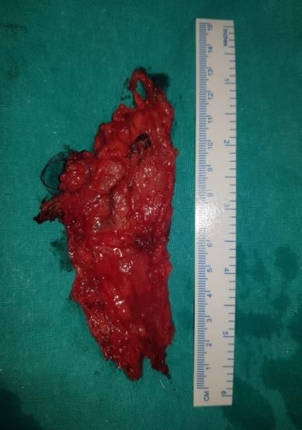
Shukla S et al. Int Surg J. 2021 Jan;8(1):391-394
complication rate, but applies only to masses within the
cervicofacial region.19 Diagnosis is often aided by the
employment of fine needle aspiration for cytology, tissue
histology, and ultrasound, magnetic resonance imaging
(MRI) or computed tomography (CT) for definition of the
mass.
CASE REPORT
A 23-year-old, otherwise healthy female was referred to
general surgery with complain of swelling in left axilla and
breast which was sudden onset and non-tender. She had no
history of trauma or surgery to that area. She had no
comorbidities, no history of any substance abuse and Figure 2: MRI STIR sequence showing the lesion of
family history was unremarkable for any congenital size 12.4×9.4×5 cm in left axilla extending till D8
masses or chromosomal abnormalities. vertebral level.
On physical examination (Figure 1), she was well-
nourished and well developed, with normal temperature,
vital signs and normal cardiopulmonary examination. She
was found to have a swelling of size around 13×10×5 cm
in the left axilla extending till the outer lower compartment
of left breast. The swelling was fluctuant, and nontender.
Magnetic resonance imaging (MRI) demonstrated a well-
defined T1 hypointense, T2/STIR hyperintense multi-
spectated cystic mass in left axilla closely abutting the left
breast parenchyma. USG guided fine needle aspiration
(FNA) was done and patient underwent surgical excision
of the tumour and histopathology confirmed the diagnosis
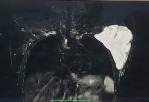
of lymphangioma. Figure 3: MRI T2 axial view showing lesion involving
left breast parenchyma.
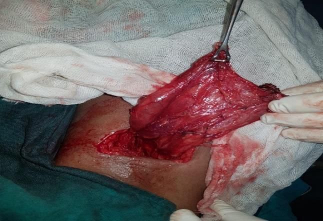
As the dissection continued cephalad, medial pectoral,
long thoracic, and thoracodorsal nerves were identified
and spared. Where the cyst become embedded in the axilla,
gentle blunt dissection was done to remove the remaining
mass (Figure 5). The skin was closed in two layers of
absorbable sutures and drained with 12 mm closed suction
drains.
Figure 1: Mass as demonstrated on physical
examination.
A full excision of the mass was performed under general
anaesthesia. The patient was positioned in the left lateral
decubitus position and an elliptical incision was given in
the axilla. The tumour was dissected away from the
pectoralis major and pectoralis minor anteriorly and
serratus anterior and subclavius muscle posteriorly. Where
the capsule wall was thin and friable suture ligation was Figure 4: Intra-operative picture showing tumour
done to prevent spillage of contents and to maintain freed from adhesions and being taken out.
continuity of the structure for its complete removal.
International Surgery Journal | January 2021 | Vol 8 | Issue 1 Page 392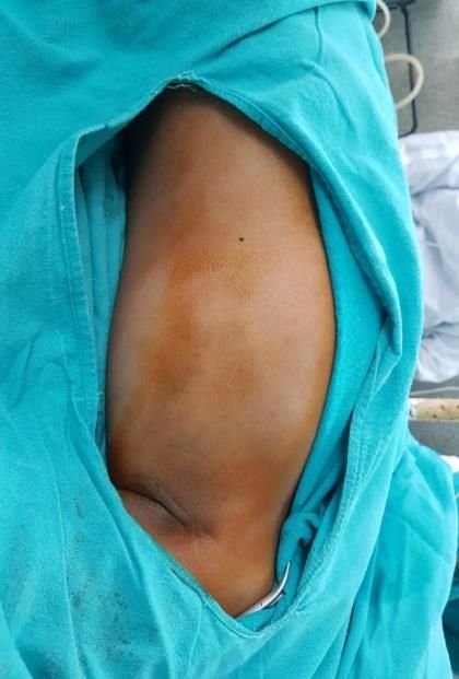
Shukla S et al. Int Surg J. 2021 Jan;8(1):391-394
mass less clear.20 However, one study demonstrated that
patients having partial resection, just one in nine had
recurrence.21 Several case reports have discussed the
inefficacy of a straightforward aspiration and
antibiotics.1,9,13 Long duration follow-up is suggested as a
vital aspect of management, as recurrence has occurred as
late as 6 years after excision.15
Some authors have highlighted the role of sclerotherapy
for the management of lymphngiomas, using agents such
as OK-432, bleomycin, doxycycline, acetic acid, alcohol,
and hypertonic saline.2,5 sclerotherapy wasn’t employed in
this case for several reasons given the proximity of the
mass to vital neurovascular structures and multicystic
Figure 5: Post-op picture of the excised tumour.
anatomy. First, the success of sclerotherapy has
occasionally been measured in terms of sufficient mass
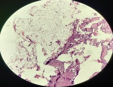
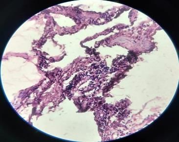
Cytological analysis of the initial fine needle cytology
reduction without complete resolution.1 Further, it’s been
showed large number of small lymphocytes against
recognized that sclerotherapy might not be effective
mucoid background suggestive of lymphangioma. The full
against multiloculated masses, or those of mixed or
thickness biopsy of the mass showed the presence of loose
microcystic anatomy.1,2,5 Sclerotherapy may induce a
fibrofatty tissue interspersed with spaces lined by
localized immune response which causes a short lived but
endothelium and at places filled with lymphocytes,
dramatic increase within the size of the mass. Perkins et al
polymorphs and red blood cells. The stroma at places
has also recognized the potential for shock-like reaction in
showing lymphoid nodule.
the setting of penicillin allergy and OK-432 use.5
CONCLUSION
Continued reporting of adult-onset cystic lymphangioma
is of particular importance, as the nature and management
of these rare masses are elucidated. The role of
chromosomal abnormalities in cases presenting in the
adult is not yet understood and may be of interest as such
masses continue to be reported. This case contributes to
the body of evidence supporting the role of cystic
lymphangioma in a differential diagnosis for masses in the
adult, especially in the acute setting. Further, the future
management of cystic hygromas in the axilla, with
proximity to important neurovascular structures, is better
informed with our addition to examples of uncomplicated
resection.
Funding: No funding sources
Figure 6: Microscopic section.
Conflict of interest: None declared
Ethical approval: Not required
DISCUSSION
REFERENCES
Differential diagnosis of the lymphangioma includes soft
tissue sarcoma, abscess, synovial cyst, and hematoma.18 1. Gow L, Gulati R, Khan A, Mihaimeed F. Adult-onset
Surgical excision has been the treatment of cystic hygroma: a case report and review of
lymphangiomas historically, which is believed to be management. Grand Rounds. 2011;11(1):5-11.
preferred in adults because the lesion is circumscribed.1- 2. Bloom DC, Perkins JA, Manning SC. Management
3,5,15
During this case, surgical excision was felt to be of lymphatic malformations. Curr Opin Otolaryngol
particularly uncomplicated with thin encapsulation of the Head Neck Surg. 2004;12:500-4.
mass, weak adhesions to surrounding tissue, with minimal 3. Naidu SI, McCalla MR. Lymphatic malformations of
neurovascular sacrifice. Further, the potential for the head and neck in adults: a case report and review
spontaneous bacterial infection heightens the hazard of of the literature. Ann Otol Rhinol Laryngol.
delaying therapy in hopes of achieving the spontaneous 2004;113:218-22.
regression sometimes seen in children.3 Some concern 4. Sarin YK. Cystic hygroma. Indian Pediatr.
regarding the surgical therapy exist with the possibility of 2000;37:1139-40.
recurrence. Some reports have mentioned the role of
intraoperative cyst rupture making the boundaries of the
International Surgery Journal | January 2021 | Vol 8 | Issue 1 Page 393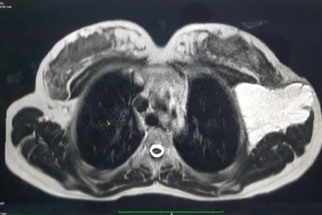
Shukla S et al. Int Surg J. 2021 Jan;8(1):391-394

5.    Perkins JA, Manning SC, Tempero RM,                            14. Smith RC, Sherk HH, Kollmer C, Javitt MC. Cystic
      Cunningham MJ, Edmonds JL, Hoffer FA.                              lymphangioma in the adult: an unusual axillary mass.
      Lymphatic malformations: review of current                         Magn Reson Imaging. 1989;7:561-3.
      treatment. Otolaryngol Head Neck Surg.                         15. Schefter RP, Olsen KD, Gaffey TA. Cervical
      2010;142:795-803.                                                  lymphangioma in the adult. Otolaryngol Head Neck
6.    Adams MT, Saltzman B, Perkins JA. Head and neck                    Surg. 1985;93(1):65-9.
      lymphatic malformation treatment: a systematic                 16. Kennedy TL, Whitaker M, Pellitteri P, Wood WE.
      review.      Otolaryngol      Head     Neck     Surg.              Cystic hygroma/lymphangioma: a rational approach
      2012;147(4):627-39.                                                to management. Laryngoscope. 2001;111(11):1929-
7.    Philippakis GE, Manoloudakis N, Marinakis A. A                     37.
      rare case of a giant cavernous lymphangioma of the             17. Smith RJ, Burke DK, Sato Y, Poust RI, Kimura K,
      chest wall and axilla in an adult patient. Int J Surg              Bauman NM. OK-432 therapy for lymphangiomas.
      Case Rep. 2013;4(2):164-6.                                         Arch       Otolaryngol    Head      Neck       Surg.
8.    Krainick-strobel U, Krämer B, Walz-Mattmüller R,                   1996;122(11):1195-9.
      Kaiserling E, Rohm C, Bergmann A. Massive                      18. Mulliken JB, Glowacki J. Hemangiomas and
      cavernous lymphangioma of the breast and thoracic                  vascular malformations in infants and children: a
      wall: case report and literature review. Lymphology.               classification based on endothelial characteristics.
      2006;39(3):147-51.                                                 Plast Reconstr Surg. 1982;69(3):412-22.
9.    Michail O, Michail P, Kyriaki D, Kolindou A,                   19. De Serres LM, Sie KC, Richardson MA. Lymphatic
      Klonaris C, Griniatsos J. Rapid development of an                  malformations of the head and neck. A proposal for
      axillary mass in an adult: a case of cystic hygroma.               staging. Arch Otolaryngol Head Neck Surg.
      South Med J. 2007;100:845-9.                                       1995;121(5):577-82.
10.   Stoss S, Kalbermatten DF, Robertson A, Bruder E,               20. Sherman BE, Kendall K. A unique case of the rapid
      Rasmus M, Gambazzi F. Large cystic tumour at the                   onset of a large cystic hygroma in the adult. Am J
      chest wall mimicking an echinococcosis: a case                     Otolaryngol. 2001;22(3):206-10.
      report. J Plast Reconstr Aesthet Surg. 2008;61:13-6.           21. Riechelmann H, Muehlfay G, Keck T, Mattfeldt T,
11.   Nguyen K, Karsif K, Lee S, Chorny K, Chen M.                       Rettinger G. Total, subtotal, and partial surgical
      Lymphangioma in an elderly patient: an unusual                     removal of cervicofacial lymphangiomas. Arch
      cause of axillary mass. Breast. 2011;17:416-26.                    Otolaryngol Head Neck Surg. 1999;125(6):643-8.
12.   Gebrim LH, de Lima GR, Tanaka CI. Axillary cystic
      lymphangioma in pregnancy. Int J Gynaecol Obstet.
      1995;48:327-8.                                                    Cite this article as: Shukla S, Shivhare R, Lal V,
13.   Katharina C, Quack L, Abdul-Raham J, Elisabeth G,                 Rathore D. Cystic lymphangioma of breast and
      Judit P, Ernst B. Axillary cavernous lymphangioma                 axillary region in an adult: a rare presentation. Int Surg
      in pregnancy and puerperium. Gynecol Obstet Invest.               J 2021;8:391-4.
      2005;60:108-11.

                                                              International Surgery Journal | January 2021 | Vol 8 | Issue 1   Page 394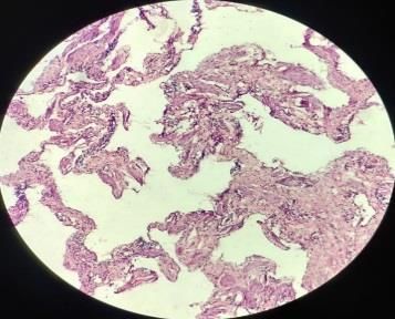

You can also read