A rare case report of sinonasal undifferentiated carcinoma of paranasal sinuses
←
→
Page content transcription
If your browser does not render page correctly, please read the page content below
International Journal of Otorhinolaryngology and Head and Neck Surgery
Ratti J et al. Int J Otorhinolaryngol Head Neck Surg. 2021 Jan;7(1):177-179
http://www.ijorl.com                                                                         pISSN 2454-5929 | eISSN 2454-5937

                                                           DOI: https://dx.doi.org/10.18203/issn.2454-5929.ijohns20205645
Case Report

           A rare case report of sinonasal undifferentiated carcinoma of
                                 paranasal sinuses
                     Jasmine Ratti*, Vishav Yadav, Sanjeev Bhagat, Dinesh K. Sharma

  Department of ENT, Government Medical College, Patiala, Punjab, India

  Received: 23 October 2020
  Accepted: 03 December 2020

  *Correspondence:
  Dr. Jasmine Ratti,
  E-mail: jasmineratti7992@gmail.com

  Copyright: © the author(s), publisher and licensee Medip Academy. This is an open-access article distributed under
  the terms of the Creative Commons Attribution Non-Commercial License, which permits unrestricted non-commercial
  use, distribution, and reproduction in any medium, provided the original work is properly cited.

   ABSTRACT

   Sino nasal malignancies account for only 0.2-0.8% of all malignancies and undifferentiated carcinoma is rare
   malignant tumour of sinonasal tract, with extremely poor prognosis. We report a case of sinonasal undifferentiated
   carcinoma which we managed by surgery followed by post-operative radiotherapy with concomitant platinum-based
   chemotherapy. Although the overall survival is about 20% at 5 years and there are frequent recurrences combined
   modality treatment is the best management option available at present.

   Keywords: Sinonasal undifferentiated carcinoma, Maxillectomy, Blue small round cell tumors

INTRODUCTION                                                           nature. History of left sided nasal bleed, spontaneous,
                                                                       intermittent, 2-3 episodes in a week self-limiting in
Sino nasal malignancies are rare malignancies with an                  nature. Patient had associated symptom of blood-tinged
incidence of 0.5-1 per 100 000 per year and account for                foul smelling anterior nasal discharge and posterior nasal
only 0.2-0.8% of all malignancies and 3% of upper aero                 discharge. There was associated occasional dull aching
digestive tract neoplasm.1,2 Mostly develop in the fifth               pain on left side of face and excessive watering from left
and sixth decades of life.3 Sinonasal undifferentiated                 eye also.
carcinoma was described relatively recently by Frierson
et al.4 It is otherwise known as anaplastic carcinoma and              On DNE- left nasal cavity-A single firm friable yellowish
is hard to distinguish from high-grade olfactory                       mass was present in left nasal cavity between the septum
esthesioneuroblastoma. It is a highly aggressive and                   and middle turbinate arising from middle meatus, which
invasive tumour produces rather subtle symptoms                        bled on touch, probe could not be passed superiorly and
initially despite its extensive nature.5 Sino nasal                    laterally. Deviated nasal septum to left with septal spur
undifferentiated carcinoma is believed to originate from               inferiorly was noted.
Schneiderian epithelium or from the nasal ectoderm of
the paranasal sinuses.4 It typically presents as a rapidly             Visual acuity and extra ocular movements were normal,
enlarging tumor mass involving multiple (sinonasal tract)              Bilateral pupillary reflex (direct and consensual) were
sites, often with an evidence of extension beyond the                  equally reactive to light.
anatomic confines of the sinonasal tract.
                                                                       Non contrast computer tomogram (NCCT NOSE PNS)
CASE REPORT                                                            both coronal and axial cuts. Findings were, soft tissue
                                                                       density filling left nasal cavity, anterior and posterior
A 42-year-old female presented to ENT OPD with                         ethmoids, left maxillary sinus, blocking left osteomeatal
complaint of left sided nasal obstruction and bleeding                 complex, erosions were noted at the level of horizontal
from left nasal cavity since past 3 months. On left side               and vertical lamella of cribriform plate, left lamina
nasal obstruction was insidious in onset progressive in                erosion was noted but no involvement of periorbita.

                    International Journal of Otorhinolaryngology and Head and Neck Surgery | January 2021 | Vol 7 | Issue 1   Page 177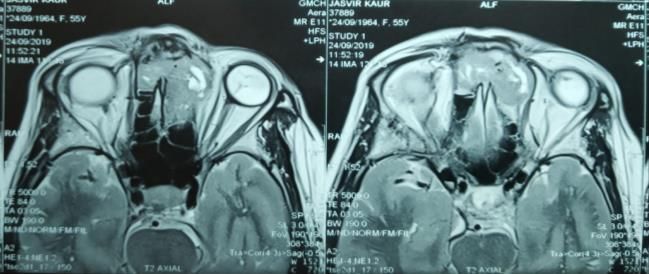
Ratti J et al. Int J Otorhinolaryngol Head Neck Surg. 2021 Jan;7(1):177-179
Excised tissue and margin sent for HPE. Haemostasis
was secured, post-operative period was uneventful.
HPE showed invasive tumor arranged in diffused sheets
and in perivascular pattern. Tumor cells showing
moderate to marked anaplasia with vesicular to
hyperchromatic nuclei with prominent nucleoli with high
atypical mitotic activity. Tumor giant cells were also
seen. Features suggestive of undifferentiated malignant
tumor.
Immunohistochemistry was reactive for CD56, CK and
Figure 1: NCCT nose PNS axial sections of soft tissue CDX-2, suggestive of sino-nasal undifferentiated
density material filling left nasal cavity ethmoids and carcinoma.
eroding lamina papyracea.
Patient then underwent external beam radiotherapy 66 Gy
Patient underwent diagnostic biopsy under GA, tissue in 30 fractions in 6 weeks by volumetric modulated arc
was sent for HPE. radiotherapy. PET CT was after radiotherapy and was
suggestive of FDG avid soft tissue thickening left
HPE showed tumor cells with cylindrical basaloid ethmoid sinus but no metabolically active lymph nodes.
morphology lying in papillary, ribbon like pattern with Re-surgery was not possible was hence planned with
central necrosis. Cells exhibit marked nuclear atypia and chemotherapy with cisplatin and etoposide. Patient was
brisk mitotic activity. Tumor is devoid of significant symptomatically relieved.
keratinisation. HPE features are those of Non keratinizing
transitional cell carcinoma of nasal cavity Later patient developed cutaneous fistula communicating
from left nasolabial fold to nasal cavity.
In view of malignant nature of lesion, patient was
planned for excision via lateral rhinotomy approach
followed by post op radiotherapy. Pre-operative MRI was
done and was s/o tumor extending intracranially but
extradural.
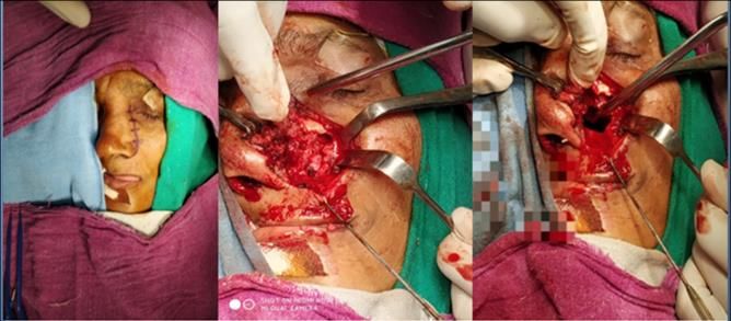
Figure 3: Left medial maxillectomy being performed
via lateral rhinotomy approach.
DISCUSSION
Figure 2: MRI of heterogenous mass which is Sinonasal undifferentiated carcinoma of para nasal
hypointense on T2WI axial sections with multiple sinuses is extremely rare accounting for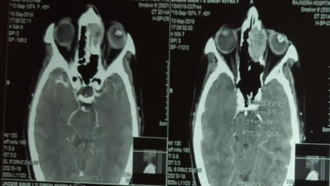
Ratti J et al. Int J Otorhinolaryngol Head Neck Surg. 2021 Jan;7(1):177-179

crucial for differentiating between these entities.                      head      and       neck      tumors.      2nd      ed.
Radiologic findings show a more anterior involvement of                  Philadelphia: Saunders. 1999;558-81.
the nasal cavity and ethmoids by sinonasal                           3. Goldenberg D, Golz A, Fradis M, Netzer A,
undifferentiated    carcinoma    as     compared     to                  Joachims HZ. Malignant tumors of the nose and
nasopharyngeal carcinoma.6                                               paranasal sinuses: az retrospective review of 291
                                                                         cases. Ear Nose Throat J. 2001;80:272-7.
Multimodality therapy (surgery, radiotherapy and                     4. Frierson HF, Mills SE, Fechner RE, Taxy JB, Levine
chemotherapy) has generally been demonstrated to be the                  PA. Sinonasal undifferentiated carcinoma. Am J
most effective approach in the treatment of squamous cell                Surg Pathol. 1986;10:771-9.
carcinoma and sinonasal undifferentiated carcinoma. Due              5. Su SY, Bell D, Hanna EY. Esthesioneuroblastoma,
to its extension outside the natural anatomical boundaries               neuroendocrine       carcinoma,     and      sinonasal
of the nasal cavity and PNS, results in morbid surgery                   undifferentiated carcinoma: differentiation in
which include maxillectomy, orbital exenteration and                     diagnosis and treatment. Int Arch Otorhinolaryngol.
craniotomies.7-9                                                         2014;18:S149-56.
                                                                     6. Smith SR, Som P, Fahmy A, Lawson W, Sacks S,
If operable surgery followed by post-operative                           Brandwein M. A Clinicopathological Study of
radiotherapy      with    concomitant      platinum-based                Sinonasal Neuroendocrine Carcinoma and Sinonasal
chemotherapy is the most common approach. In large                       Undifferentiated.      Carcinoma       Laryngoscope.
volume tumours initial non-surgical treatment with                       2000;110:1617-22.
(chemo)radiotherapy or chemotherapy alone followed by                7. Yoshida E, Aouad R, Fragoso R, Farwell DG,
chemoradiotherapy appears to give better results.10,11 Now               Gandour-Edwards R, Donald PJ et al. Improved
a day’s treatment with proton beam radiotherapy where                    clinical outcomes with multimodality therapy for
available is also coming into picture, with the possibility              sinonasal undifferentiated carcinoma of the head and
of reducing radiation-related morbidity.12                               neck. Am J Otolaryngol. 2013;34(6):658-63.
                                                                     8. Reiersen DA, Pahilan ME, Devaiah AK. Meta-
Similarly, our case also underwent surgical resection                    analysis of treatment outcomes for sinonasal
followed    by    postoperative  radiotherapy    and                     undifferentiated carcino- ma. Otolaryngol Head Neck
chemotherapy sessions.                                                   Surg, 2012;147:7-14.
                                                                     9. Kim BS, Vongtama R, Juillard G. Sinonasal
CONCLUSION                                                               undifferentiated carcinoma: case series and literature
                                                                         review. Am J Otolaryngol. 2004;25:162-6.
Sino nasal undifferentiated carcinoma is rare malignant              10. National Institute for Health and Care Excellence.
tumour of sinonasal tract, with extremely poor prognosis.                Cancer of the upper aerodigestive tract: assessment
The overall survival is about 20% at 5 years. There is                   and management in people aged 16 and over.
frequent recurrence with metastasis to lymph nodes and                   London:      NICE;      2016.      Available     from:
distant sites. Combined modality treatment is the best                   https://www.nice. org.uk/Guidance/NG36/evidence.
management option which is Surgical excision followed                    Accessed on 10/10/2020.
by adjuvant chemotherapy or radiotherapy.                            11. Lund VJ, Clarke PM, Swift AC. Nose and paranasal
                                                                         sinus tumours: United Kingdom National
Funding: No funding sources                                              Multidisciplinary Guidelines. J Laryngol Otol.
Conflict of interest: None declared                                      2016;130(S2):S111-8.
Ethical approval: Not required                                       12. Christopherson K, Werning JW, Malyapa RS.
                                                                         Radiotherapy      for    sinonasal    undifferentiated
REFERENCES                                                               carcinoma. Am J Otolaryngol. 2014;35(2):141-6.

1.   Dulguerov P, Jacobsen MS, Allal AS, Lehmann W,
     Calcaterra T. Nasal and paranasal sinus carcinoma:                Cite this article as: Ratti J, Yadav V, Bhagat S,
     are we making progress? Cancer. 2001;92:3012-29.                  Sharma DK. A rare case report of sinonasal
2.   Rice DH, Stanley RB. Surgical therapy of tumors of                undifferentiated carcinoma of paranasal sinuses. Int J
     the nasal cavity, ethmoid sinus, and maxillary sinus.             Otorhinolaryngol Head Neck Surg 2021;7:177-9.
     In: Panje W (ed). Comprehensive management of

                  International Journal of Otorhinolaryngology and Head and Neck Surgery | January 2021 | Vol 7 | Issue 1   Page 179
You can also read