Meningioma infiltrating into porous polymethylmethacrylate cranioplasty-report of a unique case
←
→
Page content transcription
If your browser does not render page correctly, please read the page content below
Journal of Surgical Case Reports, 2020;6, 1–3

                                                                               doi: 10.1093/jscr/rjaa149
                                                                               Case Report

                                                                                                                                                             Downloaded from https://academic.oup.com/jscr/article/2020/6/rjaa149/5859610 by Universitaetsbibliothek Regensburg user on 30 October 2020
CASE REPORT

Meningioma infiltrating into porous
polymethylmethacrylate cranioplasty—report
of a unique case
Karl-Michael Schebesch*, Martin Proescholdt, Nils Ole Schmidt, and
Julius Höhne
Department of Neurosurgery, University Medical Center Regensburg, Regensburg, Germany
*Correspondence address. Department of Neurosurgery, University Medical Center Regensburg, Franz-Josef-Strauss Allee 11, 93053 Regensburg, Germany.
Tel: ++49 +941 944 9001; Fax: ++49 +941 944 9002; E-mail: karl-michael.schebesch@ukr.de

Abstract
Implantation of a cranioplasty after osteoclastic craniotomy or craniectomy is one of the most common neurosurgical
procedures, and polymethylmethacrylate (PMMA) is one of the most frequently applied materials for cranioplasty. This report
describes the unique case of a patient with recurrent transitional meningioma WHO I that infiltrated the PMMA cranioplasty
7 years after primary surgery. We propose to restrict the use of porous PMMA as cranioplasty after the removal of convexity
meningioma.

INTRODUCTION                                                                   CASE REPORT
Remodeling the calvarial bone with freehand polymethyl-                        In 2012, the 45-year-old female patient presented at a neuro-
methacrylate (PMMA) cranioplasty after craniectomy is a                        surgical department with aphasia and facial palsy. Magnetic
very common neurosurgical procedure. Apart from freehand                       resonance imaging (MRI) showed a large space-occupying
PMMA application after decompressive hemicraniectomy or                        tumor infiltrating the frontal bone. Consequently, the tumor
postoperative osteomyelitis in stroke patients, some authors                   was removed, and histological workup confirmed a benign
have advocated the use of computer-assisted design and                         meningioma WHO I. Two years later, a CAD/CAM non-resorbable
computer-assisted manufacturing (CAD/CAM) cranioplasties                       biocompatible cranioplasty (BIOMET, Germany) composed
[1, 2], also after the removal of infiltrated skull or hyperostotic            of PMMA spherical macro beads, coated and fused with
bone in meningioma surgery [3, 4]. PMMA is one of the most                     polyhydroxyethylmethacrylate, was implanted, see Fig. 1. Until
frequently applied materials for cranioplasty. To the best of                  February 2018, consecutive MRIs had shown a tumor-free
our knowledge, tumorous invasion of PMMA in a patient with                     area, and the clinical course had been uneventful. The MRI
recurrent meningioma has never been reported. We describe                      conducted in February 2018 and the subsequent MRI in June 2019
the case of a patient with recurrent meningioma WHO I that                     (Fig. 2A and B) showed a progressive contrast-enhancing mass
infiltrated the implanted PMMA cranioplasty 7 years after                      along the falx cerebri that was strongly suspicious of recurrent
primary surgery.                                                               meningioma. Therefore, revision surgery was recommended

Received: April 9, 2020. Accepted: April 27, 2020
Published by Oxford University Press and JSCR Publishing Ltd. All rights reserved. © The Author(s) 2020.
This is an Open Access article distributed under the terms of the Creative Commons Attribution Non-Commercial License (http://creativecommons.org/
licenses/by-nc/4.0/), which permits non-commercial re-use, distribution, and reproduction in any medium, provided the original work is properly cited. For
commercial re-use, please contact journals.permissions@oup.com

                                                                                                                                                    1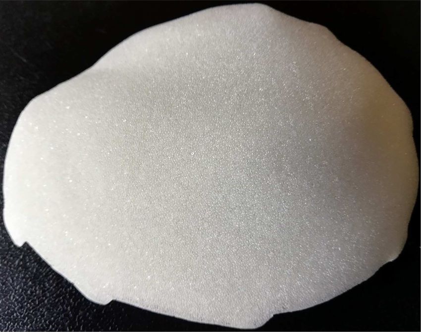
2 K.-M. Schebesch et al.
were cryosected and sections were fixed in 4% paraformalde-
hyde and washed in phosphate buffered saline. To detect menin-
gioma cell infiltration, cryosections were immunostained using
a human epithelial membrane antigen (EMA) specific mouse
monoclonal antibody (clone E 29, Agilent DAKO, Santa Clara, CA,
USA). To exclude glial cell infiltration, adjacent sections were
stained with a polyclonal rabbit anti-human glial fibrillary acidic
protein (GFAP) antibody (Agilent DAKO, Santa Clara, CA, USA).
Downloaded from https://academic.oup.com/jscr/article/2020/6/rjaa149/5859610 by Universitaetsbibliothek Regensburg user on 30 October 2020
To test for unspecific antibody binding, control sections were
incubated either in non-immunized mouse IgG (EMA staining)
or non-immunized rabbit IgG (GFAP staining) at identical protein
concentrations.
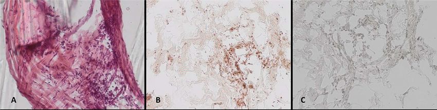
Pieces of the infiltrated cranioplasty were microscopically
examined after topical staining as described above. Immuno-
histology clearly identified meningioma cell formations inside
the cranioplasty (Fig. 3B and C), and topographical microscopy
showed meningioma formations along the preformed caverns
(Fig. 3A).
Figure 1: Model of a CAD/CAM non-resorbable biocompatible cranioplasty
(Biomet, Germany) composed of polymethylmethacrylate (PMMA) spherical
macro beads coated and fused with polyhydroxyethylmethacrylate (PMHA). DISCUSSION
Only a few articles in the neurosurgical literature have focused
to remove the tumorous mass along the falx. Before surgery, on tumor infiltration into the cranial flap. In 1994, Wester
computed tomography (CT) was carried out to visualize the described six patients undergoing reimplantation of tumor-
bony attachments of the PMMA cranioplasty (Fig. 2C). Neither infiltrated autologous bone flaps after autoclaving. In 1997,
imaging modality had depicted any tumorous tissue inside Vanaclocha et al. published their experiences in reimplanting
the cranioplasty. Thus, preoperatively, the cranioplasty was not autoclaved bone flaps in 62 patients with various benign
considered an area of tumor infiltration. and malignant skull-infiltrating tumors [5]. In the histological
Revision surgery with the intent to remove the recurrent evaluation of bone samples after the autoclaving procedure,
meningioma was conducted in November 2019. The initial the authors found no living tumor cells inside the bone but
PMMA cranioplasty that moderately adhered to the dura was preserved mineral matrices. This finding, in combination with
removed in one piece. After splitting of the cranioplasty, several the low rate of bone resorption during follow-up, resulted in
pieces were sent to the department of neuropathology because the authors’ conclusion that autoclaved calvarial bone flaps are
of the surgeon’s strong impression that tumor tissue had safe and feasible. It is noteworthy that no long-term follow-up
infiltrated the porous material of the cranioplasty. Further focusing on tumor regrowth or reinfiltration was provided in
surgery was uneventful: the tumor was dissected and completely this large series.
removed, the convexity dura was reconstructed, and a preformed On the other hand, many large studies show a very high rate
titanium cranioplasty was inserted. Histologically, the tumor of up to 25% of surgical revision after reimplantation of autolo-
was confirmed as transitional meningioma WHO I. gous bone flaps due to bone resorption [6, 7]. The main reason for
replacing bones with artificial materials, such as PMMA, hydrox-
Immunohistochemical analysis of meningioma cell
yapatite bioceramics, titanium and others, is aseptic bone necro-
infiltration
sis [8, 9] that can be gradually visualized as a shrinking bone flap
The fragments were frozen in isopentane and embedded in Tis- in CT. Osteoconductive materials such as hydroxyapatite have
sue Tek® OCT compound (Sakura, Staufen, Germany). Samples been intensively examined histologically. Bruno et al. reported
Figure 2: (A–C) Preoperative neuroimaging shows recurrent meningioma along the falx cerebri and under the cranioplasty (A: native CT scan, axial plane; B: contrast-
enhanced MRI, coronal plane; C: contrast-enhanced MRI, sagittal plane).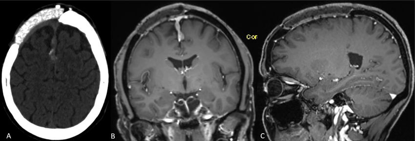
Meningioma infiltrating into porous polymethylmethacrylate cranioplasty                       3

                                                                                                                                                                        Downloaded from https://academic.oup.com/jscr/article/2020/6/rjaa149/5859610 by Universitaetsbibliothek Regensburg user on 30 October 2020
Figure 3: (A–C) Histological work-up of the explanted cranioplasty: (A) hematoxylin/eosin staining, immunohistochemical staining for (B) epithelial membrane antigen,
and (C) GFAP (all ×20 magnification).

that neo-formed lamellar and trabecular bone tissue fragments                              preformed titanium versus freehand molded Polymethyl-
that are accompanied by amorphous reticular tissue showed                                  methacrylate implants. J Neurol Surg A Cent Eur Neurosurg
diffuse ossification in explanted bioceramic cranioplasties [10].                          2018;79:200–5.
This intense osteo-conductive effect obviously facilitates the                        2.   Marbacher S, Andereggen L, Erhardt S, Fathi AR, Fandino J,
osseous integration of artificial material into the skull; however,                        Raabe A, et al. Intraoperative template-molded bone flap
invasive neoplastic engraftment may be potentially devastating,                            reconstruction for patient-specific cranioplasty. Neurosurg
particularly in recurrent skull-infiltrating bone tumors.                                  Rev 2012;35:527–35.
    Currently, most neurosurgeons prefer the primary removal                          3.   Eppley BL, Kilgo M, Coleman JJ 3rd. Cranial reconstruc-
of the potentially tumor-infiltrated autologous bone flap and                              tion with computer-generated hard-tissue replacement
replacement with a CAD/CAM cranioplasty [1] or, at least, with                             patient-matched implants: indications, surgical technique,
a simple PMMA reconstruction.                                                              and long-term follow-up. Plast Reconstr Surg 2002;109:
    The most common skull-infiltrating tumor is meningioma,                                864–71.
most of which are graded WHO I. To the best of our knowledge,                         4.   Frassanito P, De Bonis P, Mattogno PP, Mangiola A, Novello M,
no study has ever evaluated whether recurrent meningioma                                   Brinchi D, et al. The fate of a macroporous hydroxyapatite
has the propensity to infiltrate artificial materials covering and                         cranioplasty four years after implantation: macroscopical
integrating into the osteoclastic defect and, if so, which materials                       and microscopical findings in a case of recurrent atypical
have a higher risk of meningioma growth. However, one cannot                               meningioma. Clin Neurol Neurosurg 2013;115:1496–8.
necessarily assume that tumor growth inside the cranioplasty                          5.   Vanaclocha V, Saiz-Sapena N, Garcia-Casasola C, De Alava
is significant or results in more extensive tumor regrowth. It is                          E. Cranioplasty with autogenous autoclaved calvarial bone
noteworthy that our findings in this unique case are in contrast                           flap in the cases of tumoural invasion. Acta Neurochir
to the case report of Frassanito and co-workers who presented                              1997;139:970–6.
the case of recurrent atypical meningioma that recurred without                       6.   Wachter D, Reineke K, Behm T, Rohde V. Cranioplasty
infiltrating the hydroxyapatite cranioplasty [4].                                          after decompressive hemicraniectomy: underestimated
    However, we report this unique observation that resulted in                            surgery-associated complications? Clin Neurol Neurosurg
the departmental decision to stop reconstructing the skull with                            2013;115:1293–7.
any porous, bone-like, or osteoconductive material after menin-                       7.   Zanaty M, Chalouhi N, Starke RM, Clark SW, Bovenzi CD,
gioma surgery. We now exclusively use titanium or a preformed                              Saigh M, et al. Complications following cranioplasty:
calcium phosphate composition whenever necessary and do not                                incidence and predictors in 348 cases. J Neurosurg
reinsert the autoclaved autologous skull. Further in-vitro and in-                         2015;123:182–8.
vivo studies are mandatory to shed light on the potential of                          8.   Piitulainen JM, Kauko T, Aitasalo KM, Vuorinen V, Vallittu PK,
meningioma invasion into cranioplasty.                                                     Posti JP. Outcomes of cranioplasty with synthetic materials
                                                                                           and autologous bone grafts. World Neurosurg 2015;83:708–14.
                                                                                      9.   Reddy S, Khalifian S, Flores JM, Bellamy J, Manson PN,
CONFLICT OF INTEREST STATEMENT                                                             Rodriguez ED, et al. Clinical outcomes in cranioplasty: risk
None declared.                                                                             factors and choice of reconstructive material. Plast Reconstr
                                                                                           Surg 2014;133:864–73.
                                                                                     10.   Bruno Z, Angelo N, Riccardo S, Nicola Z, Stefano P, Camillo PP,
REFERENCES                                                                                 et al. Custom-made hydroxyapatite cranioplasty: radiologi-
 1. Hohne J, Werzmirzowsky K, Ott C, Hohenberger C, Has-                                   cal and histological evidence of bone-biomaterial osteointe-
    sanin BG, Brawanski A, et al. Outcomes of Cranioplasty with                            gration in five patients. Asian J Neurosurg 2020;15:198–203.
You can also read