Effectiveness of Multidetector Computed Tomography in Prosthetic Valve Endocarditis - Scientific Research Publishing
←
→
Page content transcription
If your browser does not render page correctly, please read the page content below
Open Journal of Thoracic Surgery, 2021, 11, 31-35
                                                                                                 https://www.scirp.org/journal/ojts
                                                                                                             ISSN Online: 2164-3067
                                                                                                               ISSN Print: 2164-3059

Effectiveness of Multidetector Computed
Tomography in Prosthetic Valve Endocarditis

Kayo Sugiyama, Hirotaka Watanuki, Masaho Okada, Yasuhiro Futamura, Masayuki Saito,
Satoshi Makino, Katsuhiko Matsuyama

Department of Cardiac Surgery, Aichi Medical University Hospital, Nagakute, Japan

How to cite this paper: Sugiyama, K., Wa-       Abstract
tanuki, H., Okada, M., Futamura, Y., Saito,
M., Makino, S. and Matsuyama, K. (2021)         Background: Redo aortic valve replacement for prosthetic valve endocarditis
Effectiveness of Multidetector Computed         is a challenge for surgeons. Echocardiography is occasionally not an effective
Tomography in Prosthetic Valve Endocardi-
                                                modality for the detection of infectious signs in prosthetic valve endocarditis.
tis. Open Journal of Thoracic Surgery, 11,
31-35.
                                                Case presentation: Herein, we report the case of a patient whose prosthetic
https://doi.org/10.4236/ojts.2021.111005        valve endocarditis was detected by multidetector computed tomography and
                                                who successfully underwent redo aortic valve replacement. Preoperative
Received: January 6, 2021
                                                echocardiography revealed no remarkable findings related to endocarditis
Accepted: March 15, 2021
Published: March 18, 2021
                                                such as perivalvular leakage or vegetation; however, multidetector computed
                                                tomography revealed a thickened right coronary cusp. Intraoperatively, the
Copyright © 2021 by author(s) and               right coronary cusp was confirmed to be covered with thick infected tissue.
Scientific Research Publishing Inc.
                                                The pathological findings revealed broad destruction due to infection of the
This work is licensed under the Creative
Commons Attribution International
                                                right coronary cusp. Conclusion: Multidetector computed tomography was
License (CC BY 4.0).                            useful in detecting infectious signs in prosthetic valves.
http://creativecommons.org/licenses/by/4.0/
                Open Access                     Keywords
                                                Prosthetic Valve Endocarditis, Multidetector Computed Tomography,
                                                Compromised Host

                                              1. Introduction
                                              Echocardiography is a well-known, effective modality for diagnosing infective
                                              endocarditis (IE); however, it can occasionally be difficult to evaluate signs of
                                              infection in prosthetic valve endocarditis (PVE). Echocardiography is the “gold
                                              standard” for assessing the anatomy of cardiac valves and peri-valvular appara-
                                              tus; however, the effectiveness may be limited by the patient’s morphology and
                                              artifacts due to valvular calcifications or prosthetic material [1] [2]. Multidetec-
                                              tor computed tomography (MDCT) may be effective for the structural evalua-

DOI: 10.4236/ojts.2021.111005        Mar. 18, 2021                 31                              Open Journal of Thoracic Surgery
K. Sugiyama et al.

                                tion of prosthetic valves because it can eliminate artifacts caused by artificial
                                structures that are peculiarly seen on echocardiography [2]. By synchronizing
                                the imaging acquisition with the cardiac pulsation, a more detailed image of the
                                intracardiac structure can be obtained in this modality.
                                   Herein, we report a case of a patient who successfully underwent redo aortic
                                valve replacement after an accurate diagnosis of PVE by MDCT.

                                2. Case Presentation
                                The patient was a 64-year-old woman who had undergone ventricular septal de-
                                fect closure at the age of 22. She underwent residual shunt closure and pulmo-
                                nary valve repair for pulmonary valve stenosis at the age of 45. One year pre-
                                viously, she was diagnosed with bicuspid aortic valve induced progression of
                                aortic valve stenosis and ascending aortic dilatation and underwent the follow-
                                ing procedures: 1) aortic valve replacement using a 23-mm INSPIRIS RESILIA
                                aortic valveTM (Edwards Lifesciences, Irvine, USA) for aortic valve stenosis of the
                                bicuspid aortic valve; 2) graft replacement for dilated ascending aorta using a
                                30-mm Triplex one-branched graftTM (Terumo Corporation, Tokyo, Japan); and
                                3) left atrial appendage closure for chronic atrial fibrillation. She was also suf-
                                fering from idiopathic pancytopenia, with laboratory data as follows: white
                                blood cell count, 2.3 × 10³/μL; neutrophils, 56%; hemoglobin level, 9.1 mg/dL;
                                platelet count, 5.0 × 10⁵/μL; C-reactive protein level, 0.04 mg/dL; procalcitonin,
                                0.06 ng/mL; and N-terminal pro-brain natriuretic peptide, 49 pg/mL. Four
                                months previously, she had developed a high fever due to sepsis. Antibiotic
                                therapy was initiated because methicillin-resistant coagulase-negative staphylo-
                                cocci were detected in the blood culture. After antibiotic treatment with teicop-
                                lanin for 4 weeks, she was discharged with a prescription for oral tedizolid
                                phosphate; however, it was discontinued because of severe nausea. Transthoracic
                                and transesophageal echocardiography during hospitalization did not detect any
                                significant signs related to PVE.
                                   Four months later, she was readmitted in an emergency, complaining of fever
                                and general fatigue. Transthoracic and transesophageal echocardiography did
                                not detect any signs related to PVE (Figure 1(a)); however, MDCT revealed
                                thickening of the right coronary cusp (Figure 1(b), Figure 1(c)). Preoperative
                                laboratory data indicated inflammatory changes and pancytopenia, with the fol-
                                lowing results: white blood cell count, 2.5 × 10³/μL; neutrophils, 63%; hemoglo-
                                bin level, 10.5 mg/dL; platelet count, 5.6 × 10⁵/μL; C-reactive protein level, 2.3
                                mg/dL; procalcitonin, 0.28 ng/mL; and N-terminal pro-brain natriuretic peptide,
                                187 pg/mL.
                                   Therefore, surgical treatment was performed through a median re-sternotomy.
                                Cardiopulmonary bypass was instituted via bicaval drainage and ascending aor-
                                tic cannulation. After cardiac arrest induced through antegrade cardioplegia, the
                                exposure of the prosthetic valve was obtained through the ascending aorta. The
                                right coronary cusp was covered with thick vegetation (Figure 2(a)); however,
                                the remaining leaflets appeared intact, and there was no evidence of an obvious

DOI: 10.4236/ojts.2021.111005                        32                             Open Journal of Thoracic Surgery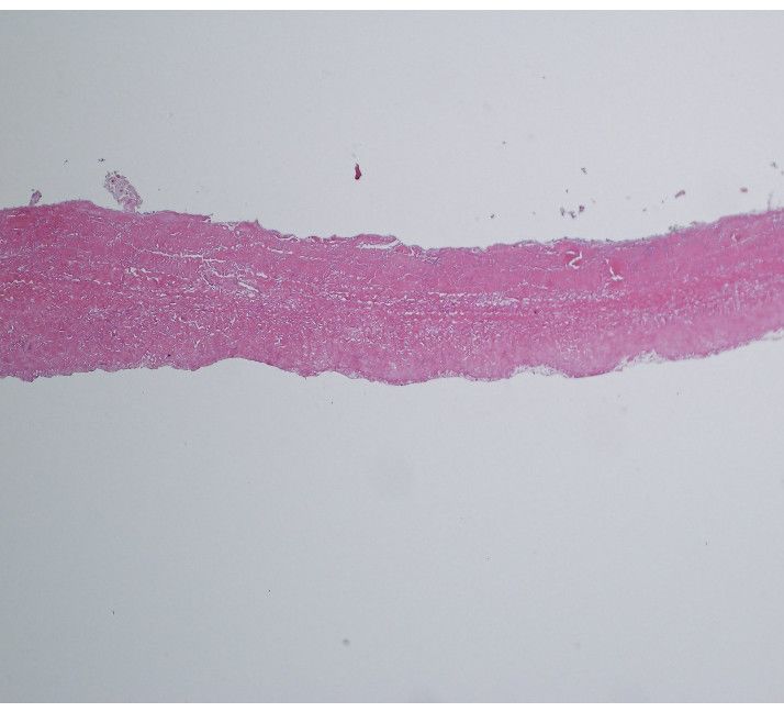
K. Sugiyama et al.

                                              (a)                          (b)                           (c)

                                Figure 1. (a) Preoperative transesophageal echocardiography showing no remarkable in-
                                fectious signs in the bioprosthesis; (b) Preoperative multidetector computed tomography
                                showing thickening of the right coronary cusp in the bioprosthesis (axial view); (c) Preo-
                                perative multidetector computed tomography showing thickening of the right coronary
                                cusp in the bioprosthesis (sagital view).

                                              (a)                          (b)                           (c)

                                Figure 2. (a) Resected prosthetic valve with thickened vegetation in one leaflet without
                                destruction of the annulus; (b) Postoperative pathological findings showing broad de-
                                struction with a thickened mass of bacteria and dense collection of neutrophils and ma-
                                crophages in the leaflet; (c) Postoperative pathological findings showing an almost intact
                                leaflet without any infectious signs.

                                annular abscess or detachment of the prosthetic valve. After complete removal of
                                the previous prosthesis, redo aortic valve replacement with a 23-mm INSPIRIS
                                RESILIA aortic valveTM (Edwards Lifesciences, Irvine, USA) was performed. The
                                patient was weaned from cardiopulmonary bypass without difficulty, and the
                                postoperative course was uneventful. As the intraoperative culture also showed
                                the growth of methicillin-resistant coagulase-negative staphylococci, injections
                                of tedizolid phosphate were initiated. After 4 weeks of intravenous tedizolid
                                phosphate administration, oral intake of clindamycin was initiated at the time of
                                discharge. Pathological findings revealed broad destruction due to infection in
                                the right coronary cusp (Figure 2(b)); however, the remaining coronary cusps
                                and prosthetic annulus were intact (Figure 2(c)). The patient is currently reco-
                                vering well and has not presented any symptoms related to the recurrence of in-
                                fection for 12 months.

                                3. Discussion
                                This report presents a case with PVE in aortic valve bioprosthesis that has been
                                successfully treated by surgery. Transthoracic and transesophageal echocardio-
                                graphy did not detect any significant findings related to infection, and MDCT

DOI: 10.4236/ojts.2021.111005                          33                               Open Journal of Thoracic Surgery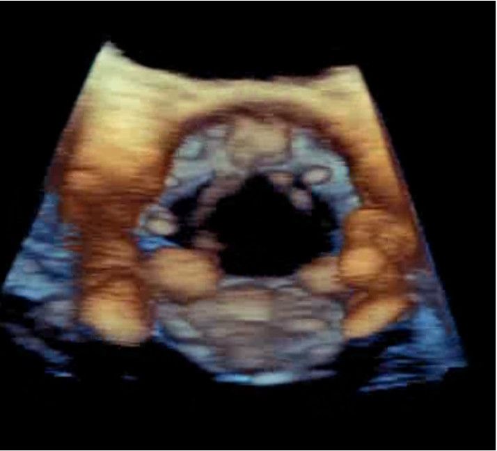
K. Sugiyama et al.

                                was effective for the evaluation of prosthetic valve leaflets.
                                  Although echocardiography is known to be an effective modality for diagnos-
                                ing infective endocarditis, it sometimes cannot detect the signs of infection in
                                PVE [1] [2]. The diagnosis of infectious endocarditis is based on the modified
                                Duke criteria, as well as clinical, biological, and imaging findings. Despite being
                                the “gold standard” for assessing cardiac valves and perivalvular apparatus,
                                echocardiography may be ineffective due to the patient’s morphology and arti-
                                facts caused by valvular calcifications or prosthetic material [1] [2]. Echocardio-
                                graphy has limited ability for perivalvular complications, especially for PVE [3].
                                Furthermore, echocardiography requires a highly trained operator, and the re-
                                sults are, to a certain degree, operator-dependent. For patients with high clinical
                                suspicion of IE, multimodality imaging such as MDCT should be considered to
                                confirm or rule out IE [3]. Although its relative limitations imposed by radiation
                                exposure and the risk of nephrotoxicity associated with the injection of iodi-
                                nated contrast [3], MDCT can effectively evaluate the structure of prosthetic
                                valves by synchronizing the imaging acquisition with the cardiac pulsation to
                                eliminate artifacts caused by artificial structure and provide a more detailed im-
                                age of the intracardiac structure. MDCT has demonstrated promising results in
                                valvular and perivalvular damage, providing high-resolution anatomic informa-
                                tion and affording multiplanar reformations [2]. In accordance with Sollini et
                                al., the “3M” approach consisting of multimodality, multitracers, and multidis-
                                ciplinary is essential for the detection of cardiovascular infections [4]. In the case
                                of PVE, three other imaging-based findings are now included as either major or
                                minor criteria to be considered: 1) cardiac computed tomography (CT); 2) fluo-
                                rodeoxyglucose (FDG) positron emission tomography (PET)/CT or white blood
                                cell (WBC) single-photon emission CT (SPECT)/CT; and 3) embolic events or
                                infectious aneurysms [4].
                                  The present case revealed severe destruction due to infection in the right co-
                                ronary cusp, intraoperatively and pathologically; however, there were no evident
                                infectious signs in the annulus. There have been few reports describing the du-
                                rability against infection of INSPIRIS RESILIA aortic valve. Shang et al. reported
                                that the aggregation of macrophages and giant cells was reduced in an in vitro
                                rabbit model with novel bovine pericardial tissue [5]. This study revealed a re-
                                duced inflammatory response in a novel bovine pericardial tissue treated with
                                aldehyde capping chemistry and glycerolization [5]. Although further studies
                                regarding durability are warranted, the INSPIRIS RESILIA aortic valve showed
                                durability against infection in the annulus in the present case.

                                4. Conclusion
                                The patient successfully underwent redo aortic valve replacement for prosthetic
                                valve endocarditis. While echocardiography did not detect infectious signs be-
                                cause of artifacts, MDCT was useful for detecting infectious signs in prosthetic
                                valves.

DOI: 10.4236/ojts.2021.111005                         34                              Open Journal of Thoracic Surgery
K. Sugiyama et al.
Acknowledgements
We would like to thank the Honyaku Center for reviewing and editing the ma-
nuscript and our colleagues for their insightful comments.
Conflicts of Interest
The authors declare no conflicts of interest regarding the publication of this
paper.
References
[1] Habib, G., Lancellotti, P., Antunes, M.J., Bongiorni, M.G., Casalta, J.P., Del Zotti, F.,
et al. (2015) ESC Guidelines for the Management of Infective Endocarditis. Euro-
pean Heart Journal, 36, 3075-3123. https://doi.org/10.1093/eurheartj/ehv319
[2] Grob, A., Thuny, F., Villacampa, C., Flavian, A., Gaubert, J.Y., Raoult, D., et al.
(2014) Cardiac Multidetector Computed Tomography in Infective Endocarditis: A
Pictorial Essay. Insights Imaging, 5, 559-570.
https://doi.org/10.1007/s13244-014-0353-1
[3] Erba, P.A., Pizzi, M.N., Roque, A., Salaun, E., Lancellotti, P., Tornos, P., et al. (2019)
Multimodality Imaging in Infective Endocarditis. An Imaging Team within the En-
docarditis Team. Circulation, 140, 1753-1765.
https://doi.org/10.1161/CIRCULATIONAHA.119.040228
[4] Sollini, M., Berchiolli, R., Bolton, R.C.D., Rossi, A., Kirienko, M., Boni, R., et al.
(2017) The “3M” Approach to Cardiovascular Infections: Multimodality, Multitrac-
ers, and Multidisciplinary. Seminars in Nuclear Medicine, 12, 199-224.
https://doi.org/10.1053/j.semnuclmed.2017.12.003
[5] Shang, H., Claessens, S.M., Tian, B. and Wright, G.A. (2017) Aldehyde Reduction in
a Novel Pericardial Tissue Reduces Calcification Using Rabbit Intramuscular Mod-
el. Journal of Materials Science: Materials in Medicine; 28, 16.
https://doi.org/10.1007/s10856-016-5829-8
DOI: 10.4236/ojts.2021.111005 35 Open Journal of Thoracic SurgeryYou can also read