Morphologic changes of diving-related barotrauma lung injury in the setting of Birt-Hogg-Dubé
←
→
Page content transcription
If your browser does not render page correctly, please read the page content below
http://css.sciedupress.com                                Case Studies in Surgery                                        2017, Vol. 3, No. 4

                                                                                                          CASE REPORTS

Morphologic changes of diving-related barotrauma
lung injury in the setting of Birt-Hogg-Dubé
Maria J. Merino∗1 , Marston Linehan2 , Sara Gil1
1
  Translational Surgical Pathology, Laboratory of Pathology, National Cancer Institute, National Institutes of Health, United
States
2
  Urologic Oncology Branch, Center for Cancer Research, National Cancer Institute, National Institutes of Health, United States

Received: May 30, 2017                    Accepted: August 24, 2017           Online Published: November 20, 2017
DOI: 10.5430/css.v3n4p25                  URL: https://doi.org/10.5430/css.v3n4p25

A BSTRACT
Manifestations of barotrauma-associated lung injury with scuba diving are rarely reported, aside from anecdotic historical
accounts. The pathology of the lung changes has not been adequately characterized. We report the histologic findings of
diving-related chronic lung injury in a patient with Birt-Hogg-Dube (BHD). The histologic findings included bullae, chronic
pleuritis, edema, vascular and interstitial thickening. In addition, there were multiple necrotizing granulomas containing fungal
organisms. BHD is a hereditary condition characterized by skin, lung, and renal lesions. This is to our knowledge the first report
of the histologic findings of diving-related pulmonary barotrauma (PBT) injury, albeit in the setting of BHD.

Key Words: Barotrauma, Pulmonary, Spontaneous pneumothorax, Birt-Hogg-Dubé, Histopathology

1. I NTRODUCTION                                                       diagnosis to backing appropriate clinical management. The
Despite the increasing popularity of diving, pulmonary baro-           number of case reports describing pathological findings of
trauma (PBT) with or without decompression sickness re-                PBT of the lung is limited by its unusual and clinical diag-
mains a relatively infrequent medical complication. Educa-             nosis. In the incoming report we depict the morphologic
tion and technological advancements have made this condi-              changes of PBT chronic lung injury, in the setting of Birt-
tion rare, and the clinical history and presentation typically         Hogg-Dube (BHD).
does not warrant pathology evaluation of tissue to ascertain a
diagnosis. Patients present with symptoms characteristic of            2. C ASE REPORT
pneumothorax, pneumomediastinum, or signs suggestive of               An apparently wholesome 32-year-old man member of a
arterial emboli. This is usually corroborated by conventional         family with known BHD came for an initial evaluation and
imaging in the acute or sub-acute period, showing subpleu-            screening at the National Institutes of Health, in the BHD
ral bullae and blebs, interstitial emphysema, lung cysts and          protocol. On physical examination, he had numerous fibro-
ectopic air.[1] However, chronic PBT changes in the lung              folliculomas in the skin consistent with BHD. He gave a
may not be distinguishable from other lesions by traditional          history of pneumothoraces; a pneumothorax at the age of
imaging. Needle core biopsy of the lung in general supply             15 after some trauma, and in June 2008 experienced a com-
an chance to set up a relatively non-invasive pathological            plete right pneumothorax with air embolism during ascent of
     ∗ Correspondence: Maria J. Merino; Email: mjmerino@mail.nih.gov; Address: Laboratory of Pathology, National Cancer Institute, National

Institutes of Health, 10 Center Drive, Building 10, Office 3S235C, Bethesda, United States.

Published by Sciedu Press                                                                                                               25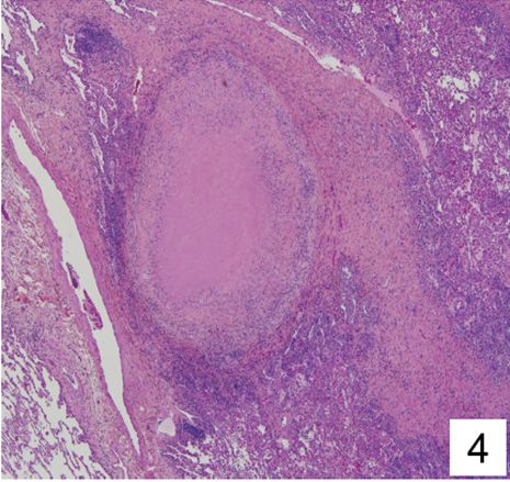
http://css.sciedupress.com Case Studies in Surgery 2017, Vol. 3, No. 4
compressed air diving. Otherwise he was in good medical The medical team warned the patient of the dangers of con-
health. An abdominal computed tomograph (CT) did not tinuing to practice diving, especially due to pulmonary com-
show evidence of any renal lesions. However, CT imaging plications such as Pneumothorax, and gave information to
of the lungs showed pulmonary parenchyma with numerous him and his family about increased risk of renal neoplasms.
emphysematous bullae, blebs and pulmonary cysts. In ad-
dition, a well-circumscribed lesion was noted contiguous, Macroscopic and histologic detection
but external to the bullae with hyperattenuation Proposing a The specimen consisted of a left lower lobe wedge resection
firm mass (see Figure 1). The differential diagnosis included measuring 15 cm × 4 cm × 3.5 cm. The pleural surface was
infection, abscess, pulmonary cyst with solid component, opaque and showed multiple blebs. Subsequent bivalving
hamartoma and neoplasm. The mass estimated to measure revealed a 2.5-cm white firm circumscribed nodule. The
3 cm × 2 cm. Given the patient’s diagnosis of BHD, a lesion was focally necrotic and friable. The surrounding pul-
neoplastic lesion could not be excluded and sampling was monary parenchyma showed numerous dilated airways and
warranted. A needle-core biopsy showed only necrotic tissue. bullae. Histologically, the nodule showed extensive necrosis
The possibility of necrotic tumor or an infectious process was with chronic inflammation in the periphery. An abnormal
considered and it was decided to excise to further elucidate vessel was discerned in the center of the lesion. Diffuse
the lesion. The patient had an uneventful recuperation and pleural bullae with associated chronic pleuritis were present.
follow up. The lung parenchyma showed scattered dilated cystic spaces
consistent with emphysematous changes. Pulmonary edema
was focally present (see Figure 2). The vessels showed
prominent thickening of their wall and some showed par-
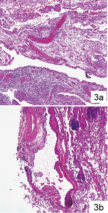
tial occlusion of their lumen (see Figure 3a and 3b). In the
remaining parenchyma, small non-necrotizing granulomata
were identified, containing fungal organisms consistent with
Histoplasma capsulatum (see Figure 4).
3. D ISCUSSION
In 1942, Jacques-Yves Cousteau and Emile Gagnan invented
a device coined the “Aqua Lung” the predecessor to modern
scuba apparati allowing divers to plunge to unforeseen depths.
In the advent of technology, PBT became an infrequent but
important complication, due to tissue injury resulting from
expanding gas during rapid ascent, precluding sufficient or
Figure 1. Bullae with hyperattenuation, hematoxylin and
appropriate exhalation. According to Boyle’s Law, the vol-
eosin stain (H&E)
ume of a given mass of gas is inversely proportional to the
absolute pressure, whereas during ascent as the ambient pres-
sure decreases, gas inside the lungs expands. The expansion
of compressed air in the lungs can lead to pulmonary tissue
damage, pneumothorax, pneumomediastinum, and most se-
riously arterial air emboli. Persons with history of trauma,
previous pneumothorax,[2] lung cysts,[3] emphysema,[4] or
genetic conditions like cystic fibrosis, Ehlers Danlos, and
Marfans[5] are predisposed to develop sequelae, and pre-
cluded from diving. Our patient was diagnosed, secondary
to family history and clinical constellation with Birt-Hogg-
Dubé, a genodermatosis characterized by fibrofolliculomas,
renal tumors, and spontaneous pneumothorax, a condition
also obviating fitness to dive.[6] CT imaging has shown
that pulmonary injury from PBT tends to resolve quickly,
however persistence may remain in patients with preexist-
ing lung injury,[7] akin to our case. Despite utilization of
Figure 2. Pulmonary edema (H&E) radiographic techniques, these findings are not diagnostic in
26 ISSN 2377-7311 E-ISSN 2377-732X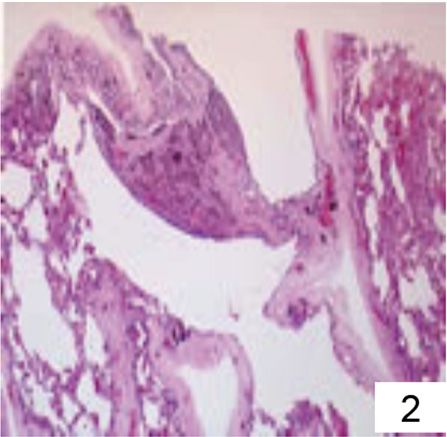
http://css.sciedupress.com                                    Case Studies in Surgery                                           2017, Vol. 3, No. 4

patients with delayed presentation, and may be suggestive
of a more aggressive lesion. Published case reports of the
histologic findings of PBT injury identified rarely describe
or illustrate the morphologic changes associated with PBT
lung injury in compressed air diving.[8–10]

                                                                            Figure 4. Small non-necrotizing granulomata with fungal
                                                                            organisms consistent with Histoplasma capsulatum (H&E)

                                                                            In our case, histologic findings revealed bullae, chronic pleu-
                                                                            ritis, focal edema, vascular and interstitial thickening. The
                                                                            presence of bullae, could be attributed to either his long-
                                                                            standing history of diving or his hereditary condition. The
                                                                            longstanding history of diving and early symptomatology
                                                                            suggested barotrauma as the most likely cause of the pneu-
                                                                            mothorax. This case illustrates the limitations sometimes
                                                                            encountered in patients which complex medical histories,
                                                                            allowing an exceptionally rare case of compressed air diving
                                                                            associated PBT injury in the setting of BHD to be eluci-
                                                                            dated by histological examination of the resected wedge.
                                                                            The histopathologic features of PBT injury in compressed air
                                                                            diving such as air emboli and vascular damage are distinct.
                                                                            The presence of necrotic granulomatous inflammation and
                                                                            fungi was most likely an unrelated event.

                                                                            C ONFLICTS OF I NTEREST D ISCLOSURE
                                                                            The authors declare they have no conflicts of interest.
Figure 3. Vessels with prominent thickening of their wall
and some with partial occlusion of their lumen (H&E, 3a
and 3b)

R EFERENCES                                                                       https://doi.org/10.1136/thx.52.9.805
  [1] Reuter M, Tetzlaff K, Warninghoff V, et al. Computed tomogra-
                                                                              [3] Simpson G. Primary lung bullae and scuba diving. SPUMS J. 1998;
      phy of the chest in diving-related pulmonary barotrauma. British
                                                                                  28: 10-12.
      Journal of Radiology. 1997; 70(833): 440-5. PMid: 9227223.
      https://doi.org/10.1259/bjr.70.833.9227223                              [4] Mellem H, Emhjellen S, Horgen O. Pulmonary barotrauma and arte-
  [2] Sadikot RT, Greene T, Meadows K, et al. Recurrence of primary spon-         rial gas embolism caused by an emphysematous bulla in a SCUBA
      taneous pneumothorax. Thorax. 1997; 52: 805-809. PMid: 9371212.             diver. Aviat Space Environ Med. 1990; 61: 559-562. PMid: 2369396.

Published by Sciedu Press                                                                                                                      27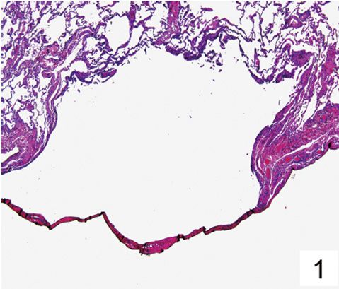
http://css.sciedupress.com                                       Case Studies in Surgery                                             2017, Vol. 3, No. 4

  [5] Godden D, Currie G, Denison D, et al. British Thoracic Society            [8] Chambers HM, van Velzen D. Ventilator-related pathology in the
      guidelines on respiratory aspects of fitness for diving. Thorax. 2003;        extremely immature lung. Pathology. 1989; 21(2): 79-83. https:
      58: 3-13. https://doi.org/10.1136/thorax.58.1.3                               //doi.org/10.3109/00313028909059539
                                                                                [9] Rouby JJ, Lherm T, Martin de Lassale E, et al. Histologic aspects of
  [6] Schmidt LS, Warren MB, Nickerson ML, et al. Birt-Hogg-Dubé
                                                                                    pulmonary barotrauma in critically ill patients with acute respiratory
      syndrome, a genodermatosis associated with spontaneous pneu-
                                                                                    failure. Intensive Care Med. 1993; 19(7): 383-389. PMid: 8270717.
      mothorax and kidney neoplasia, maps to chromosome 17p11.2.
                                                                                    https://doi.org/10.1007/BF01724877
      Am J Hum Genet. 2001; 69: 876-882. PMid: 11533913. https:
      //doi.org/10.1086/323744                                                 [10] Tsokos M, Paulsen F, Petri S, et al. Histologic, immunohistochem-
                                                                                    ical, and ultrastructural findings in human blast lung injury. Am J
  [7] Tetzlaff K, Reuter M, Leplow B, et al. Risk Factors for Pulmonary             Respir Crit Care Med. 2003; 168(5): 549-555. PMid: 12842857.
      Barotrauma in Divers Chest. 1997; 112: 654-659. PMid: 9315797.                https://doi.org/10.1164/rccm.200304-528OC

28                                                                                                               ISSN 2377-7311      E-ISSN 2377-732X
You can also read