Deep Learning-Based Positron Emission Tomography Molecular Imaging in the Assessment of Cognitive Dysfunction in Patients with Epilepsy - Hindawi.com
←
→
Page content transcription
If your browser does not render page correctly, please read the page content below
Hindawi
Scientific Programming
Volume 2021, Article ID 2714222, 8 pages
https://doi.org/10.1155/2021/2714222

Research Article
Deep Learning-Based Positron Emission Tomography Molecular
Imaging in the Assessment of Cognitive Dysfunction in
Patients with Epilepsy

 Mayila Tuerxun ,1 Lixin Yin ,1 Huiqun Chen ,2 and Jingqian Lin 3

 1
 Department of Neurology, The First Affiliated Hospital of Xinjiang Medical University, Urumqi 830000, Xinjiang, China
 2
 Digestive Medicine, The 967th Hospital of Joint Logistics Support Force of Chinese People’s Liberation Army, Dalian 116000,
 Liaoning, China
 3
 Department of Neurology, Yantai Laiyang Central Hospital, Laiyang 265200, Shandong, China

 Correspondence should be addressed to Jingqian Lin; 150511133@stu.sxit.edu.cn

 Received 14 July 2021; Revised 12 September 2021; Accepted 14 September 2021; Published 6 October 2021

 Academic Editor: Gustavo Ramirez

 Copyright © 2021 Mayila Tuerxun et al. This is an open access article distributed under the Creative Commons Attribution
 License, which permits unrestricted use, distribution, and reproduction in any medium, provided the original work is
 properly cited.

 This work aimed to investigate the application of positron emission tomography (PET) molecular imaging based on the deep
 learning algorithm in the assessment of cognitive dysfunction in patients with epilepsy. In this study, 52 hospitalized patients with
 epilepsy were selected as the epilepsy group and treated with different kinds of antiepileptic drugs, and 52 volunteers were selected
 as the control group. A U-net optimized network structure algorithm based on deep learning was proposed in this study and
 compared with a fully convolutional neural network (FCNN). Besides, it was applied in the PET molecular imaging of patients
 with epilepsy, and the segmentation effect of the U-net optimized network structure was good. According to event-related
 potential examinations, the proportion of patients with cognitive dysfunction in the epilepsy group (74.19%) was higher than the
 proportion of the control group (7.46%) (P < 0.05). The patients with cognitive dysfunction (57.89%) who took one antiepileptic
 drug were lower than those with two antiepileptic drugs (84.61%) (P < 0.05). The difference was statistically obvious in the overall
 quality of life of patients with epilepsy (P < 0.05). The occurrence of cognitive dysfunction in patients with epilepsy was related to
 the type of seizures. In addition, the quality of life of patients who suffered from cognitive dysfunction was low.

1. Introduction 0.8–4.5%, and there are about 500,000 new epilepsy patients
 each year [4]. With the continuous updating of electronic
Epilepsy is a group of chronic diseases and syndromes of medical equipment, humans’ understanding of epilepsy has
central nervous system dysfunction caused by abnormal gradually deepened. The cognitive dysfunction caused by
excessive discharge of brain neurons [1]. Epilepsy occurs in epilepsy has a major impact on the health and quality of life
people of any age, region, and ethnicity. In recent years, the of patients, and long-term application of antiepileptic drugs
incidence of cerebrovascular disease, dementia, and neu- brings patients’ families severe economic pressure. There-
rodegenerative diseases has increased with the aging of the fore, it has attracted the attention of many scholars [5].
Chinese population. Moreover, the incidence of epilepsy in Cognitive function means that the information is simply
the elderly shows an upward trend [2]. According to the processed and transformed into its own psychological
location of abnormal discharge of neurons and the range of changes when the brain receives information from the
discharge, the main clinical manifestations are the different outside world. In the process of receiving information, it
dysfunctions, including different motor, sensory, cognitive, mainly includes language, vision, hearing, comprehension,
and autonomic nerve [3]. According to epidemiological memory, and other aspects. Cognitive dysfunction refers to
research data, the annual prevalence of epilepsy is about the abnormality of one or more of the above functions [6].
2 Scientific Programming

Kim et al. [7] pointed out that the intelligence score was and 0.1 g carbamazepine tablet was also taken three times a
generally lower than 75 points after the onset of epilepsy. day. The drug dose was adjusted reasonably according to the
Research data shows that about 35% of epilepsy patients will condition of epilepsy patients, and the highest dose was 0.3 g.
be accompanied by cognitive dysfunction, and patients will The treatment lasted for 6 months, and the outcome indi-
show obvious defects at the onset. cators were observed.
 PET is considered to be one of the most promising Before surgery, each patient should be asked about the
imaging technologies. It can be applied to observe the re- cause of disease, age at first onset, clinical manifestations,
lationship between brain functional activities and changes in time of onset, frequency of onset, past history (birth status,
blood flow metabolism in a living body. It is called “phys- history of brain surgery, stroke, and brain nerve tissue
iological tomography” and has been widely applied in the disease), the administration of antiepileptic drugs (in-
positioning of preoperative epilepsy lesions in recent years cluding the type of drugs, administration time, and dosage
[8]. Deep learning is a type of machine learning algorithm taken), family history of epilepsy, craniocerebral PET ex-
that uses multiple layers to gradually extract higher-level amination, and the assessment results of Quality of Life in
features from the original input. In image processing, the Epilepsy-31 (QOLIE-31). According to the PET scanning
lower layer can identify the edges, and the higher layer can and the number and type of epilepsies a year ago, the
identify the parts that are meaningful to humans [9]. Deep patients were classified as mild, moderate, and severe based
learning has achieved very marked effects in the computer on their specific conditions. The mild symptom meant that
vision. Besides, the U-net network structure is a biomedical patients had no seizures within one year, and the moderate
image segmentation method obtained by further optimizing symptom referred to patients who were between mild and
the full convolutional network algorithm. This network severe. What is more, the severe symptom was that patients
structure is divided into an upsampling path and a down- suffered from more than 15 partial episodes within a year,
sampling path and distributed in a symmetrical “U” shape, more than 3 complex seizures, and more than 1 tonic
so it was named “U-net” [10]. spasm.
 The U-net optimized network structure algorithm was
proposed based on deep learning, compared with the fully
convolutional neural network (FCNN) structure, and ap- 2.3. Event-Related Potential Measurement. The Viking Quest
plied to the PET molecular images of 52 patients with ep- (produced by Nicol, USA) evoked potential meter was
ilepsy. The objective of this study was to explore the PET adopted, and the electrodes were placed in accordance with
molecular imaging to assess the influencing factors of the international system method. The electrodes were placed
cognitive dysfunction in patients with epilepsy and the at three positions (Ez, Dz, and Qz). The reference electrode
quality of life of patients with epilepsy. was set at the base of the nose, and the ground wire was
 placed at the right earlobe. The skin impedance was less than
 5 gigabytes, and three sets of data were recorded at the same
2. Materials and Methods
 time. During the examination, the patient should maintain a
2.1. Selection of Research Objects. In this study, 52 patients quiet and relaxed mood and sit on the examination chair.
with epilepsy who were treated in hospital from November Each patient needed to conduct a pretest in advance and
17, 2017, to May 19, 2019, were selected as the epilepsy understood the purpose and requirements of this experi-
group. Besides, 52 healthy volunteers were taken as the ment, and the short-, medium-, and high-frequency tones
control group. There were 53 males and 51 females, with an were recorded. The test was started after the patient could
average age of 31.32 ± 11.87 years. This experiment had been distinguish clearly. The target stimulation frequency was
authorized by the Medical Ethics Committee of Hospital, 3 KHz, the stimulation intensity was 85 dB, and the random
and the patients and their family members had understood probability was set to 25%. The nontarget stimulation fre-
the situation of this experiment and signed the informed quency was 1 KHz, the intensity was 85 dB, and the random
consent forms. probability was set to 75%. Furthermore, the target stimulus
 The criteria for inclusion were defined to include patients was superimposed 450 times, the sensitivity was 10 μv, the
who met the clinical and pathological diagnostic criteria of bandpass range was set to 2–50 Hz, and the analysis time was
the International Antiepilepsy League’s classifications of 1 second.
epilepsy and epilepsy syndrome and were 18–75 years old.
 The criteria for exclusion were defined to include pa-
tients who suffered from cognitive dysfunction before the 2.4. Assessment of Quality of Life in Patients with Epilepsy.
onset of epilepsy, had other cognitive dysfunctions, had The patients were assessed by the Chinese translation version
mental retardation and were unable to cooperate with the of QOLIE-31 version 1.0. The total score of each question
examination, and suffered from major mental illnesses. was divided by the corresponding number of questions,
 which was recorded as the initial score of each item. The
 product of the initial score of each item in the item table and
2.2. Treatment, Observation, and Classification of Patients the sum of the weight scores was taken as the total score. The
with Epilepsy. The epilepsy group was treated with carba- higher the score was, the higher the patient’s quality of life
mazepine combined with butylphthalide soft capsules; 0.2 g was. The cognitive function included 6 items that had the
butylphthalide soft capsules were taken three times a day, serial numbers 12, 17, 18, 21, 25, and 29 in the scale, and the
Scientific Programming 3

social functions contained 5 items that had the serial the value b, thereby affecting the total loss function. In
numbers 11, 13, 15, 19, and 22 in the scale. general, the range of value b was [0.5–1] to increase the
 weight of the positive sample loss function.
2.5. PET Scanning of Patients with Epilepsy. The Siemens
PET/magnetic resonance (MR) all-in-one machine was 2.7. Evaluation Indexes of Segmentation Results. 3 indexes
used for imaging data collection. Before the scanning, all were applied in this study to evaluate the segmentation
patients fasted for 6–8 hours, and their blood glucose effect, namely, Dice similarity coefficient (DSC), sensitivity
levels were controlled at 3.8–6.2 mmol/L. β-2-[18F]-Flu- (SEN), and positive predictive value (PPV). DSC was a
oro-2-deoxy-D-glucose (18F-FDG) was intravenously comprehensive index, which was often adopted to determine
injected with a dose of 0.2 mCi/kg. The patient rested for the coincidence between automatic segmentation results and
30 minutes in a calm environment and then was actual values. Thus, DSC could be calculated as follows:
examined by the cranial PET/MR. Before the PET 2TPV
scanning, MR imaging (MRI) equipment was adopted to DSC � . (7)
 FPV + FNV + 2TPV
scan and locate the examined brain area. During the
examination, the examination procedure should be In equation (7), TPV stood for the number of true
explained to the patient. The patient was lying on the positive pixels, FPV represented the number of false positive
scanning table in a supine position and the head was pixels, and FNV expressed the number of false negative
fixed. The data collection time was set to 8 minutes, and pixels. PPV was employed to calculate the ratio of the
the data was collected in 3D mode. During the scanning number of true positive pixels to the number of false positive
process, the physician needed to pay attention to the vital pixels, and it could be expressed in the following equation:
characteristics of the patient at any time. TPV
 PPV � . (8)
 TPV + FPV
2.6. U-Net Optimized Network Structure Algorithm. On the SEN was applied to calculate the ratio of the number of
basis of the structure of U-net itself, data amplification
 true positive pixels to the number of false negative pixels,
methods were added so that overfitting would not occur
 which could be expressed as follows:
when the data was small. U-net optimized network structure
could be added to the cross-entropy loss function for two TPV
 SEN � . (9)
classification problems. The cross-entropy loss function TPV + FNV
(CLF) was calculated as follows:
 The range of the above three evaluation indexes was [0, 1].
 m
 1 The closer the calculation result was to 1, the better the seg-
 CLF(q, y) � − y log qi + 1 − yi , (1) mentation effect was; the closer the calculation result was to 0,
 m i�1 i
 the worse the segmentation effect was.
or
 −log(q) if y � 1, 2.8. Statistical Methods. In this study, SPSS 20.0 statistical
 CLF(q, y) � (2) analysis software was used for data processing. The standard
 −log(1 − q), else.
 deviation ± variance was used for normal distribution; the
 In equations (1) and (2), q represented the estimated average age of epidemiology, the quality of life score of
value, and its value range was [0, 1]. The value of y was 0 or 1, patients, and the calculation data were expressed by per-
and q was substituted by qs , as shown in equation (3). centage (%). The DSC, SEN, and PPV of the two algorithms
Besides, equation (2) was transformed into equation (4). were compared, as well as the seizure type proportion and
 seizure frequency cases. In addition, P < 0.05 meant that the
 q if y � 1, difference was statistically substantial.
 qs � (3)
 1 − q, else.
 3. Results
 CLF(q, y) � CLF qs
 (4) 3.1. Comparison on the Segmentation Effect of the Two Al-
 � −log qs . gorithms in the Reconstruction Model. The U-net optimized
 The coefficient λs was added on the basis of the cross- network structure algorithm was proposed based on deep
entropy loss function to get equations (5) and (6). learning and compared with the FCNN structure. Moreover,
 it was applied in the PET molecular images of 52 patients
 CLF qs � −λs log qs . (5) with epilepsy. The U-net optimized network structure
 contained 4 densely connected blocks in the upsampling and
 b if y � 1, downsampling paths, and there was a densely connected
 λs � (6) block between the two. The number of densely connected
 1 − b, else.
 blocks was different from the incremental rate. If the number
 According to the above equations, the positive and of layers was denser inside the densely connected blocks,
negative of the sample were adjusted by adjusting the size of there were more parameters to be tested for the entire
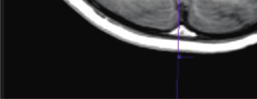
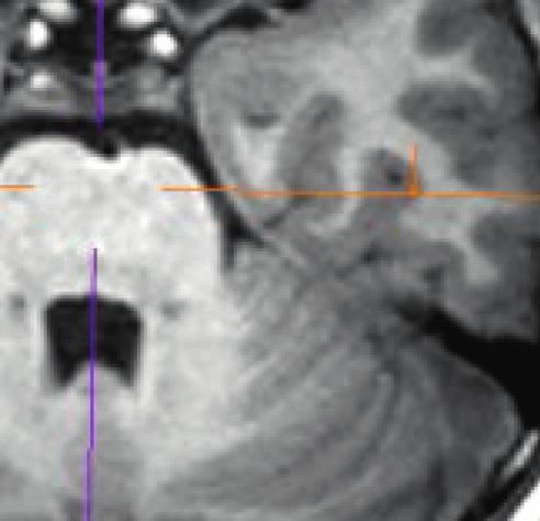
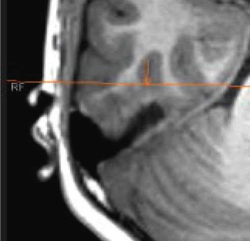
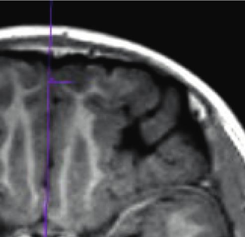
4 Scientific Programming

network structure. The DSC (89.47%) of the U-net opti- ∗
mized network structure was higher than the DSC (84.56%) 90
of the FCNN structure, indicating that the two image seg- 88

 Proportoion (%)
mentation methods had a higher degree of overlap between 86
the segmentation results of this research algorithm and the
 84
actual value. The PPV of the U-net optimized network
structure (88.97%) was higher than the PPV of the FCNN 82
structure (85.15%), and the number of true positive pixels 80
optimized by U-net was more. What is more, the SEN of 78
U-net optimized network structure (89.51%) was higher DSC PPV SEN
than the SEN (82.79%) of the FCNN structure, and the FCNN
difference was statistically obvious (P < 0.05) (Figure 1). Optimized U-net
Therefore, the U-net optimized network structure was better
 Figure 1: Comparison on the segmentation effects of the two
than the segmentation effect of the FCNN.
 algorithms in the reconstruction model.

3.2. Epidemiological Data Statistics. There were 52 epilepsy
patients included in this study, aged between 18 and 50
years, with an average age of 31.97 ± 13.47 years, including Epilepsy group
28 males and 24 females. According to educational levels, 15
patients were undergraduate and above, and 37 cases were
under high school. There were 52 patients (25 males and 27
females) in the control group, and they were 17–52 years old, Control group
with an average age of 32.16 ± 12.04 years. According to
education levels, 18 patients were undergraduate and above,
and 34 were under high school. The above differences were 0 5 10 15 20 25 30 35 40 45 50

not statistically marked (P > 0.05) (Figures 2 and 3). Average age (years old)

 Figure 2: Comparison on the average age of subjects from the
 control group and the epilepsy group.
3.3. Characteristics of Clinical Seizures in Patients with
Epilepsy. In the 52 patients with epilepsy, 12 had partial
seizures, which specifically included simple partial seizures,
complex partial seizures, and partial seizures extending to
generalized seizures; 24 patients had generalized seizures, Epilepsy group
including general absence seizures, generalized tonic spasm,
and generalized myoclonus; and 16 patients had partial and
generalized seizures (Figure 4). In the enrolled patients, 42 Control group
patients had monthly seizures less than or equal to 1 within
one year, accounting for 80.76%, and 10 patients had
 0 10 20 30 40 50 60
monthly seizures greater than 1, accounting for 19.24%. The Proportion (%)
difference between the two was statistically substantial
(P < 0.05) (Figure 5). Women
 Man

 Figure 3: Comparison on the gender of subjects from the control
3.4. PET/MRI Molecular Imaging Characteristics of Patients group and the epilepsy group.
with Epilepsy. The tuberous sclerosis 2 (TSC2) gene was
mutated at c.600–2 A > G. The images of brain MRI showed
that cortical or subcortical nodules presented the high signals on
 46.15
T2-weighted imaging (T2WI), T2 FLAIR, and double inversion
 50
recovery sequence (DIR), as well as the low signals on T1WI 45 30.77
sequence. 18F-fluorodeoxyglucose (18F-FDG) PET/MRI 40
indicated that the aforementioned abnormal signal areas had
 Proportion (%)

 35 23.08
 30
low-metabolism areas matching the size of the nodules
 25
themselves. At the left anterior temporal area where the scalp 20
EEG indicated the epileptic area, PET/MRI showed that there 15
was extensive low metabolism, and the metabolic area was 10
 5
dramatically larger than the nodule itself. For the non- 0
epileptogenic nodules, the left posterior temporal area and the Partial seizure Comprehensive Other types
 attack
left occipital area of the nodule had the same low-metabolic
range as the nodule itself. The above results suggested that Figure 4: Types of seizures in patients with epilepsy.
Scientific Programming 5

 50

 40

 30
Cases

 ∗
 20

 10

 0
 ≤1time/month >1time/month

Figure 5: Comparison on frequency of epilepsy patients (note: ∗
indicated that the difference was statistically obvious in contrast to
less than or equal to 1 time/month (P < 0.05)).

epileptogenic nodules could be combined with abnormal low-
metabolic cortex. The aforementioned imaging features were
helpful to identify epileptogenic nodules. There was excessive
discharge of the lesion, rapid and repeated depolarization of a
large number of neuron cell membranes, and a significant
increase in energy consumption, resulting in an obvious growth
of local blood flow and glucose metabolism; 18F-FDG PET
imaging showed a high metabolic focus (Figures 6 and 7).
 Figure 6: CT image of polyps (note: the image showed that the
 polyps protruded to the inside of the intestinal cavity, which was in
3.5. PET Examination Results of Patients with Epilepsy. In a large amount and dense).
this study, 52 patients with epilepsy completed the PET
molecular imaging examinations, and it was found that there
were no obvious abnormal changes in the brains of 12
patients. Obvious lesions were found in 40 patients, ac-
counting for 76.9%. Among them, there were 10 patients
with hippocampal lesions (19.23%), 16 patients with frontal
lobe lesions (30.77%), and 8 patients with temporal lobe
lesions (15.38%). There were 6 cases of parietal disease,
accounting for 11.54% (Figure 8).

3.6. Results of Event-Related Potentials in Patients with
Epilepsy. The 52 epilepsy patients enrolled in the group
completed the event-related potential examinations, show-
ing that there were 14 patients with normal cognitive
function (25.81%). However, there were 48 patients with
normal cognitive function in the control group, accounting
for 92.54%. The difference between the two was statistically
great (P < 0.05). There were 38 patients with cognitive Figure 7: An image of polyp cancer (note: the local tube wall was
dysfunction in the epilepsy group, occupying 74.19%, and 4 thickened, with irregular edges).
patients in the control group with cognitive dysfunction,
accounting for 7.46%. Thus, the difference was statistically
substantial in the number of cases with cognitive dysfunc- were 8 patients with normal cognitive function and 14
tion from the two groups (P < 0.05) (Figure 9). patients with cognitive dysfunction who were with high
 school degrees or below. The above differences were not
 statistically obvious (P > 0.05) (Figure 10).
3.7. Influence of Education Levels of Epilepsy Patients on
Cognitive Function. In the 52 patients with epilepsy, the
highest degree was postgraduate, and the lowest degree was 3.8. Medications and Cognitive Function of Epilepsy Patients.
primary school. Based on the degree of education, there were In the 52 patients, 19 patients took a kind of antiepileptic
15 patients with bachelor degree or above and 37 patients drug, among which 11 cases (57.89%) developed cognitive
with high school degree or below. Moreover, 11 patients dysfunction. There were 26 patients taking two or more
with normal cognitive function and 19 patients with cog- antiepileptic drugs, among which 22 (84.61%) had cognitive
nitive dysfunction had the bachelor degrees or above. There dysfunction. 7 patients were not given antiepileptic drugs,
6 Scientific Programming

 ∗
 35 90
 30 80
 70
 Proportion (%)

 Proportion (%)
 25 60
 20 50
 40 #
 15 30 ∗
 10 20
 10
 5
 0
 0 A drug Two or more Did not take drugs
 A B C D drugs
 Lesion type
 Figure 11: Influences of medication on cognitive function in
Figure 8: Brain PET examination results of patients with epilepsy patients with epilepsy (note: ∗ meant that the difference was sta-
(note: A, B, C, and D stood for the hippocampal lesion, frontal lobe tistically huge in contrast to taking one drug (P < 0.05); # indicated
lesion, temporal lobe lesion, and parietal lobe lesion, respectively). that there was a statistically remarkable difference in contrast to
 taking two or more drugs (P < 0.05)).

 versus 3.68 ± 0.79; 3.56 ± 0.23 points versus 3.74 ± 0.26
 100 ∗
 points), and the difference was statistically significant
 80
 (P < 0.05). It was found that the quality of life of patients
Proportion (%)

 60 with cognitive dysfunction was relatively poor (Figure 12).
 40
 ∗ 4. Discussion
 20

 0 Epilepsy is a common neurological disease. More than 30%
 Control group Epilepsy group
 of patients with diagnosed epilepsy will have cognitive
 Normal cognitive function dysfunction [11]. In the past, people mainly paid attention to
 Cognitive disorder how to control epileptic seizures but ignored the cognitive
Figure 9: Results of event-related potentials in patients with dysfunction caused by epilepsy and the quality of life of
epilepsy. patients with epilepsy. With the continuous development of
 medical high-tech equipment, people’s attention to cognitive
 dysfunction caused by epilepsy has gradually increased [12].
 Hasan et al. [13] explored a variety of factors that caused
 20 cognitive dysfunction in patients with epilepsy, including
 factors such as gender, age, seizure frequency, seizure type,
 15 and antiepileptic drug administration. They found that the
 age, frequency of seizures, type of seizures, and drug use
 Cases

 10
 were all related to cognitive dysfunction. In particular, the
 5 effect of frequency and type of seizures on cognitive function
 was very obvious, which was consistent with the results of
 0 this study. The event-related potential examinations were
 Control group Epilepsy group
 applied in this study, and the results suggested that seizure
 Bachelor and above frequency, seizure type, brain PET molecular imaging, and
 Under the high school drug use were all influencing factors of cognitive dysfunc-
Figure 10: Influence of education levels on cognitive function in tion, which were in line with the above research viewpoints.
epilepsy patients. Chen et al. [14] retrospectively analyzed the cognitive
 dysfunction of 129 patients with epilepsy. The most serious
 damage to cognitive function was generalized tonic spasm,
and only 1 case had cognitive dysfunction. The difference followed by complex partial seizures. If the frequency of
between any two groups was statistically marked (P < 0.05) complex partial seizures was more than 2 times/month, it
(Figure 11). would cause a serious decline in the patient’s memory and
 mobility. Postulart et al. [15] believed that complex partial
 seizures and extension of partial seizures to generalized
3.9. Quality of Life Scores of Patients with Epilepsy. The seizures were likely to cause damage to cognitive function. It
quality of life in 52 patients with epilepsy was scored by indicated that cognitive dysfunction was highly correlated
QOLIE-31, and the standard score method was adopted to with the type of seizure, which was in accordance with the
improve the score and total score value of each project. The viewpoint of this study.
cognitive and social function scores in epilepsy group were The most common way to treat epilepsy is to take an-
significantly higher than those in control group (3.12 ± 0.34 tiepileptic drugs, which can reduce cognitive dysfunction
Scientific Programming 7

 5 samples collected within a certain period of time, which may
 4.5 ∗
 ∗ ∗
 cause certain deviations in the results of this study. Later, it is
 4
 3.5
 considered to expand the sample range for further im-
Life quality score

 3 provement. In short, the treatment of epilepsy cannot be
 2.5 through a single drug. It was also necessary to consider the
 2
 adverse effects of cognitive dysfunction caused by epilepsy.
 1.5
 1
 The patients’ attention to cognitive function has been in-
 0.5 creased to a certain extent, and their overall quality of life has
 0 been improved.
 Total points Cognitive function Social function Drugs affect

 Normal
 Obstacle
 Data Availability
Figure 12: Quality of life score and cognitive function in patients The data used to support the findings of this study are
with epilepsy (note: ∗ showed that the difference was statistically available from the corresponding author upon request.
substantial compared with normal cognitive function (P < 0.05)).

 Conflicts of Interest
caused by epilepsy through antiepileptic drugs. However,
antiepileptic drugs have side effects, and the dosage and The authors declare no conflicts of interest.
frequency of administration will also have a certain impact
on cognitive function [16]. Liguori et al. [17] pointed out that References
the more types of antiepileptic drugs were applied, the more
obvious the degree of cognitive impairment in patients was. [1] V. K. Pilli, J. W. Jeong, P. Konka, A. Kumar, H. T. Chugani,
Oldan et al. [18] thought that antiepileptic drugs could and C. Juhász, “Objective PET study of glucose metabolism
 asymmetries in children with epilepsy: implications for
inhibit the conduction of neuronal excitement and hinder
 normal brain development,” Human Brain Mapping, vol. 40,
the transmission of neurotransmitters, thereby affecting the no. 1, pp. 53–64, 2019.
cognitive function of patients. It was found that patients [2] C. E. Popescu, R. Mai, R. Sara et al., “The role of FDG-PET in
taking two or more antiepileptic drugs could promote the patients with epilepsy related to periventricular nodular
occurrence of cognitive dysfunction, indicating that the type heterotopias: diagnostic features and long-term outcome,”
of drug taken was one of the independent risk factors that Journal of Neuroimaging: Official Journal of the American
affected cognitive dysfunction. The QOLIE-31 was adopted Society of Neuroimaging, vol. 29, no. 4, pp. 512–520, 2019.
to evaluate the quality of life of patients with epilepsy, so that [3] S. Desarnaud, C. Mellerio, F. Semah et al., “18F-FDG PET in
the results of this study had good credibility. Ogundare et al. drug-resistant epilepsy due to focal cortical dysplasia type 2:
[19] used QOLIE-31 to assess 85 patients with epilepsy and additional value of electroclinical data and coregistration with
healthy controls and found that the scores of epilepsy pa- MRI,” European Journal of Nuclear Medicine and Molecular
 Imaging, vol. 45, no. 8, pp. 1449–1460, 2018.
tients were sharply lower than the scores of the control
 [4] S. Elwan, A. Alexopoulos, D. C. Silveira, and P. Kotagal,
group. In addition, patients with cognitive dysfunction had “Lateralizing and localizing value of seizure semiology:
the lowest scores, indicating that epilepsy-induced cognitive comparison with scalp EEG, MRI and PET in patients suc-
dysfunction could reduce the quality of life of patients. The cessfully treated with resective epilepsy surgery,” Seizure,
results of this study revealed that the differences in the vol. 61, pp. 203–208, 2018.
overall quality of life, cognitive function, and social function [5] Y.-H. Wang, Y. An, X.-T. Fan et al., “Comparison between
of patients with epilepsy were statistically obvious (P < 0.05), simultaneously acquired arterial spin labeling and 18F-FDG
which was consistent with the results of the above research. PET in mesial temporal lobe epilepsy assisted by a PET/MR
 system and SEEG,” NeuroImage: Clinic, vol. 19, pp. 824–830,
 2018.
5. Conclusion [6] K. Shang, J. Wang, X. Fan et al., “Clinical value of hybrid TOF-
 PET/MR imaging-based multiparametric imaging in local-
The U-net optimized network structure algorithm was izing seizure focus in patients with MRI-negative temporal
proposed based on deep learning. Besides, it was compared lobe epilepsy,” American Journal of Neuroradiology, vol. 39,
with the FCNN structure and applied to the PET molecular no. 10, pp. 1791–1798, 2018.
images of 52 patients with epilepsy. U-net optimized net- [7] J. A. Kim, J. W. Jeong, M. E. Behen et al., “Metabolic correlates
work structure had better segmentation effect than FCNN. of cognitive function in children with unilateral Sturge-Weber
The occurrence of cognitive dysfunction in patients with syndrome: evidence for regional functional reorganization
epilepsy was related to the type of seizures, the frequency of and crowding,” Human Brain Mapping, vol. 39, no. 4,
 pp. 1596–1606, 2018.
seizures, the consumption of antiepileptic drugs, and brain
 [8] X.-Q. Wu, Y.-N. Zhao, J. Ding et al., “Decreased vesicular
PET molecular imaging. The total score of quality of life, acetylcholine transporter related to memory deficits in epi-
cognitive function, and social function scores of patients lepsy: a [18 F] VAT positron emission tomography brain
with cognitive dysfunction were lower compared to patients imaging study,” Epilepsia, vol. 59, no. 9, pp. 1655–1666, 2018.
with normal cognitive function, and their quality of life was [9] M. Hu, Y. Zhong, S. Xie, and L. Haibin, L. Zhihan, “Fuzzy
low. The limitation of this study is that there are fewer case system based medical image processing for brain disease
8 Scientific Programming

 prediction,” Frontiers in Neuroscience, vol. 15, Article ID
 714318, 2021.
[10] Z. Yu, S. U. Amin, M. Alhussein, and L. Zhihan, “Research on
 disease prediction based on improved DeepFM and IoMT,”
 IEEE Access, vol. 9, no. 99, p. 1, 2021.
[11] H. Seo, C. Huang, M. Bassenne, R. Xiao, and L. Xing,
 “Modified U-net (mU-Net) with incorporation of object-
 dependent high level features for improved liver and liver-
 tumor segmentation in ct images,” IEEE Transactions on
 Medical Imaging, vol. 39, no. 5, pp. 1316–1325, 2020.
[12] Y. Li, J. Zhao, Z. Lv, and J. Li, “Medical image fusion method
 by deep learning,” International Journal of Cognitive Com-
 puting in Engineering, vol. 2, pp. 21–29, 2021.
[13] S. M. Kamrul Hasan and C. A. Linte, “A modified U-Net
 convolutional network featuring a nearest-neighbor re-sam-
 pling-based elastic-transformation for brain tissue charac-
 terization and segmentation,” in Proceedings of the 2018 IEEE
 Western New York Image and Signal Processing Workshop
 (WNYISPW), Rochester, NY, USA, October 2018.
[14] S. Chen, A. Qin, D. Zhou, and D. Yan, “Technical note: U-net-
 generated synthetic CT images for magnetic resonance im-
 aging-only prostate intensity-modulated radiation therapy
 treatment planning,” Medical Physics, vol. 45, no. 12,
 pp. 5659–5665, 2018.
[15] D. M. IJff, D. Postulart, D. A. J. E. Lambrechts et al., “Cog-
 nitive and behavioral impact of the ketogenic diet in children
 and adolescents with refractory epilepsy: a randomized
 controlled trial,” Epilepsy and Behavior, vol. 60, pp. 153–157,
 2016.
[16] D. Chen, P. Wawrzynski, and Z. Lv, “Cyber security in smart
 cities: a review of deep learning-based applications and case
 studies,” Sustainable Cities and Society, vol. 66, Article ID
 102655, 2020.
[17] C. Liguori, C. Costa, F. Franchini et al., “Cognitive perfor-
 mances in patients affected by late-onset epilepsy with un-
 known etiology: a 12-month follow-up study,” Epilepsy and
 Behavior: E & B, vol. 101, Article ID 106592, 2019.
[18] J. D. Oldan, H. W. Shin, A. H. Khandani, C. Zamora,
 T. Benefield, and V. Jewells, “Subsequent experience in hybrid
 PET-MRI for evaluation of refractory focal onset epilepsy,”
 Seizure, vol. 61, pp. 128–134, 2018.
[19] T. Ogundare, T. O. Adebowale, and O. A. Okonkwo, “Quality
 of life among patients with epilepsy in Nigeria: predictors and
 barriers to routine clinical use of QOLIE-31,” Quality of Life
 Research, vol. 30, no. 2, pp. 487–496, 2020.
You can also read