Deep learning in biomedical optics - arXiv
←
→
Page content transcription
If your browser does not render page correctly, please read the page content below
Deep learning in biomedical optics
                                                  Lei Tian,1* Brady Hunt,2 Muyinatu A. Lediju Bell,3,4,5 Ji Yi,4,6 Jason T. Smith,7 Marien Ochoa,7
                                                  Xavier Intes,7 Nicholas J. Durr3,4*
arXiv:2105.11046v1 [physics.optics] 23 May 2021

                                                  1
                                                    Department of Electrical and Computer Engineering, Boston University, Boston, MA, USA
                                                  2
                                                    Thayer School of Engineering, Dartmouth College, Hanover, NH, USA
                                                  3
                                                    Department of Electrical and Computer Engineering, Johns Hopkins University, Baltimore, MD, USA
                                                  4
                                                    Department of Biomedical Engineering, Johns Hopkins University, Baltimore, MD, USA
                                                  5
                                                    Department of Computer Science, Johns Hopkins University, Baltimore, MD, USA
                                                  6
                                                    Department of Ophthalmology, Johns Hopkins University, Baltimore, MD, USA
                                                  7
                                                    Center for Modeling, Simulation, and Imaging in Medicine, Rensselaer Polytechnic Institute
                                                  *
                                                    Corresponding author: L. Tian, leitian@bu.edu; N.J. Durr, ndurr@jhu.edu

                                                  This article reviews deep learning applications in biomedical
                                                  optics with a particular emphasis on image formation. The
                                                  review is organized by imaging domains within biomedical
                                                  optics and includes microscopy, fluorescence lifetime imag-
                                                  ing, in vivo microscopy, widefield endoscopy, optical coher-
                                                  ence tomography, photoacoustic imaging, diffuse tomogra-
                                                  phy, and functional optical brain imaging. For each of these
                                                  domains, we summarize how deep learning has been ap-
                                                  plied and highlight methods by which deep learning can
                                                  enable new capabilities for optics in medicine. Challenges
                                                  and opportunities to improve translation and adoption of
                                                                                                                    Fig 1: Number of reviewed research papers which utilize DL
                                                  deep learning in biomedical optics are also summarized.
                                                                                                                    in biomedical optics stratified by year and imaging domain.
                                                  Key Words: biomedical optics; biophotonics; deep learn-
                                                  ing; machine learning; computer aided detection; mi-              ious forms of data analysis [2, 3]. As a result, DL is increas-
                                                  croscopy; fluorescence lifetime; in vivo microscopy; optical      ingly being utilized within biomedical optics as a data-driven
                                                  coherence tomography; photoacoustic imaging; diffuse to-          approach to perform image processing tasks, solve inverse
                                                  mography; functional optical brain imaging; widefield en-         problems for image reconstruction, and provide automated
                                                  doscopy                                                           interpretation of downstream images. This trend is high-
                                                                                                                    lighted in Fig. 1, which summarizes the articles reviewed in
                                                  INTRODUCTION                                                      this paper stratified by publication year and image domain.
                                                  Biomedical optics is the study of biological light-matter in-        This review focuses on the use of DL in the design and
                                                  teractions with the overarching goal of developing sensing        translation of novel biomedical optics systems. While im-
                                                  platforms that can aid in diagnostic, therapeutic, and sur-       age formation is the main focus of this review, DL has also
                                                  gical applications [1]. Within this large and active field of     been widely applied to the interpretation of downstream im-
                                                  research, novel systems are continually being developed to        ages, as summarized in other review articles [4, 5]. This re-
                                                  exploit unique light-matter interactions that provide clini-      view is organized as follows. First, a brief introduction to
                                                  cally useful signatures. These systems face inherent trade-       DL is provided by answering a set of questions related to
                                                  offs in signal-to-noise ratio (SNR), acquisition speed, spatial   the topic and defining key terms and concepts pertaining
                                                  resolution, field of view (FOV), and depth of field (DOF).        to the articles discussed throughout this review. Next, re-
                                                  These trade-offs affect the cost, performance, feasibility, and   cent original research in the following eight optics-related
                                                  overall impact of clinical systems. The role of biomedical op-    imaging domains is summarized: (1) microscopy, (2) fluores-
                                                  tics developers is to design systems which optimize or ideally    cence lifetime imaging, (3) in vivo microscopy, (4) widefield
                                                  overcome these trade-offs in order to appropriately meet a        endoscopy, (5) optical coherence tomography, (6) photoa-
                                                  clinical need.                                                    coustic imaging, (7) diffuse tomography, and (8) functional
                                                     In the past few decades, biomedical optics system design,        This work was supported by the National Science Foun-
                                                  image formation, and image analysis have primarily been           dation 1813848, 1846784 (L.T.); the National Institutes of
                                                  guided by classical physical modeling and signal processing       Health R21EB024700 (N.J.D.), R21EB025621 (M.A.L.B),
                                                  methodologies. Recently, however, deep learning (DL) has          R01NS108464 (J.Y.), R01EB019443, R01CA207725,
                                                  become a major paradigm in computational modeling and             R01CA237267, and R01CA250636 (X.I., J.T.S., M.O.);
                                                  demonstrated utility in numerous scientific domains and var-      MTEC:20-05-IMPROVE-004 and W912CG2120001 (X.I.).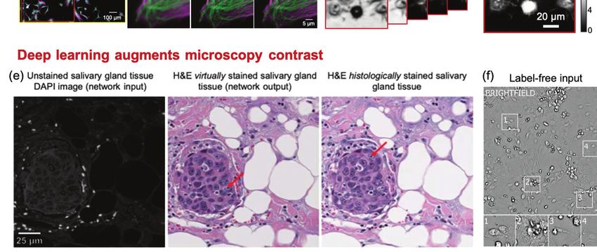
DEEP LEARNING IN BIOMEDICAL OPTICS                                                        1

                                                                  tions. In the case of images, low-level representations could
                                                                  be textures and edges of the objects, whereas higher level
                                                                  representations would be object-like compositions of those
                                                                  features. The joint optimization of both feature representa-
                                                                  tion at multiple levels of abstraction and predictive model
                                                                  parameters is what makes DNNs so powerful.

                                                                  How is deep learning implemented?
                                                                  The majority of the existing DL models in biomedical optics
                                                                  are implemented using the supervised learning strategy. At a
Fig 2: (a) Classical machine learning uses engineered fea-
                                                                  high-level, there are three primary components to implement
tures and a model. (b) Deep learning uses learned features
                                                                  a supervised DL model: 1) labeled data, 2) model architec-
and predictors in an “end-to-end” deep neural network.
                                                                  ture, and 3) optimization strategy. Labeled data consist of
                                                                  the raw data inputs as well as the desired model output.
optical brain imaging. Within each domain, state-of-the-art       Large amounts of labeled data are often needed for effective
approaches which can enable new functionality for optical         model optimization. This requirement is currently one of
systems are highlighted. We then offer our perspectives on        the main challenges for utilizing DL on small-scale biomed-
the challenges and opportunities across these eight imaging       ical data sets, although strategies to overcome this are an
domains. Finally, we provide a summary and outlook of ar-         active topic in the literature, such as unsupervised [8], self-
eas in which DL can contribute to future development and          supervised [9], and semi-supervised learning [10]. For a typ-
clinical impact biomedical optics moving forward.                 ical end-to-end DL model, model architecture defines the
                                                                  hypothesis class and how hierarchical information flows be-
DEEP LEARNING OVERVIEW                                            tween each layer of the DNN. The selection of a DNN archi-
                                                                  tecture depends on the desired task and is often determined
What is deep learning?                                            empirically through comparison of various state-of-the-art
To define DL, it is helpful to start by defining machine learn-   architectures. Three of the most widely used DNN architec-
ing (ML), as the two are closely related in their historical      tures in current biomedical optics literature are illustrated
development and share many commonalities in their prac-           in Fig. 3.
tical application. ML is the study of algorithms and statis-         The encoder-decoder network [11] shown in Fig. 3(a) aims
tical models which computer systems use to progressively          to establish a mapping between the input and output images
improve their performance on a specified task [6]. To ensure      using a nearly symmetrically structure with a contracting
the development of generalizable models, ML is commonly           “encoder” path and an expanding “decoder” path. The en-
broken in two phases: training and testing. The purpose of        coder consists of several convolutional blocks, each followed
the training phase is to actively update model parameters to      by a down-sampling layer for reducing the spatial dimension.
make increasingly accurate predictions on the data, whereas       Each convolutional block consists of several convolutional
the purpose of testing is to simulate a prospective evaluation    layers (Conv2D) that stacks the processed features along
of the model on future data.                                      the last dimension, among which each layer is followed by a
   In this context, DL can be considered a subset of ML, as       nonlinear activation function, e.g. the Rectified Linear Unit
it is one of many heuristics for development and optimiza-        (ReLU). The intermediate output from the encoder has a
tion of predictive, task-specific models [7]. In practice, DL     small spatial dimension but encodes rich information along
is primarily distinguished from ML by the details of the un-      the last dimension. These low-resolution “activation maps”
derlying computational models and optimization techniques         go through the decoder, which consists of several additional
utilized. Classical ML techniques rely on careful develop-        convolutional blocks, each connected by a upsampling con-
ment of task-specific image analysis features, an approach        volutional (Up-Conv) layer for increasing the spatial dimen-
commonly referred to as “feature engineering” (Fig. 2(a)).        sion. The output of the network typically has the same di-
Such approaches typically require extensive manual tuning         mension as the input image.
and therefore have limited generalizability. In contrast, DL         The U-Net [12] architecture shown in Fig. 3(b) can be
applies an “end-to-end” data-driven optimization (or “learn-      thought of as an extension to the encoder-decoder network.
ing”) of both feature representations and model predictions       It introduces additional “skip connections” between the en-
[2] ((Fig. 2(b)). This is achieved through training a type        coder and decoder paths so that information across different
of general and versatile computational model, termed deep         spatial scales can be efficiently tunneled through the net-
neural network (DNN).                                             work, which has shown to be particularly effective to pre-
   DNNs are composed of multiple layers which are con-            serve high-resolution spatial information [12].
nected through computational operations between layers, in-          The generative adversarial network (GAN) [13] shown in
cluding linear weights and nonlinear “activation” functions.      Fig. 3(c) is a general framework that involves adversarially
Thereby, each layer contains a unique feature representa-         training a pair of networks, including the “generator” and
tion of the input data. By using several layers, the model        “discriminator”. The basic idea is to train the generator
can account for both low-level and high-level representa-         to make high-quality image predictions that are indistin-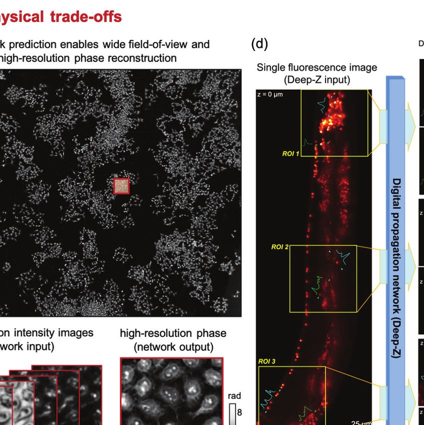
DEEP LEARNING IN BIOMEDICAL OPTICS                                                       2

                                                                  propagation” [15]. Given labeled data, a model architecture,
                                                                  and the optimization guides the model parameters towards
                                                                  a local minimum of the cost function, thereby optimizing
                                                                  model performance.
                                                                     With the recent success of DL, several software frame-
                                                                  works have been developed to enable easier creation and
                                                                  optimization of DNNs. Many of the major technology com-
                                                                  panies have been active in this area. Two of the front-runners
                                                                  are TensorFlow and PyTorch, which are open-source frame-
                                                                  works published and maintained by Google and Facebook,
                                                                  respectively [16, 17]. Both frameworks enable easy construc-
                                                                  tion of custom DNN models, with efficient parallelization
                                                                  of DNN optimization over high-performance graphics com-
                                                                  puting units (GPUs). These frameworks have enabled non-
                                                                  experts to train and deploy DNNs and have played a large
                                                                  role in the spread of DL research into many new applica-
                                                                  tions, including the field of biomedical optics.

                                                                  What is deep learning used for in biomedical optics?
                                                                  There are two predominant tasks for which DL has been uti-
                                                                  lized in biomedical optics: 1) image formation and 2) image
                                                                  interpretation. Both are important applications; however,
                                                                  image formation is a more central focus of biomedical optics
Fig 3: Three of the most commonly-used DNN architectures
                                                                  researchers and consequently is the focus of this review.
in biomedical optics: (a) Encoder-decoder, (b) U-Net, and
                                                                     With regards to image formation, DL has proven very use-
(c) GAN.
                                                                  ful for effectively approximating the inverse function of an
                                                                  imaging model in order to improve the quality of image re-
                                                                  constructions. Classical reconstruction techniques are built
guishable from the real images of the same class (e.g. H&E        on physical models with explicit analytical formulations. To
stained lung tissue slices). To do so, the discriminator is       efficiently compute the inverse of these analytical models,
trained to classify that the generator’s output is fake, while    approximations are often needed to simplify the problem,
the generator is trained to fool the discriminator. Such al-      e.g. linearization. Instead, DL methods have shown to be
ternating training steps iterate until a convergence is met       very effective to directly “learn” an inverse model, in the
when the discriminator can hardly distinguish if the images       form of a DNN, based on the training input and output
produced from the generator are fake or real. When apply-         pairs. This in practice has opened up novel opportunities to
ing to biomedical optics techniques, the generator is often       perform image formation that would otherwise be difficult
implemented by the U-Net. The discriminator is often im-          to formulate an analytical model. In addition, the directly
plemented using an image classification network. The input        learned DL inverse model can often better approximate the
image is first processed by several convolutional blocks and      inverse function, which in turn leads to improved image qual-
downsampling layers to extract high-level 2D features. These      ity as shown in several imaging domains in this review.
2D features are then “flattened” to a 1D vector, which is            Secondly, DL has been widely applied for modeling im-
then processed by several fully connected layers to perform       age priors for solving the inverse problems across multiple
additional feature synthesis and make the final classification.   imaging domains. Most image reconstruction problems are
   Once labeled data and model architecture have been de-         inherently ill-posed in that the reconstructed useful image
termined, optimization of model parameters can be under-          signal can be overwhelmed by noise if a direct inversion is
taken. Optimization strategy includes two aspects: 1) cost        implemented. Classical image reconstruction techniques rely
function, and 2) training algorithm. Definition of a cost func-   on regularization using parametric priors for incorporating
tion (a.k.a. objective function, error, or loss function) is      features of the expected image. Although being widely used,
needed to assess the accuracy of model predictions relative       such models severely limit the type of features that can
to the desired output and provide guidance to adjust model        be effectively modeled, which in turn limit the reconstruc-
parameters. The training algorithm iteratively updates the        tion quality. DL-based reconstruction bypasses this limita-
model parameters to improve model accuracy. This train-           tion and does not rely on explicit parameterization of image
ing process is generally achieved by solving an optimization      features, but instead represents priors in the form of a DNN
problem, using variants of the gradient descent algorithm,        which is optimized (or “learned”) from a large data set that
e.g. stochastic gradient descent and Adam [14]. The opti-         is of the same type of the object of interest (e.g. endoscopic
mizer utilizes the gradient of the cost function to update        images of esophagus). By doing so, DL enables better quality
each layer of the DNN through the principle of “error back-       reconstructions.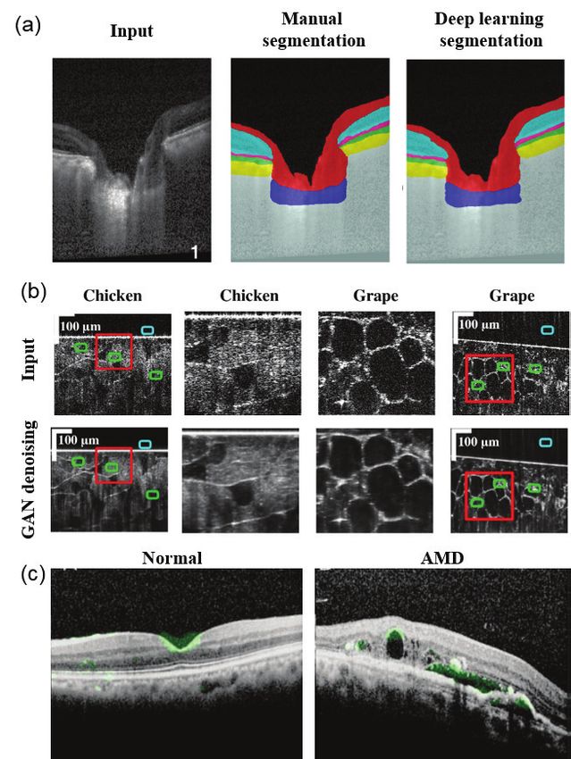
DEEP LEARNING IN BIOMEDICAL OPTICS 3
Beyond achieving higher quality reconstructions, there are and shallow DOF. This section summarizes recent achieve-
other practical benefits of DNNs in image formation. Clas- ments in leveraging DL strategies to overcome various phys-
sical inversion algorithms typically require an iterative pro- ical tradeoffs and expand the imaging capabilities.
cess that can take minutes to hours to compute. Further- 1) Denoising. Enhancing microscopy images by DL-
more, they have stringent sampling requirements, which if based denoising has been exploited to overcome the tradeoffs
lessened, make the inversion severely ill-posed. Due to more between light exposure, SNR, and imaging speed, which in
robust “learned” priors, DL-based techniques can accom- turn alleviates photo-bleaching and photo-toxicity. The gen-
modate highly incomplete or undersampled inputs while eral strategy is to train a supervised network that takes a
still providing high-quality reconstructions. Additionally, al- noisy image as the input and produces the SNR-enhanced
though DNNs typically require large datasets for training, image output. Weigert et al. [18] demonstrated a practical
the resulting models are capable of producing results in real training strategy of a U-Net on experimental microscopy
time with a GPU. These combined capabilities allow DL- data that involves taking paired images with low and high
based techniques to bypass physical trade-offs (e.g., acquisi- light exposures as the noisy input and high-SNR output
tion speed and imaging quality) and enable novel capabilities of the network (Fig. 4(a)). This work showed that the
beyond existing solutions. DNN can restore the same high-SNR images with 60-fold
By leveraging these unique capabilities of DL methods, in- fewer photons used during the acquisition. Similar strategies
novative techniques have been broadly reported across many have been applied to several microscopy modalities, includ-
imaging domains in biomedical optics. Examples include im- ing widefield, confocal, light-sheet [18], structured illumina-
proving imaging performance, enabling new imaging func- tion [19], and multi-photon microscopy [20].
tionalities, extracting quantitative microscopic information, 2) Image reconstruction. Beyond denoising, the imag-
and discovering new biomarkers. These and other technical ing capabilities of several microscopy techniques can be
developments have the potential to significantly reduce sys- much expanded by performing image reconstruction. To per-
tem complexity and cost, and may ultimately improve the form reconstruction by DL, the common framework is to
quality, affordability, and accessibility of biomedical imaging train a DNN, such as the U-Net and GAN, that takes the
in health care. raw measurements as the input and the reconstructed image
as the output. With this DL framework, three major bene-
DEEP LEARNING APPLICATIONS IN BIOMED- fits have been demonstrated. First, Wang et al. showed that
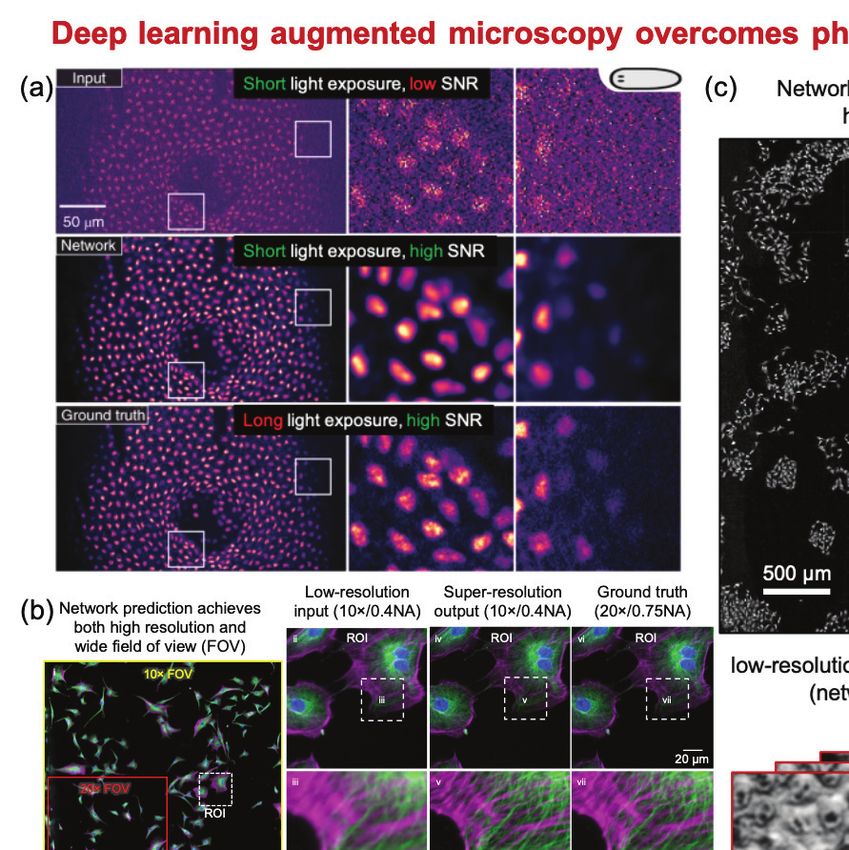
ICAL OPTICS GAN-based super-resolution reconstruction allows recover-
ing high-resolution information from low-resolution mea-
Microscopy surements, which in turn provides an enlarged FOV and
Overview. Microscopy is broadly used in biomedical an extended DOF [21] (Fig. 4(b)). For widefield imaging,
and clinical applications to capture cellular and tissue struc- [21] demonstrated super-resolution reconstruction using in-
tures based on intrinsic (e.g. scattering, phase, and autoflu- put images from a 10×/0.4-NA objective lens and pro-
orescence) or exogenous contrast (e.g. stains and fluores- ducing images matching a 20×/0.75-NA objective lens. In
cent labels). Fundamental challenges exist in all forms of a cross-modality confocal-to-STED microscopy transforma-
microscopy because of the limited information that can be tion case, [21] showed resolution improvement from 290 nm
extracted from the instrument. Broadly, the limitations can to 110 nm. Similar results have also been reported in label-
be categorized based on two main sources of origin. The free microscopy modalities, including brightfield [26], holog-
first class is due to the physical tradeoffs between multiple raphy [27], and quantitative phase imaging [22] (Fig. 4(c)).
competing performance parameters, such as SNR, acquisi- Second, DL-based 3D reconstruction technique allows
tion speed, spatial resolution, FOV, and DOF. The second drastically extending the imaging depth from a single-shot
class is from the intrinsic sensitivity and specificity of dif- and thus bypasses the need for physical focusing. In [23],
ferent contrast mechanisms. DL-augmented microscopy is a Wu et al. demonstrated 20× DOF extension in widefield flu-
fast-growing area that aims to overcome various aspects of orescence microscopy using a conditional GAN (Fig. 4(d)).
conventional limitations by synergistically combining novel Recent work on DL-based extended DOF has also shown
instrumentation and DL-based computational enhancement. promising results on enabling rapid slide-free histology [28].
This section focuses on DL strategies for bypassing the phys- Third, DL significantly improves both the imaging acqui-
ical tradeoffs and augmenting the contrast in different mi- sition and reconstruction speeds and reduces the number
croscopy modalities. of measurements for microscopy modalities that intrinsi-
Overcoming physical tradeoffs. An ideal microscopy cally require multiple measurements for the image forma-
technique often needs to satisfy several requirements, such tion, as shown in quantitative phase microscopy [22, 29, 30]
as high resolution in order to resolve the small features in (Fig. 4(c)), single molecule localization microscopy [31–33],
the sample, low light exposure to minimize photo-damage, and structured illumination microscopy [19]. For example,
and a wide FOV in order to capture information from a in [22], a 97% data reduction as compared to the con-
large portion of the sample. Traditional microscopy is fun- ventional sequential acquisition scheme was achieved for
damentally limited by the intrinsic tradeoffs between various gigapixel-scale phase reconstruction based on a multiplexed
competing imaging attributes. For example, a short light ex- acquisition scheme using a GAN.
posure reduces the SNR; a high spatial resolution requires a
high-magnification objective lens that provides a small FOV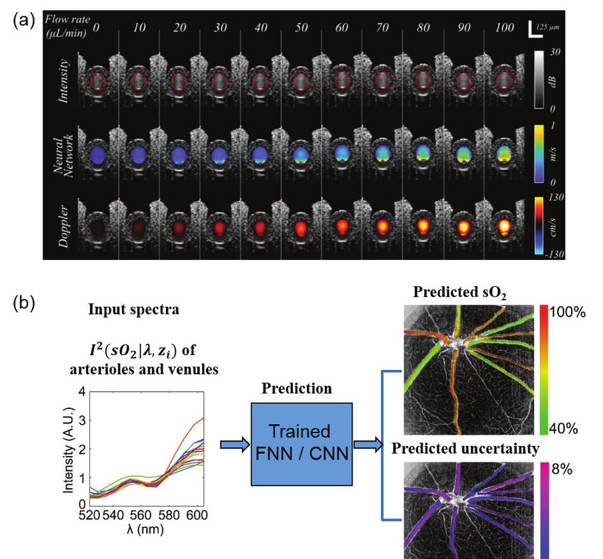
DEEP LEARNING IN BIOMEDICAL OPTICS                                                        4

Fig 4: DL overcomes physical tradeoffs and augments microscopy contrast. (a) CARE network achieves higher SNR with
reduced light exposure (with permission from the authors [18]). (b) Cross-modality super-resolution network reconstructs
high-resolution images across a wide FOV [21] (with permission from the authors). (c) DL enables wide-FOV high-resolution
phase reconstruction with reduced measurements (adapted from [22]). (d) Deep-Z network enables digital 3D refocusing
from a single measurement [23] (with permission from the authors). (e) Virtual staining GAN transforms autofluorescence
images of unstained tissue sections to virtual H&E staining [24] (with permission from the authors) (f) DL enables predicting
fluorescent labels from label-free images [25] (Reprinted from Cell, 2018 Apr 19;173(3):792-803.e19, Christiansen et al., In
Silico Labeling: Predicting Fluorescent Labels in Unlabeled Images, Copyright (2020), with permission from Elsevier).

   Augmenting contrasts. The image contrast used in                1) Virtual staining/labeling. The main idea of vir-
different microscopy modalities can be broadly categorized       tual staining/labeling is to digitally transform the captured
into endogenous and exogenous. For example, label-free mi-       label-free contrast to the target stains/labels. DL has been
croscopy captures endogenous scattering and phase contrast,      shown to be particularly effective to perform this “cross-
and is ideal for imaging biological samples in their natural     modality image transformation” task. By adapting this idea
states, but suffers from lack of molecular specificity. Speci-   to different microscopy contrasts, two emerging applications
ficity is often achieved by staining with absorbing or fluo-     have been demonstrated. First, virtual histological staining
rescent labels. However, applications of exogenous labeling      has been demonstrated for transforming a label-free image
are limited by the physical staining/labeling process and po-    to the brightfield image of the histologically-stained sample
tential perturbation to the natural biological environment.      (Fig. 4(e)). The label-free input utilized for this task in-
Recent advances in DL-augmented microscopy have the po-          clude autofluorescence [24, 34], phase [35, 36], multi-photon
tential to achieve the best of both label-free and labeled       and fluorescence lifetime [37]. The histological stains include
microscopy. This section summarizes two most widely used         H&E, Masson’s Trichrome and Jones’ stain. Notably, the
frameworks for augmenting microscopy contrast with DL.           quality of virtual staining on tissue sections from multiple
                                                                 human organs of different stain types was assessed by board-
DEEP LEARNING IN BIOMEDICAL OPTICS                                                         5

certified pathologists to show superior performance [24]. A        the physical process and speed up the diagnosis. By fur-
recent cross-sectional study has been carried out for clini-       ther advancing the digital fluorescence labeling techniques,
cal evaluation of unlabeled prostate core biopsy images that       it can enable high-throughput and highly multiplexed single-
have been virtually stained [38]. The main benefits of the         cell profiling and cytometry. Beyond clinical diagnoses, this
virtual staining approach include saving time and cost [24],       may find applications in drug screening and phenotyping.
as well as facilitating multiplexed staining [34]. Interested         In addition, several emerging DL techniques can fur-
readers can refer to a recent review on histopathology using       ther enhance the capabilities of microscopy systems. First,
virtual staining [39].                                             DL can be applied to optimize the hardware parameters
   Second, digital fluorescence labeling has been demon-           used in microscopy experiments. In quantitative phase mi-
strated for transforming label-free contrast to fluorescence       croscopy, DL was applied to optimized the illumination
labels [25, 40–43] (Fig. 4(f)). In the first demonstration [25],   patterns to reduce the data requirement [30, 56]. In single
Christiansen et al. performed 2D digital labeling using trans-     molecule localization microscopy, DL was used to optimize
mission brightfield or phase contrast images to identify cell      the point spread functions to enhance the localization accu-
nuclei (accuracy quantified by Pearson correlation coeffi-         racy [33, 57]. DL has also been used to optimize the illumi-
cient PCC = 0.87–0.93), cell death (PCC = 0.85), and               nation power [58] and focus positions [59–61].
to distinguish neuron from astrocytes and immature di-                Second, new DL frameworks are emerging to significantly
viding cells (PCC = 0.84). A main benefit of digital flu-          reduce the labeled data requirements in training, which is
orescence labeling is digital multiplexing of multiple sub-        particularly useful in biomedical microscopy since acquir-
cellular fluorescence labels, which is particularly appealing      ing a large-scale labeled training data set is often imprac-
to kinetic live cell imaging. This is highlighted in [40], 3D      tical. For example, a novel denoising approach, known as
multiplexed digital labeling using transmission brightfield or     Noise2Noise [62], has been developed that can be trained
phase contrast images on multiple subcellular components           using only independent pairs of noisy images, and by-
are demonstrated, including nucleoli (PCC∼0.9), nuclear            passes the need for ground-truth clean images. Following
envelope, microtubules, actin filaments (PCC∼0.8), mito-           this work, self-supervised denoising DL approaches have
chondria, cell membrane, Endoplasmic reticulum, DNA+               been advanced to further alleviate the training data re-
(PCC∼0.7), DNA (PCC∼0.6), Actomyosin bundles, tight                quirement. Techniques, such as Noise2Void, Noise2Self and
junctions (PCC∼0.5), Golgi apparatus (PCC∼0.2), and                their variants, can be directly trained on noisy data set
Desmosomes (PCC∼0.1). Recent advances further exploit              without the need for paired noisy images [63–65]. In addi-
other label-free contrasts, including polarization [41], quan-     tion, semi-supervised and unsupervised DL approaches have
titative phase map [43], and reflectance phase-contrast mi-        also been developed to reduce or completely remove the
croscopy [42]. Beyond predicting fluorescence labels, recent       need for labeled training data during training, which have
advances further demonstrate multiplexed single-cell profil-       been demonstrated for vessel segmentation [66, 67]. Lastly,
ing using the digitally predicted labels [42].                     physics-embedded DL opens up a new avenue for reducing
   In both virtual histopathological staining and digital flu-     training requirements by incorporating the physical model
orescence labeling, the U-Net forms the basic architecture         of the microscopy technique [68, 69].
to perform the image transformation. GAN has also been                Finally, uncertainty quantification techniques address the
incorporated to improve the performance [24, 38].                  need for assessing the reliability of the DL model by quan-
   2) Classification Instead of performing pixel-wise vir-         tifying the confidence of the predictions, and has recently
tual stain/label predictions, DL is also very effective in         been applied in quantitative phase reconstruction [22].
holistically capturing complex ‘hidden’ image features for
classification. This has found broad applications in aug-
menting the label-free measurements and provide improved           Fluorescence Lifetime Imaging
specificity and classify disease progression [44, 45] and can-        Overview. Fluorescence imaging has become a central
cer screening [46–48], as well as detect cell types [49, 50],      tool in biomedical studies with high sensitivity to observe en-
cell states [44, 51], stem cell lineage [52–54], and drug re-      dogenous molecules [70,71] and monitor important biomark-
sponse [55]. For example, in [44], Eulenberg et al. demon-         ers [72]. Increasingly, fluorescence imaging is not limited to
strated a classification accuracy of 98.73% for the G1/S/G2        intensity-based techniques but can extract additional infor-
phase, which provided 6× improvement in error rate as com-         mation by measuring fluorophore lifetimes [73–75]. Fluores-
pared to the previous state-of-the-art method based on clas-       cence lifetime imaging (FLI) has become an established tech-
sical ML techniques.                                               nique for monitoring cellular micro-environment via analysis
                                                                   of various intracellular parameters [76], such as metabolic
   Opportunities and challenges. By overcoming the                 state [77, 78], reactive oxygen species [79] and/or intracel-
physical tradeoffs in traditional systems, DL-augmented mi-        lular pH [80]. FLI is also a powerful technique for study-
croscopy achieves unique combinations of imaging attributes        ing molecular interactions inside living samples, via Förster
that are previously not possible. This may create new op-          Resonance Energy Transfer (FRET) [81], enabling applica-
portunities for diagnosis and screening. By augmenting the         tions such as quantifying protein-protein interactions [82],
contrast using virtual histological staining techniques, DL        monitoring biosensor activity [83] and ligand-receptor en-
can open up unprecedented capabilities in label-free and           gagement in vivo [84]. However, FLI is not a direct imaging
slide-free digital pathology. This can significantly simplify      technique. To quantify lifetime or lifetime-derived parame-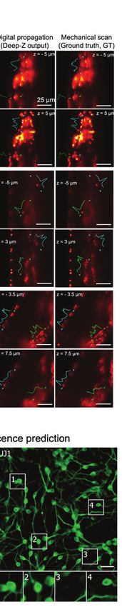
DEEP LEARNING IN BIOMEDICAL OPTICS                                                      6

ters, an inverse solver is required for quantification and/or
interpretation.
   To date, image formation is the main utility of DL in
FLI. Contributions include reconstructing quantitative life-
time image from raw FLI measurements, enabling enhanced
multiplexed studies by leveraging both spectral and lifetime
contrast simultaneously, and facilitating improved instru-
mentation with compressive measurement strategies.
   Lifetime quantification, representation, and re-
trieval. Conventionally, lifetime quantification is obtained
at each pixel via model-based inverse-solvers, such as least-
square fitting and maximum-likelihood estimation [85], or
the fit-free phasor method [86, 87]. The former is time-
consuming, inherently biased by user-dependent a priori set-
tings, and requires operator expertise. The phasor method
is the most widely-accepted alternative for lifetime repre-
sentation [87]. However, accurate quantification using the
phasor method requires careful calibration, and when con-
sidering tissues/turbid-media in FLI microscopy (FLIM) ap-        Fig 5: Example of quantitative FLI metabolic imaging as
plications, additional corrections are needed [87, 88]. There-    reported by NADH tm for a breast cancer cell line (AU565)
fore, it has largely remained qualitative in use.                 as obtained (a) with SPCImage and (b) FLI-Net. (c) Linear
   Wu et al. [89] demonstrated a multilayer perceptron            regression with corresponding 95% confidence band (gray
(MLP) for lifetime retrieval for ultrafast bi-exponential         shading) of averaged NADH Tm values from 4 cell line data
FLIM. The technique exhibited an 180-fold faster speed then       (adapted from [90]).
conventional techniques, yet it was unable to recover the
true lifetime-based values in many cases due to ambiguities
caused by noise. Smith et al. [90] developed an improved
3D-CNN, FLI-Net, that can retrieve spatially independent
                                                                  when higher dimensional data are sought [93] (e.g., hyper-
bi-exponential lifetime parameter maps directly from the 3D
                                                                  spectral imaging [94]). However, these computational meth-
(x, y, t) FLI data. By training with a model-based approach
                                                                  ods are based on more complex inverse models that require
including representative noise and instrument response func-
                                                                  user expertise and input.
tions, FLI-Net was validated across a variety of biologi-
                                                                     Yao et al. [95] developed a CNN, NetFLICS, capable of re-
cal applications. These include quantification of metabolic
                                                                  trieving both intensity and lifetime images from single-pixel
and FRET FLIM, as well as preclinical lifetime-based stud-
                                                                  compressed sensing-based time-resolved input. NetFLICS
ies across the visible and near-infrared (NIR) spectra. Fur-
                                                                  generated superior quantitative results at low photon count
ther, the approach was generalized across two data acquisi-
                                                                  levels, while being four orders of magnitude faster than ex-
tion technologies – Time-correlated Single Photon Counting
                                                                  isting approaches. Ochoa-Mendoza et al. [96] further devel-
(TCSPC) and Intensified Gated CCDs (ICCD). FLI-Net has
                                                                  oped the approach to increase its compression ratio to 99%
two advantages. First, it outperformed classical approaches
                                                                  and the reconstruction resolution to 128×128 pixels. This
i‘n the presence of low photon counts, which is a common
                                                                  dramatic improvement in compression ratio enables signif-
limitation in biological applications. Second, FLI-Net can
                                                                  icantly faster imaging protocols and demonstrates how DL
output lifetime-based whole-body maps at 80 ms in wide-
                                                                  can impact instrumentation design to improve clinical utility
field pre-clinical studies, which highlights the potential of
                                                                  and workflow [97].
DL methods for fast and accurate lifetime-based studies. In
                                                                     Recent developments have made hyperspectral FLI imag-
combination with DL in silico training routines that can be
                                                                  ing possible across microscopic [98] and macroscopic set-
crafted for many applications and technologies, DL is ex-
                                                                  tings [99]. Traditionally, combining spectral and lifetime
pected to contribute to the dissemination and translation
                                                                  contrast analytically is performed independently or sequen-
of FLI methods as well as to impact the design and imple-
                                                                  tially using spectral decomposition and/or iterative fit-
mentation of future-generation FLI instruments. An exam-
                                                                  ting [100]. Smith et al. [101] proposed a DNN, UNMIX-
ple FLI-Net output for metabolic FLI is shown in Fig. 5.
                                                                  ME, to unmix multiple fluorophore species simultaneously
   Emerging FLI applications using DL. The technolo-              for both spectral and temporal information. UNMIX-ME
gies used in FLI have not fundamentally shifted over the          takes a 4D voxel (x, y, t, λ) as the input and outputs spa-
last two decades. One bottleneck for translation is a lack        tial (x, y) maps of the relative contributions of distinct
of sensitive, widefield NIR detectors. Advances in computa-       fluorophore species. UNMIX-ME demonstrated higher per-
tional optics have sparked development of new approaches          formance during tri- and quadri-abundance coefficient re-
using structured light [91], such as single-pixel methods [92].   trieval. This method is expected to find utility in applica-
These methods are useful when widefield detectors are lack-       tions such as autofluorescence imaging in which unmixing of
ing, such as in applications with low photon budget and           metabolic and structural biomarkers is challenging.
DEEP LEARNING IN BIOMEDICAL OPTICS                                                        7

   Although FLI has shown promise for deep tissue imaging          lenges, a variety of strategies have been applied which range
in clinical scenarios, FLI information is affected by tissue op-   from simple classification schemes (benign vs malignant) us-
tical properties. Nonetheless, there are several applications      ing pre-trained CNNs [110] to more complicated tasks, such
that would benefit from optical property-corrected FLI with-       as cross-domain feature learning and multi-scale encoder-
out solving the full 3D inverse problem. For optical guided        decoder networks [112, 117]. The following section contrasts
surgery, Smith et al. [102] proposed a DNN that outputs            recent reports and methods utilizing DL for diagnostic im-
2D maps of the optical properties, lifetime quantification,        age analysis of pCLE and RCM image datasets.
and the depth of fluorescence inclusion (topography). The             1) CNNs and transfer learning approaches. Early
DNN was trained using a model-based approach in which              reports on DL-based image classification for CLE and
a data simulation workflow incorporated “Monte Carlo eX-           RCM have demonstrated that transfer learning using pre-
treme” [103] to account for light propagation through tur-         trained CNNs can outperform conventional image analysis
bid media. The method was demonstrated experimentally,             approaches, especially when data is limited as is often the
with real-time applicability over large FOVs. Both widefield       case for CLE and RCM [110, 118–120].
time-resolved fluorescence imaging and Spatial Frequency              Aubreville et al. published an early and impactful study
Domain Imaging (SFDI) in its single snapshot implementa-           comparing the performance of two CNN-based approaches
tion were performed with fast acquisition [91] and process-        to a textural feature-based classifier (random forest) on
ing speeds [104]. Hence, their combination with DL-based           pCLE video sequences acquired during surgical resection of
image processing provides a possible future foundation for         oral squamous carcinoma (Fig. 6b) [110]. Of their two CNN-
real-time intraoperative use.                                      based approaches, one was a LeNet-5 architecture and was
   While recent advances in FLI-based classification and seg-      trained to classify sub-image patches whereas the other uti-
mentation are limited to using classical ML techniques [105–       lized transfer learning of a pre-trained CNN (Fig. 6c) for
107], Sagar et al. [108] used MLPs paired with bi-exponential      whole image classification. Using leave-one-out cross valida-
fitting for label-free detection of microglia. However, DL ap-     tion on 7,894 frames from 12 patients, the two CNN-based
proaches often outperform such “shallow learning” classi-          approaches both outperformed the textural classifier.
fiers. Although reports using DL for classification based on          Transfer learning is one strategy to overcome limited
FLI data are currently absent from the literature, it is ex-       dataset sizes, which remains a common challenge for CLE
pected that DL will play a critical role in enhancing FLI          and RCM. As larger CLE and RCM datasets are obtainable
classification and semantic segmentation tasks in the near         in the future, transfer learning is unlikely to be an optimal
future.                                                            strategy for image classification; however, it can remain a
                                                                   useful benchmark for the difficulty of image classification
In vivo microscopy                                                 tasks on novel, small-scale datasets moving forward. The
Overview. In vivo microscopy (IVM) techniques enable               subsequent sections introduce alternatives to transfer learn-
real-time assessment of intact tissue at magnifications sim-       ing which utilize video data as well as cross-domain learning.
ilar to that of conventional histopathology [113]. As high-           2) Recurrent convolutional approaches. CLE and
resolution assessment of intact tissue is desirable for many       RCM are typically used in video recording while the optical
biomedical imaging applications, a number of optical tech-         probe is physically or optically scanned to obtain images
niques and systems have been developed which have trade-           over a larger tissue area or at varying depths. Some reports
offs in FOV, spatial resolution, achievable sampling rates,        have utilized recurrent convolutional networks to account
and practical feasibility for clinical deployment [113]. How-      for spatial and/or temporal context of image sequences [121–
ever, a commonality of IVM systems used for clinical imag-         123]. The additional spatial/temporal modeling provided by
ing is the need for image analysis strategies to support in-       recurrent networks is one promising approach to leverage
traoperative visualization and automated diagnostic assess-        video data. [121–123].
ment of the high-resolution image data. Currently, three of           3) Cross-domain learning. A novel approach, termed
the major IVM techniques for which DL is being utilized are        “transfer recurrent feature learning”, was developed by Gu
optical coherence tomography (OCT) [114], confocal laser           et al. which leveraged cross-domain feature learning for clas-
endomicroscopy (CLE, Fig. 6) [115], and reflectance con-           sification of pCLE videos obtained from 45 breast tissue
focal microscopy (RCM) [116]. This section focuses on DL           specimens [112]. Although this method relied on data ac-
approaches for CLE and RCM. More specifically, endoscopic          quired ex vivo, the data itself is not qualitatively differ-
imaging using probe-based CLE (pCLE) and dermal imag-              ent from other pCLE datasets and still provides a proof-
ing for RCM. OCT is discussed in a subsequent section.             of-principle. Their model utilized a cycle-consistent GAN
                                                                   (CycleGAN) to first learn feature representations between
   Automated diagnosis. Automated diagnostic classifi-             H&E microscopy and pCLE images and to identify visually
cation has been the earliest and most frequent application         similar images (Fig. 6e). The optimized discriminator from
of DL within IVM. Most commonly, histopathology analysis           the CycleGAN is then utilized in conjunction with a recur-
of imaged specimens provides a ground truth categorization         rent neural network to classify video sequences (Fig. 6f).
for assessing diagnostic accuracy. The limited size of pCLE        The method outperformed other DL methods and achieved
and RCM datasets and logistical challenges in precisely cor-       84% accuracy in classifying normal, benign, and malignant
relating them with histopathology remain two ongoing chal-         tissues.
lenges for training robust classifiers. To address these chal-
DEEP LEARNING IN BIOMEDICAL OPTICS                                                                                               8

  (a)   Confocal laser endomicrosopy        (b)   Example CLE images acquired in the oral cavity         (e)    Style transfer between CLE and H&E image domains using a
        (CLE) optical probes                                                                                    cycle consistency GAN

                                                                                                                            Histology (H&E) image                   H&E    CLE
                                                                                                                                                                 Synthesis via GANs

                                                                                                                            CLE video mosaic

  (c)   Fine-tuning approach for training classifiers on CLE datasets
                                                                                                         (f)   Transfer recurrent feature learning (GAN-LSTM) for training diagnostic
                                                                                                               classifiers using CLE and histology images

  (d)   Super-resolution network for sparse, irregular CLE data

Fig 6: DL approaches to support real-time, automated diagnostic assessment of tissues with confocal laser endomicroscopy.
(a) Graphical rendering of two confocal laser endomicroscopy probes (left: Cellvizio, right: Pentax) (adapted from [109]).
(b) Example CLE images obtained from four different regions of the oral cavity (adapted from [110]) (c) Fine-tuning of
CNNs pre-trained using ImageNet is utilized in the majority of CLE papers reported since 2017 (adapted from [110]). (d)
Super-resolution networks for probe-based CLE images incorporate novel layers to better account for the sparse, irregular
structure of the images (adapted from [111]). (e) Example H&E stained histology images with corresponding CLE images.
Adversarial training of GANs to transfer between these two modalities has been successful (adapted from [112]). (f) Transfer
recurrent feature learning utilizes adversarially trained discriminators in conjunction with an LSTM for state-of-the-art
video classification performance (adapted from [112]).

   4) Multiscale segmentation. Kose et al. [117] de-                                                 Super-resolution. Several IVM techniques, including
veloped a novel segmentation architecture, “multiscale                                             pCLE, utilize flexible fiber-bundles as contact probes to il-
encoder-decoder network” (MED-Net), which outperformed                                             luminate and collect light from localized tissue areas [127].
other state-of-the-art network architectures for RCM mosaic                                        Such probes are needed for minimally invasive endoscopic
segmentation . In addition to improving accuracy, MED-Net                                          procedures and can be guided manually or via robotics. The
produced more globally consistent, less fragmented pixel-                                          FOV of fiber-optic probes is typically
DEEP LEARNING IN BIOMEDICAL OPTICS                                                        9

   Others have taken a more computational approach to            ages from laser-illumination endoscopy images by training
pCLE super-resolution by using synthetic datasets. For ex-       on image pairs acquired of the same tissue with both coher-
ample, Ravı̀ et al. [129] demonstrated super-resolution of       ent and incoherent illumination sources [141].
pCLE images using unpaired image data via a CycleGAN,               Improving image quality. In widefield endoscopy, wet
and Szczotka et al. [111] introduced a novel Nadaraya-           tissue is often imaged in a perpendicular orientation to the
Watson layer to account for the irregular sparse artifacts       optical axis, and the close positioning of the camera and light
introduced by the fiber-bundle (Fig. 6d).                        sources leads to strong specular reflections that mask un-
   Future directions. Beyond automatic diagnosis and             derlying tissue features. GANs have been applied to reduce
super-resolution approaches in IVM, recent advances also         these specular reflections [142]. In this case, unpaired train-
highlight ways in which DL can enable novel instrumenta-         ing data with and without specular reflections were used
tion development and image reconstructions to enable new         in a CycleGAN architecture with self-regularization to en-
functionalities for compact microscopy systems. Such ex-         force similarity between the input specular and predicted
amples include multispectral endomicroscopy [130], more          specular-free images. Other work has found that specular
robust mosaicking for FOV expansion [131], and end-to-           reflection removal can be achieved in a simultaneous local-
end image reconstruction using disordered fiber-optic probes     ization and mapping elastic fusion architecture enhanced
[132, 133]. We anticipate that similarly to ex vivo mi-          by DL depth estimation [143]. Lastly, Ali et al. [144] intro-
croscopy, in the coming years DL will be increasingly utilized   duced a DL framework that identifies a range of endoscopy
to overcome physical constraints, augment contrast mecha-        artifacts (multi-class artifact detection), including specu-
nisms, and enable new capabilities for IVM systems.              lar reflection, blurring, bubbles, saturation, poor contrast,
                                                                 and miscellaneous artifacts using YOLOv3-spp with classes
Widefield endoscopy                                              that were hand-labeled on endoscopy images. These artifacts
   Overview. The largest application of optics in medical        were then removed and the image restored using GANs.
imaging, by U.S. market size, is widefield endoscopy [134]. In      Resolution enhancement. For capsule endoscopy ap-
this modality, tissue is typically imaged on the >1 cm scale,    plications, where small detectors with low pixel counts are
over a large working distance range, with epi-illumination       required, DL tools have been applied for super-resolution
and video imaging via a camera. Endoscopic and laparo-           with the goal of obtaining conventional endoscopy-like im-
scopic examinations are commonly used for screening, di-         ages from a capsule endoscope [145]. In this study, a condi-
agnostic, preventative, and emergency medicine. There has        tional GAN was implemented with spatial attention blocks,
been extensive research in applying various DL tools for an-     using a loss function that included contributions of pixel
alyzing conventional endoscopy images for improving and          loss, content loss, and texture loss. The intuition behind the
automating image interpretation [135–138]. This section in-      incorporation of spatial attention blocks is that this module
stead reviews recent DL research in image formation tasks        guides the network to prioritize the estimation of the sus-
in endoscopy, including denoising, resolution enhancement,       picious and diagnostically relevant regions. This study also
3D scene reconstruction, mapping of chromophore concen-          performed ablation studies and found that the content and
trations, and hyperspectral imaging.                             texture loss components are especially important for esti-
   Denoising. A hallmark of endoscopic applications is           mating high-spatial frequency patterns, which becomes more
challenging geometrical constraints. Imaging through small       important for larger upsampling ratios. With this frame-
lumens such as the gastrointestinal tract or “keyholes” for      work, the resolution of small bowel images was increased
minimally-invasive surgical applications requires optical sys-   by up to 12× with favorable quantitative metrics as well as
tems with compact footprints–often on the order of 1-cm di-      qualitative assessment by gastroenterologists. Though this
ameter. These miniaturized optical systems typically utilize     study demonstrated that the resolution of gastrointestinal
small-aperture cameras with high pixel counts, wide FOVs         images could be enhanced, it remains to be seen if prepro-
and even smaller illumination channels. Consequently, man-       cessing or enhancing these images provides any benefit to
aging the photon budget is a significant challenge in en-        automated image analysis.
doscopy, and there have been several recent efforts to apply        3D imaging and mapping. The three dimensional
DL to aid in high-quality imaging in these low-light condi-      shape of the tissue being imaged via endoscopy is useful
tions. A low-light net (LLNET) with contrast-enhancement         for improving navigation, lesion detection and diagnosis, as
and denoising autoencoders has been introduced to adap-          well as obtaining meaningful quality metrics for the effec-
tively brighten images [139]. This study simulated low-light     tiveness of the procedure [146]. However, stereo and time-
images by darkening and adding noise and found that train-       of-flight solutions are challenging and expensive to imple-
ing on this data resulted in a learned model that could          ment in an endoscopic form factor. Accordingly, there has
enhance natural low-light images. Other work has applied         been significant work in estimating the 3D shape of an endo-
a U-Net for denoising on high-speed endoscopic images of         scopic scene from monocular images using conditional GANs
the vocal folds, also by training on high-quality images that    trained with photo realistic synthetic data [147, 148]. Do-
were darkened with added noise [140]. Brightness can also        main adaptation can be used to improve the generalizability
be increased via laser-illumination, which allows greater cou-   of these models, either by making the synthetic data more
pling efficiency than incoherent sources, but results in laser   realistic, or by making the real images look more like the syn-
speckle noise in the image from coherent interference. Con-      thetic data that the depth-estimator is trained on [149]. Re-
ditional GANs have been applied to predict speckle-free im-      searchers have also explored joint conditional random fields
DEEP LEARNING IN BIOMEDICAL OPTICS 10
and CNNs in a hybrid graphical model to achieve state- enable quantitative spectroscopy measurements in endo-
of-the-art monocular depth estimation [150]. A U-Net style scopic imaging, it may be necessary to combine hyperspec-
architecture has been implemented for simultaneously es- tral techniques with structured illumination and 3D map-
timating depth, color, and oxygen saturation maps from a ping [104, 151, 155, 158].
fiber-optic probe that sequentially acquired structured light Future directions. Future research in endoscopy and
and hyperspectral images [151]. Lastly, DL tools have been DL will undoubtedly explore clinical applications. Imag-
applied to improve simultaneous localization and mapping ing system for guiding surgery are already demonstrating
(SLAM) tasks in endoscopic applications, both by incorpo- clinical potential for ex-vivo tissue classification: a modified
rating a monocular depth estimation prior into a SLAM al- Inception-v4 CNNs was demonstrated to effectively classify
gorithm for dense mapping of the gastrointestinal tract [143], squamous cell carcinoma versus normal tissue at the cancer
and by developing a recurrent neural network to predict margin from ex-vivo hyperspectral images with 91 spectral
depth and pose in a SLAM pipeline [152]. bands [159]. For in-vivo applications, where generalizability
may be essential and training data may be limited, future
Widefield spectroscopy. In addition to efforts to re- research in domain transfer [149] and semi-supervised learn-
construct high-quality color and 3D maps through an en- ing [160] may become increasingly important. Moreover, for
doscope, DL is also being applied to estimate bulk tissue clinical validation, these solutions must be real-time, easy-
optical properties from wide FOV images. Optical prop- to-use, and robust, highlighting the need for efficient archi-
erty mapping can be useful for meeting clinical needs in tectures [104] and thoughtful user interface design [161].
wound monitoring, surgical guidance, minimally-invasive
procedures, and endoscopy. A major challenge to estimating Optical coherence tomography
optical properties in turbid media is decoupling the effects Overview. Optical coherence tomography (OCT) is a
of absorption, scattering, and the scattering phase function, successful example of biophotonic technological translation
which all influence the widefield image measured with flood into medicine [162,163]. Since its introduction in 1993, OCT
illumination. Spatial frequency domain imaging can provide has revolutionized the standard-of-care in ophthalmology
additional inputs to facilitate solving this inverse problem around the world, and continued thriving in technical ad-
by measuring the attenuation of different spatial frequen- vances and other clinical applications, such as dermatology,
cies [153]. Researchers have demonstrated that this inverse neurology, cardiology, oncology, gastroenterology, gynecol-
model can be solved orders of magnitude faster than con- ogy, and urology [164–171].
ventional methods with a 6-layer Perceptron [154]. Others Image segmentation. The most common use of OCT is
have shown that tissue optical properties can be directly es- to quantify structural metrics via image segmentation, such
timated from structured light images or widefield illumina- as retinal anatomical layer thickness, anatomical structures,
tion images using content-aware conditional GANs [155]. In and pathological features. Conventional image processing
this application, the adversarial learning framework reduced is challenging in the case of complex pathology where tis-
errors in the optical property predictions by more than half sue structural alteration can be complex and may not be
when compared to the same network trained with an ana- fully accounted for when designing a rigid algorithm. Im-
lytical loss function. Intuitively, the discriminator learns a age segmentation is the earliest application of DL explored
more sophisticated and appropriate loss function in adver- in OCT applications. Several DNNs have been reported
sarial learning, allowing for the generation of more-realistic for OCT segmentation in conjunction with manual annota-
optical property maps. Moreover, this study found that the tions (Fig. 7(a)), including U-Net [172–174], ResNet [175],
conditional GANs approach resulted in an increased perfor- and fully-convolutional network (FCN) [176, 177]. Success-
mance benefit when data is tested from tissue types that ful implementation of DNNs have been broadly reported
were not spanned in the training set. The authors hypothe- in different tissues beyond the eye [178–180]. In all ar-
size that this observation comes from the discriminator pre- eas of applications, the DNN showed superior segmenta-
venting the generator from learning from and overfitting to tion accuracy over conventional techniques. For example,
the context of the input image. Optical properties can also Devalla et al. [175] quantified the accuracy of the proposed
be estimated more quickly using a lighter-weight twin U- DRUNET(Dilated-Residual U-Net) for segmenting the reti-
Net architecture with a GPU-optimized look-up table [104]. nal nerve fiber layer (RNFL), retinal Layers, the retinal pig-
Further, chromophores can be computed in real-time with ment epithelium (RPE), and choroid on both healthy and
reduced error compared to an intermediate optical property glaucoma subjects, and showed that the DRUNET consis-
inference by directly computing concentrations from struc- tently outperformed alternative approaches on all the tissues
tured illumination at multiple wavelengths using conditional measured by dice coefficient, sensitivity, and specificity. The
GANs [156]. errors of all the metrics between DRUNET and the observers
Going beyond conventional color imaging, researchers are were within 10% and the patch-based neural network always
also processing 1D hyperspectral measurement through an provided greater than 10% error irrespective of the observer
endoscope using shallow CNNs to classify pixels into the cor- chosen for validation. In addition, the DRUNET segmen-
rect color profiles, illustrating the potential to classify tissue tation further allowed automatic extraction of six clinically
with complex absorbance spectra [157]. The spectral resolu- relevant neural and connective tissue structural parameters,
tion can be increased in dual-modality color/hyperspectral including the disc diameter, peripapillary RNFL thickness
systems from sparse spectral signals with CNNs [151]. To (p-RNFLT), peripapillary choroidal thickness (p-CT), min-You can also read