Diagnosis and Management of Infantile Hemangioma
←
→
Page content transcription
If your browser does not render page correctly, please read the page content below

CLINICAL REPORT           Guidance for the Clinician in Rendering Pediatric Care

Diagnosis and Management of Infantile
Hemangioma
David H. Darrow, MD, DDS, Arin K. Greene, MD, Anthony J. Mancini, MD, Amy J. Nopper, MD,
the SECTION ON DERMATOLOGY, SECTION ON OTOLARYNGOLOGY–HEAD AND NECK SURGERY, and SECTION ON PLASTIC SURGERY

                                                    abstract              Infantile hemangiomas (IHs) are the most common tumors of childhood. Unlike
                                                                          other tumors, they have the unique ability to involute after proliferation, often
                                                                          leading primary care providers to assume they will resolve without
                                                                          intervention or consequence. Unfortunately, a subset of IHs rapidly develop
                                                                          complications, resulting in pain, functional impairment, or permanent
                                                                          disfigurement. As a result, the primary clinician has the task of determining
                                                                          which lesions require early consultation with a specialist. Although several
                                                                          recent reviews have been published, this clinical report is the first based on
                                                                          input from individuals representing the many specialties involved in the
                                                                          treatment of IH. Its purpose is to update the pediatric community regarding
                                                                          recent discoveries in IH pathogenesis, treatment, and clinical associations and
This document is copyrighted and is property of the American              to provide a basis for clinical decision-making in the management of IH.
Academy of Pediatrics and its Board of Directors. All authors have filed
conflict of interest statements with the American Academy of
Pediatrics. Any conflicts have been resolved through a process
approved by the Board of Directors. The American Academy of
Pediatrics has neither solicited nor accepted any commercial
involvement in the development of the content of this publication.        NOMENCLATURE
Clinical reports from the American Academy of Pediatrics benefit from      The nomenclature and classification of vascular tumors and
expertise and resources of liaisons and internal (American Academy        malformations have evolved from clinical descriptions (“strawberry
of Pediatrics) and external reviewers. However, clinical reports from
the American Academy of Pediatrics may not reflect the views of the        birthmark,” “salmon patch,” “cavernous hemangioma,” and “port wine
liaisons or the organizations or government agencies that they            stain”) to terminology based on their cellular features, natural history, and
represent.
                                                                          clinical behavior. Originally described by Mulliken and Glowacki in 1982,
The guidance in this report does not indicate an exclusive course of
treatment or serve as a standard of medical care. Variations, taking
                                                                          the most current and widely accepted classification of vascular anomalies
into account individual circumstances, may be appropriate.                is that adopted by the International Society for the Study of Vascular
All clinical reports from the American Academy of Pediatrics              Anomalies (Table 1).1 This system includes infantile hemangioma (IH)
automatically expire 5 years after publication unless reaffirmed,          among the vascular neoplasms, which are lesions characterized by
revised, or retired at or before that time.
                                                                          abnormal proliferation of endothelial cells and aberrant blood vessel
www.pediatrics.org/cgi/doi/10.1542/peds.2015-2485                         architecture. In contrast, vascular malformations are structural anomalies
DOI: 10.1542/peds.2015-2485                                               and inborn errors of vascular morphogenesis.
PEDIATRICS (ISSN Numbers: Print, 0031-4005; Online, 1098-4275).           Although IH is the most common neoplasm, this group also includes such
Copyright © 2015 by the American Academy of Pediatrics                    tumors as congenital hemangiomas, pyogenic granulomas, tufted
                                                                          angiomas (TAs), and several types of hemangioendothelioma. Congenital
FINANCIAL DISCLOSURE: The authors have indicated they do not have
a financial relationship relevant to this article to disclose.             hemangiomas are biologically and behaviorally distinct from IH. As
POTENTIAL CONFLICT OF INTEREST: The authors have indicated they
                                                                          reflected in the name, congenital hemangiomas are present and fully
have no potential conflicts of interest to disclose.                       formed at birth; they do not exhibit the postnatal proliferative phase

                                                    Downloaded from www.aappublications.org/news by guest on October 23, 2021
FROM THE AMERICAN ACADEMY OF PEDIATRICS                                                                          PEDIATRICS Volume 136, number 4, October 2015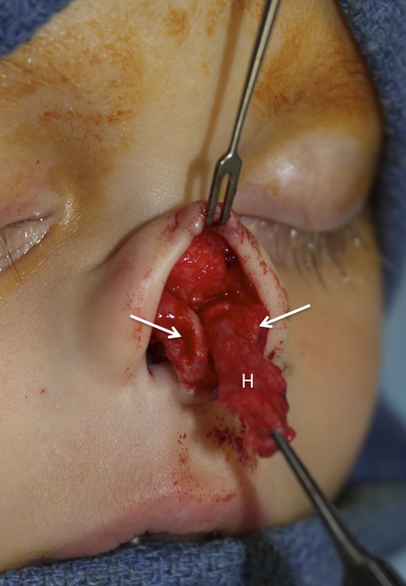
characteristic of IH. The 2 variants are                     It is a reactive proliferating vascular   and may grow slowly over the course
the noninvoluting congenital                                 lesion that is classified as a vascular    of months to years, grow rapidly,
hemangioma (NICH), which remains                             neoplasm (Table 1). This common           spontaneously regress, or remain
stable without growth or                                     acquired vascular lesion of the skin      dormant for years.14–16 Unlike KHE
involution,2,3 and the rapidly                               and mucous membranes primarily            and TA, IHs are not associated with
involuting congenital hemangioma                             affects infants and children and is       thrombocytopenia or coagulopathy.
(RICH), which undergoes a rapid                              frequently misdiagnosed as IH.            Vascular malformations are
involution phase beginning in the first                       Approximately 12% occur in infancy,       congenital lesions, but some may
year of life (Fig 1).4 RICHs, in some                        and 42% present during the first 5         become clinically apparent only later
cases, have been associated with                             years of life.10 Pyogenic granulomas      in life, presumably because of slowly
thrombocytopenia but with milder                             are most commonly located on the          progressive ectasia resulting from
and more transient coagulopathy                              head and neck, rapidly enlarge to         intraluminal flow. They exhibit
than that seen in Kasabach-Merritt                           a median size of 6.5 mm, frequently       a normal rate of endothelial cell
phenomenon (KMP; see discussion                              develop a pedunculated base, and,         turnover throughout their natural
that follows); rarely, they can be                           with erosion, are prone to bleeding       history but expand as the patient
associated with congestive heart                             that is difficult to control (Fig 2).10    grows. Vascular malformations do not
failure.5,6 Some RICHs show                                  Pyogenic granulomas are seen with         involute, and their growth may be
incomplete involution, and it is                             higher frequency within the skin          influenced by trauma, infection, and
possible that RICH and NICH lie at                           containing capillary malformations.       hormonal changes. Classification is
opposite ends of the same clinical                           Two other distinct benign vascular        based on the predominant vessel
spectrum.7,8 Both subtypes of                                neoplasms, kaposiform                     type: capillary or venulocapillary,
congenital hemangioma were initially                         hemangioendothelioma (KHE) and            venous, lymphatic, arterial, or
believed to be variants of IH that                           TA, have been confused with IH. KHE       mixed.17 As with vascular neoplasms,
exhibited prenatal growth until North                        presents primarily in infancy but with    the nomenclature of vascular
et al9 showed that, unlike IH, neither                       a far wider age range than IH, which      malformations has led to great
lesion expresses glucose transporter                         is usually apparent in the first month     confusion. Capillary or
protein isoform 1 (GLUT1).                                   of life. KHE is considered a locally      venulocapillary malformations have
Pyogenic granuloma, also known as                            aggressive neoplasm that typically        had numerous alternative
lobular capillary hemangioma, is                             appears as a deep, soft tissue mass.      designations, the most common being
neither pyogenic nor granulomatous.                          This lesion has been associated with      “port wine stain” and “nevus
                                                             KMP,11 a potentially life-threatening     flammeus.” Venous malformations
                                                             consumptive coagulopathy                  have often been mistaken for IH,
TABLE 1 Classification of Cutaneous                           characterized by severe platelet
            Vascular Anomalies, 2014                         trapping. Before KHE was described
Vascular malformations                                       in the early 1990s, KMP was
  Venous malformations                                       erroneously thought to occur in
  Lymphatic malformations
                                                             association with IH.
  Capillary malformations
  Arteriovenous malformations and fistulae                    Histopathologically, KHE shows
  Mixed (combined) malformations                             infiltrating sheets of slender, GLUT1-
Vascular tumors                                              negative endothelial cells lining
  Benign                                                     slitlike capillaries.12 TAs are benign
  Infantile hemangioma (IH)
                                                             vascular tumors that occur in infants,
  Congenital hemangioma (rapidly involuting
     [RICH]; non-involuting [NICH])                          children, or young adults and are
  Lobulated capillary hemangiomas (LCH)                      usually located on the neck or the
     (pyogenic granuloma)*                                   upper part of the thorax.13 Their
  Tufted angioma (TA)                                        clinical appearance is variable and
  Others
                                                             includes erythematous to violaceous
  Locally aggressive
  Kaposiform hemangioendothelioma (KHE)                      patches, plaques, and nodules.
  Kaposi sarcoma                                             Histopathologically, TA shows well-
  Others                                                     defined tufts of capillaries in the
  Malignant                                                  dermis that lack cellular atypia or
  Angiosarcoma
                                                             GLUT1 positivity and, like KHE, is
  Others                                                                                               FIGURE 1
                                                             associated with increased lymphatic
Adapted from the International Society for the Study of                                                RICH is fully formed at birth (A) and then
Vascular Anomalies, 2014, ref 1 (issva.org/classification).   vessels and a predisposition to KMP.      involutes, mostly during the first year of life. B,
*Reactive proliferating vascular lesion                      Both tumors behave unpredictably          The same lesion seen at 8 months of age.

                                    Downloaded from www.aappublications.org/news by guest on October 23, 2021
PEDIATRICS Volume 136, number 4, October 2015                                                                                                      e1061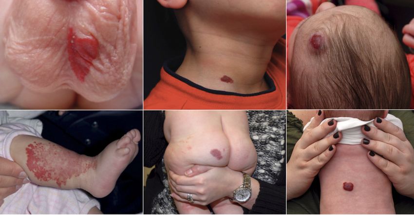
termed “cavernous hemangiomas”                     years, especially predating the                 among female infants; however,
and “venous hemangiomas” in the                    distinction between IH and the                  although older data suggest female-
literature (Fig 3A). Lymphatic                     congenital hemangiomas.                         to-male ratios ranging from 3:1 to 5:1,
malformations, which are subdivided                “Hemangioma” has also been                      more recent studies suggest a range
into microcystic and macrocystic                   inappropriately used to describe, in            of 1.4:1 to 3:1.23,24 The gender
varieties on the basis of predominant              general terms, varieties of other               discrepancy appears to be increased
lacuna size, may also be mistaken for              noninfantile hemangiomas and                    among children with PHACE
IH when there is bleeding into                     vascular malformations.                         syndrome (Posterior fossa defects,
vesicles at the surface of the skin or                                                             Hemangiomas, cerebrovascular
mucosa (Fig 3B). These lesions have                                                                Arterial anomalies, Cardiovascular
traditionally been referred to as                                                                  anomalies including coarctation of
“cystic hygromas” or                                  Highlights of This Section                   the aorta, and Eye anomalies), in
“lymphangiomas,” designations that                                                                 which studies have found a 9:1
                                                      • Infantile hemangioma (IH) is
inaccurately presume proliferative                                                                 female-to-male ratio.25 There is not
                                                        the currently accepted termi-
potential, thereby perpetuating the                                                                a definitive explanation for this
                                                        nology for the lesions that are
diagnostic confusion.                                                                              gender difference.
                                                        the focus of this clinical report.
The use of various names for IH has                                                                Most studies report a significantly
                                                      • Congenital hemangiomas are
resulted in immense diagnostic                                                                     higher incidence in white
                                                        biologically and behaviorally
confusion. For instance, the terms                                                                 infants.23,24,26 On the basis of the
                                                        distinct from IH.
“capillary hemangioma” and                                                                         success of IH treatment using
“capillary angioma” have been used to                 • Pyogenic granuloma is a re-
                                                                                                   b-blocker therapy, it has been
refer to an IH that is located primarily                active proliferating vascular
                                                                                                   proposed that black infants may
in the dermis and is bright red in                      lesion that is classified as
                                                                                                   exhibit some form of “endogenous
color. In contrast, the designations                    a vascular neoplasm and
                                                        that may occasionally be                   beta blockade,” and there are
“cavernous” or “venous” have                                                                       molecular biological data to support
inappropriately been used to define                      misdiagnosed as IH.
                                                                                                   this notion.27
an IH that, because of its depth below                • Lesions diagnosed as “cav-
the dermis, may impart a blue tinge to                  ernous hemangiomas” are                    The incidence of IH is increased
the skin surface. In addition, deep                     usually, in fact, deep IHs or              among preterm infants, affecting 22%
venous and lymphatic malformations                      venous malformations.                      to 30% of infants weighing less than
as well as arteriovenous                              • Kasabach-Merritt phenome-                  1 kg.24,28 Multivariate analysis has
malformations have been incorrectly                     non or KMP (a consumptive                  revealed that low birth weight (LBW)
diagnosed as deep IH. Finally, by                       coagulopathy) is not associ-               is the major contributor to this risk;
virtue of its sheer prevalence, the                     ated with IH but rather with               there is a 25% increase in risk of
term “hemangioma,” without the                          2 other vascular neoplasms,                developing an IH with every 500-g
adjectival descriptor “infantile” or                    kaposiform hemangioendo-                   reduction in birth weight.29 Prenatal
with the descriptor “juvenile,” has                     thelioma (KHE) and tufted                  factors have also been investigated
been used in reference to IH for many                   angioma (TA).                              for their role in IH. Studies differ
                                                                                                   regarding an increased risk resulting
                                                                                                   from maternal chorionic villus
                                                                                                   sampling24,30 or amniocentesis,30,31
                                                   EPIDEMIOLOGY                                    and any increased risk attributable to
                                                                                                   chorionic villus sampling appears to
                                                   Studies of the incidence of IH,
                                                                                                   be limited to procedures performed
                                                   including a prospective study and
                                                                                                   transcervically.31 Other possible
                                                   a review incorporating 1
                                                   retrospective study and 2 cross-                prenatal factors include older
                                                   sectional cohorts, suggest that 4% to           maternal age, multiple gestation
                                                   5% of infants are affected.18,19 Other          pregnancy, placenta previa, and
                                                   studies suggest that IH is observed in          preeclampsia.24 Placental anomalies,
                                                   1% to 3% of newborn infants20,21                such as retroplacental hematoma,
FIGURE 2                                           and 2.6% to 9.9% of older                       infarction, and dilated vascular
Pyogenic granulomas have some clinical and                                                         communications, have also been
                                                   children,22,23 but methodologic
histologic features similar to IHs, but they are
generally smaller, pedunculated, and more          shortcomings may have influenced                 associated with IH development.32 It
likely to bleed.                                   these findings. IHs are more common              is theorized that the common thread

                                    Downloaded from www.aappublications.org/news by guest on October 23, 2021
e1062                                                                                                  FROM THE AMERICAN ACADEMY OF PEDIATRICS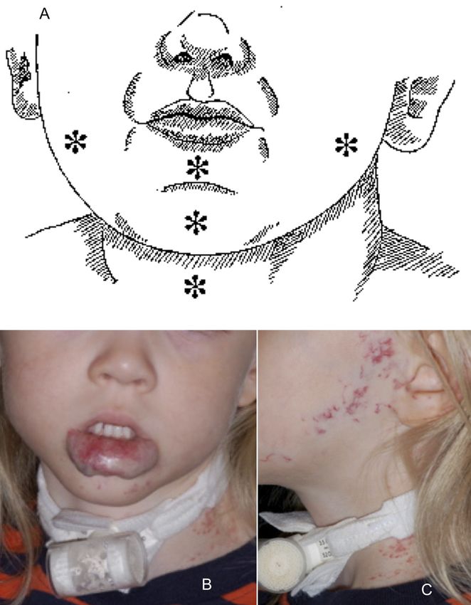
in these associations is placental
hypoxia.32,33
Although often suggested as a risk
factor, a family history of IH is
reported in only 12% of cases24;
however, familial clustering has been
reported.34,35 Associations are also
reported with maternal use of fertility
drugs,36 use of erythropoetin,37 level
of maternal education,36 breech
presentation,23 and being the first
born.23
                                                 FIGURE 3
                                                 A, The venous blood contained within a venous malformation imparts a bluish hue that may lead to
   Highlights of This Section                    misdiagnosis as a deep IH. B, Bleeding into surface vesicles of a lymphatic malformation may lead to
                                                 misdiagnosis as an IH.
   • The incidence of in the gen-
     eral population is approxi-
     mately 5%.                                  vasculogenesis, or the de novo                      This concept developed from
   • Risk factors for IH include                 formation of new blood vessels.39,40                research showing that molecular
     being white, being female,                  This theory is supported by studies                 markers characteristic of placental
     and having a low birth                      showing increased numbers of                        tissue, including GLUT1, Lewis Y
     weight.                                     circulating EPCs in blood samples                   antigen, merosin, Fc-g receptor-IIb,
   • Associations are also                       from children with IH.41 Additional                 indoleamine 2,3-deoxygenase, and
     reported with older mater-                  evidence comes from studies in which                type III iodothyronine deiodinase,
     nal age, multiple gestation                 multipotential stem cells derived                   were also present in IHs.3,9 Clinical
     pregnancy, placenta previa,                 from IH specimens (HemSCs) have                     evidence for this theory is suggested
     preeclampsia, use of fertility              shown the ability to recapitulate                   by those studies showing an
     drugs or erythropoietin,                    human IH in immunodeficient mice.42                  increased incidence of IH in
     breech presentation, and                    These HemSCs and cord blood EPCs                    association with chorionic villus
     being the first born.                        behave similarly to each other in                   sampling, placenta previa, and
                                                 several in vitro assays, suggesting                 preeclampsia.24,30,31
                                                 that circulating EPCs could be the                  A unifying theory suggests that IH
                                                 origin of IH endothelial cells.42 The               results from aberrant proliferation
                                                 concept that IHs originate from                     and differentiation of a hemogenic
PATHOGENESIS AND HISTOPATHOLOGY                  circulating multipotent progenitor                  endothelium with a neural crest
                                                 cells could explain some of the                     phenotype and a capacity for
Pathogenesis                                     features they share with placental                  endothelial, hematopoetic,
The pathogenesis of IH, despite                  blood vessels, because dysregulated                 mesenchymal, and neuronal
intensive study, has not been                    circulating EPCs have also been                     differentiation. It is hypothesized that
completely elucidated. Lines of                  implicated in many of the associated                placental chorionic villus
evidence support a cellular origin               maternal and fetal comorbid                         mesenchymal core cells embolize to
from either intrinsic endothelial                conditions (preeclampsia, retinopathy               the developing fetus and that the
progenitor cells (EPCs) or angioblasts           of prematurity, etc). HemSCs have                   timing of this embolization in relation
of placental origin, but intrinsic and           also been shown to have an                          to the migration of neural crest cells
extrinsic factors are also thought to            adipogenic potential,43 which may                   along their somitic routes determines
contribute to their development.38               explain the presence of adipocytes                  the morphology of the IH (segmental
Intrinsic factors include the influence           noted during involution. The stimulus               versus localized [focal]; see section
of angiogenic and vasculogenic                   for division of EPCs is unknown but                 entitled “Clinical Appearance”).44
factors within the IH. External factors          may be a somatic mutation or                        The cytokine niche within the IH,
include tissue hypoxia and                       abnormal signals from local tissues.                including vascular endothelial growth
developmental field disturbances.                 The theory of placental origin                      factors (VEGFs), insulin-like growth
The EPC theory holds that IHs                    suggests that fetal progenitor cells                factors, the tumor necrosis
develop from clonal expansion of                 arise from the disruption of the                    factor–related apoptosis-inducing
circulating EPCs, resulting in                   placenta during gestation or birth.                 ligand-osteoprotegerin (TRAIL-OPG)

                                    Downloaded from www.aappublications.org/news by guest on October 23, 2021
PEDIATRICS Volume 136, number 4, October 2015                                                                                                  e1063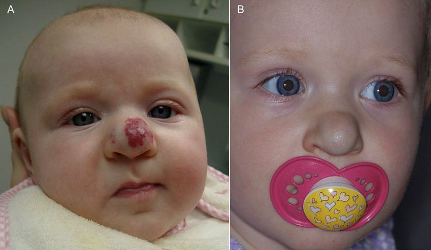
pathway, and the renin-angiotensin          is an important sensor of hypoxia.9             Histopathology
system, subsequently regulates              GLUT1 has been shown to be                      Grossly proliferative and early
growth of the IH and its response to        upregulated in hypoxic zones of                 involutive IHs are well-circumscribed,
pharmacologic therapies.44 Other            mesenchymal tumors and in                       unencapsulated masses with red-to-
authors have also embraced the              umbilical cord–derived human                    tan cut surfaces. Later involutive
“niche” concept, suggesting that            mesenchymal stem cells under                    lesions are fibrofatty in consistency
circulating EPCs find their way to           hypoxic conditions.49,50 Hypoxia-
                                                                                            and less defined. The histologic
certain locations that provide              induced factors produced by
                                                                                            features of IH change dramatically as
conditions favorable for growth into        endothelial cells appear to play an
                                                                                            they proceed through their natural
                                            important role in trafficking of
placentalike tissues.44 In tissues such                                                     course of neonatal presentation, rapid
                                            progenitor cells to ischemic tissue.
as the skin and liver, progenitor cells                                                     growth, and subsequent involution,
                                            These factors have been shown to be
may encounter the cellular signals                                                          requiring interpretation within the
                                            upregulated in the blood (VEGF-A,
and local tissue factors required to                                                        proper clinical context.52–54 There is
                                            MMP-9) and in IH tissue (stromal
stimulate their development.                                                                no sharp dividing line between
                                            cell–derived factor 1a, MMP-9, VEGF-A,
                                                                                            proliferation and involution, and
On the basis of the rapid proliferation     and hypoxia-inducible factor 1a)
                                                                                            features of involution typically coexist
of endothelial cells, earlier               from children with proliferating IH.41
                                                                                            with features of proliferation during
investigations of IH origin focused on      In addition, it has been shown that
                                                                                            much of the process. Early proliferative
angiogenesis, the sprouting of              the use of erythropoietin in preterm
                                                                                            phase IHs are composed of well-
endothelial cells from existing blood       infants increases the risk of
                                                                                            defined, unencapsulated masses of
vessels. Such studies have shown an         developing an IH.37 Thus, tissue
                                                                                            capillaries lined by plump endothelial
increased concentration of angiogenic       ischemia resulting in
                                                                                            cells rimmed by plump pericytes
factors in IH, such as basic fibroblast      neovascularization from circulating
growth factor (bFGF), VEGF-A,               EPCs has been proposed as the                   embedded within a multilaminated
insulin-like growth factor, and matrix      stimulus leading to the development             basement membrane without
metalloprotease (MMP) 9 within the          of IH.41 Clinically, an area of pallor or       associated smooth muscle cells (Fig 4).
lesion during proliferation.45 Also         decreased blood flow in the skin has             Lesions at this stage may at least
in this phase, investigators                been noted to precede the                       focally resemble other rapidly growing
have identified indoleamine                  development of IH, further                      vascular proliferations such as early
2,3-deoxygenase, a protein thought to       supporting this hypothesis.51                   pyogenic granulomas. The proliferating
slow the involution of IH by inhibiting                                                     capillaries are arranged in lobules,
cytotoxic T-lymphocyte response.46                                                          separated by delicate fibrous septae or
During involution, endothelial cell                                                         by normal intervening tissue. These
apoptosis is accompanied by                                                                 lesional capillaries, depending on tissue
downregulation of angiogenic factors,                                                       location, intermingle nondestructively
                                               Highlights of This Section                   with superficial skeletal muscle fibers,
whereas inhibitors of angiogenesis
such as interferon-b and markers of            • may develop either from in-                peripheral nerves, salivary glands, and
cell maturation such as intercellular            trinsic endothelial progenitor             adipocytes. Endothelial cells and
adhesion molecule 1 are                          cells (EPCs) or from angio-                pericytes show variably enlarged
upregulated.47 It has also been shown            blasts of placental origin.                nuclei and abundant clear cytoplasm.
that involuting IHs exhibit decreased          • IH growth is affected by in-               Normally configured mitotic figures are
production of nitric oxide,                      trinsic influences, such as                 relatively numerous (Fig 1B); and
a potentiator of the VEGF pathway, as            angiogenic and vasculogenic                widespread expression of cell
measured by reduced levels of                    factors within the IH, and by              proliferation markers, such as Ki-67,
endothelial nitric oxide synthase.48             external factors such as tis-              confirm that both pericytes and
                                                 sue hypoxia and de-                        endothelial cells are actively dividing.
It has been hypothesized that hypoxia
triggers a vascular response in                  velopmental field                           Because proliferative phase IHs are
                                                 disturbances.                              high-flow lesions, although typically
infants. As discussed above, LBW is
                                               • A unifying theory proposes                 without significant arteriovenous
a significant risk factor for IH, and in
                                                 that circulating EPCs migrate              shunting, they often contain enlarged
utero hypoxia is a common cause of
                                                 to locations in which con-                 draining veins with thick, asymmetric
LBW. Not surprisingly, there is
mounting evidence of the role of                 ditions are favorable for                  walls.
hypoxia in the development of IH.                growth into placentalike                   Involuting IHs present different
GLUT1, a facilitative glucose                    tissues.                                   diagnostic challenges. Mitotic figures
transporter used as a marker for IH,                                                        wane, and apoptotic bodies and masts

                             Downloaded from www.aappublications.org/news by guest on October 23, 2021
e1064                                                                                           FROM THE AMERICAN ACADEMY OF PEDIATRICS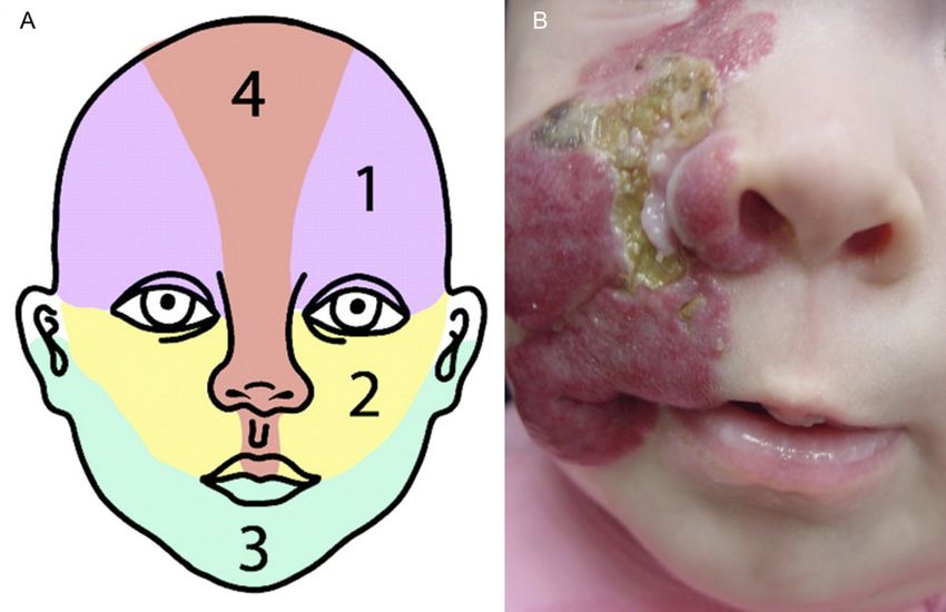
cellular linings. Epidermal atrophy
                                                                                                      Highlights of This Section
                                                     and underlying fibrous scar tissue
                                                     may be present if the lesion ulcerated           • Proliferating IHs are well
                                                     in the proliferative phase. Large                  circumscribed and lack
                                                     arteries and veins modeled during the              a capsule.
                                                     high-flow proliferative phase do not              • Involuting IHs are fibrofatty
                                                     completely regress when the capillary              and less defined.
                                                     bed drops out and thus are often
                                                                                                      • GLUT1 is a commonly used
                                                     present in involuting IH. This
                                                                                                        immunochemical marker for
                                                     phenomenon, paired with loss of
                                                                                                        IH.
FIGURE 4                                             endothelial mitotic activity, may lead
Proliferative phase IH. Well-circumscribed lobules   to mistaken histologic diagnosis as
of closely packed capillaries composed of            a vascular malformation.
plump endothelial cells and pericytes are sep-                                                     CLINICAL PRESENTATION,
arated by normal-appearing dermal stromal
                                                     Misdiagnosis can usually be avoided
                                                     by considering overall histologic             COMPLICATIONS, AND ASSOCIATIONS
elements (hematoxylin and eosin stain; original
magnification 3100; photo courtesy of Paula           appearance and clinical history.              Phases of Growth
North, MD.)                                          Ultimately, as discussed below, the
                                                     issue can be resolved by GLUT1                IHs exhibit a characteristic life cycle.
                                                     immunoreaction, because involuting            Clinical observations have suggested
                                                     infantile IHs, but not malformations,         that there are at least 2 dynamic
cells increase in number during early
                                                     will show GLUT1 immunopositivity in           evolutionary phases, namely,
involution.48,55 Lesional capillaries
                                                     residual lesion-type capillaries.9,56         proliferation and involution.
begin to disappear. There is no
                                                                                                   Proliferation occurs during early
evidence of thrombosis, and                          Histologic examination, accompanied           infancy; gradual spontaneous
inflammation is not prominent. As                     by routine immunohistochemical                involution or regression starts by
involution proceeds, lesional capillary              studies, shows that proliferative             1 year of age.58–65 An intermediate
basement membranes become thick                      phase infantile IHs are complex               period between proliferation and
and hyalinized and contain specks of                 cellular mixtures with large                  involution during mid-to-late infancy,
apoptotic debris (Fig 5). Eventually                 complements of endothelial cells,             often referred to as the “plateau”
all that remains in an end-stage lesion              pericytes, mast cells, and interstitial       phase, more likely represents
is loose fibrous or fibrofatty stroma,                 dendritic cells. Electron microscopy          a period of temporary balance
containing a few residual “ghost”                    reveals plump endothelial cells lining        between individual cells that are
vessels composed of residual,                        small lumina and resting on                   proliferating and those undergoing
thickened rinds of basement                          a multilaminated basement                     involution and apoptosis.59–66 The
membrane material containing                         membrane that envelops a cuff of              process of involution takes several
apoptotic debris and without intact                  pericytes. The endothelial cells of IH        years and varies in duration.
                                                     have been reported to immunoreact
                                                     positively for “normal” endothelial           Proliferative Phase (Up to 12 Months of
                                                     markers of the blood vasculature,             Age)
                                                     such as CD31, CD34, factor
                                                     VIII–related antigen (von Willebrand          Premonitory findings in the skin
                                                     factor), and others.57 Currently, the         during early infancy may include
                                                     most useful and widely used                   localized blanching or localized
                                                     immunohistochemical marker for the            macular telangiectatic erythema.
                                                     diagnosis of IH is GLUT1.9,57 GLUT1 is        As endothelial cell proliferation
                                                     strongly expressed by endothelial             continues, the IH enlarges, becomes
                                                     cells of IHs at all stages of their           more elevated, and develops
                                                     evolution and is not expressed by             a rubbery consistency. During this
                                                     other benign vascular anomalies and           period, IHs often show surrounding
FIGURE 5                                             reactive proliferations. GLUT1                pallor and dilatation of surrounding
Involutive phase IH. Lesional capillaries are set                                                  veins. During rapid growth periods,
within loose fibro-adipose tissue and are less        immunohistochemistry is frequently
                                                     used to distinguish IHs from other            ulceration may arise, leading to pain
densely packed than in the proliferative phase.
Note the thickened and hyalinized basement           vascular neoplasms and provides               and eventual scarring.
membranes studded with apoptotic debris,
                                                     convincing evidence that IHs are              IHs typically have their clinical onset
reflective of the involutive process. Residual
lining endothelial cells are mitotically inactive.   indeed as biologically distinctive as         before 4 weeks of age.66,67 They
(Photo courtesy of Paula North, MD)                  they are clinically distinctive.              proliferate for variable periods of

                                    Downloaded from www.aappublications.org/news by guest on October 23, 2021
PEDIATRICS Volume 136, number 4, October 2015                                                                                         e1065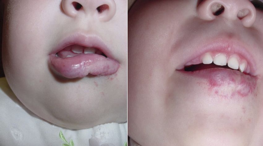
time, depending in part on their
morphology and configuration.
However, most IH growth appears to
occur between 1 and 2 months of
age.68 A large prospective study has
indicated that 80% of IH size is
generally reached by 3 months, and
most growth is completed by around
5 months of age.66 Deep IHs appear
somewhat later and grow somewhat
longer than their superficial
counterparts.66

Involution Phase
For most infants with IH, involution        FIGURE 6
begins between 6 and 12 months of           Cutaneous IHs may be classified on the basis of their depth. A, Superficial IHs are visible only at the
                                            skin surface and may be focal (as shown) or segmental. B, Deep IHs have no surface involvement. C,
age. Although the process continues
                                            Mixed, or compound, IHs have both superficial and deep components.
over years, the majority of tumor
regression occurs before age 4.48,69,70
As IHs involute, most lesions flatten
                                            soft tissue depth.61,62,72 Superficial                superficial and deep IHs. These
and shrink from the center outward.
                                            IHs (Fig 6A) are those in which the                  observations indicate that deep IHs
For those with a superficial component,
                                            surface of the tumor appears red and                 require a longer period of
this is accompanied by “central
                                            there is little to no discernible                    monitoring than those with
clearing” or graying of the surface.
                                            subcutaneous component;                              superficial morphology.
Although IHs generally undergo              historically, these IHs have been                    A specific subtype of superficial IH
spontaneous regression, observations        described as being of the                            has been variably referred to as an
of “maximal involution” do not              “strawberry” type. Deep IHs (Fig 6B)                 abortive, nonproliferative, arrested-
necessarily imply complete                  are those in which the tumor resides                 growth, minimal-growth, nascent,
resolution. Indeed, approximately           deep to the skin surface, and their                  reticular, or telangiectatic IH.73–76
50% to 70% of IHs resolve, leaving          subcutaneous location results in                     This type of IH presents as a macular,
behind residual skin changes,               a bluish surface hue or no evident                   telangiectatic patch that may be
including telangiectasia, fibrofatty         surface changes; historically, these                 accompanied by blanching of the
tissue, redundant skin, anetoderma,         have been referred to as “cavernous,”                involved skin (Fig 7). Unlike most IHs,
dyspigmentation, or scar.71                 an imprecise term that is no longer                  abortive IHs lack an obvious
                                            commonly used. Combined, mixed, or                   significant proliferative phase.
                                            compound IHs (Fig 6C) are those in                   Approximately two-thirds of these
   Highlights of This Section               which both superficial and deep                       lesions are situated on the lower
                                            components coexist.                                  extremities. Many are accompanied
   • IHs usually make their initial
     appearance before 4 weeks              Superficial IHs tend to appear earlier                by localized, small papular regions of
     of age and complete most of            and begin to involute sooner than                    vascular tissue growth, often around
     their growth by 5 months of            their deep counterparts, which,                      the periphery.77 Abortive IHs share
     age.                                   by contrast, tend to arise later                     with more typical IHs characteristic
                                            and grow for longer periods                          surface markers (eg, GLUT1),
   • Involution of IHs begins as
                                            of time before involuting (on                        confirming that they are true IHs;
     the child approaches 12
                                            average, approximately 1 month                       however, their growth phase may be
     months of age. In most cases,
                                            more).62,64,66 Investigations into                   arrested. Many of these telangiectatic
     the majority of involution is
                                            these differences confirm that these                  IHs also involute more rapidly,
     completed by age 4.
                                            timelines represent characteristic                   sometimes before 1 year of age.78
                                            growth patterns for these IHs rather                 Nevertheless, complications such as
                                            than arising out of observational                    ulceration may occur. These IHs may
                                            bias.66 As might be expected, those                  also be segmental and occasionally
Clinical Appearance                         IHs with a mixed morphology have                     have syndromic associations (see
During the proliferative phase, IHs         a growth pattern that is intermediate                section entitled “IH Syndromes and
can be classified on the basis of their      between those associated with                        Associations”).79

                             Downloaded from www.aappublications.org/news by guest on October 23, 2021
e1066                                                                                           FROM THE AMERICAN ACADEMY OF PEDIATRICS
definitively focal or segmental are                Multifocal cutaneous IHs are
                                                   considered indeterminate. Multifocal              frequently isolated to the skin but
                                                   lesions are focal lesions occurring at            may also serve as markers for
                                                   more than 1 anatomic site. One large              underlying hepatic involvement
                                                   study found that most IHs (67.5%) are             (Fig 9).88–91 Previous retrospective
                                                   localized, whereas the remainder were             reports26,81 suggested that the
                                                   segmental (13%), indeterminate                    presence of a large or segmental
                                                   (16.5%), or multifocal (3.6%).83                  (.5 cm) cutaneous IH might prove
                                                   The presence of a large, facial                   a useful marker for hepatic IHs.
FIGURE 7                                           segmental IH is a hallmark sign of                However, results from a large
Abortive IHs are macular, telangiectatic patches                                                     prospective study suggest that it is
that have failed to fully proliferate.             PHACE syndrome,25 whereas large
                                                   segmental IHs of the anogenital and               the number of cutaneous IHs rather
                                                   lumbosacral areas may be associated               than their size that is the more
IHs may also be classified on the basis             with genitourinary system and                     predictive factor.92 When 5 or more
of their anatomic configuration as                  spinal cord anomalies as part of                  IHs are present on cutaneous
either localized (focal), segmental,               other syndromes84–86 (see section                 examination, ultrasonography may
indeterminate, or multifocal.26,80,81              entitled “IH Syndromes and                        be helpful in assessing potential
Localized (focal) IHs are discrete                 Associations”). More recently, it has             hepatic involvement.84,93
lesions that seem to arise from                    been recognized that extracutaneous               Hepatomegaly and congestive heart
a single focal point, whereas                      manifestations may also arise in                  failure also suggest the presence of
segmental lesions cover a territory                association with segmental IHs                    liver IH.
that is presumed to be determined by               involving other anatomic sites, as
embryonic neuroectodermal                          part of the so-called PHACE-without-
placodes.82 Segmental IHs tend to                  face phenomenon.87 These patients                     Highlights of This Section
involve a larger surface area of skin.             may have segmental IHs of the upper                   • IHs are characterized as su-
Segmental IHs of the face have been                chest, shoulder, or arm in the                          perficial, deep, or mixed and
observed to conform to unique                      absence of facial IH involvement and                    as focal, multifocal, or
developmental units, which have                    in conjunction with structural heart                    segmental.
been mapped into 4 distinct patterns:              disease, aortic or other major vessel                 • Superficial IHs appear ear-
frontotemporal, maxillary,                         anomalies, central nervous system                       lier and begin involution
mandibular, and frontonasal                        and sternal defects, or eye                             sooner than their deeper
(Fig 8).82 Lesions that are not                    anomalies.                                              counterparts.
                                                                                                         • Segmental IHs are more
                                                                                                           commonly involved in
                                                                                                           PHACE (see text for defini-
                                                                                                           tion) and other IH syn-
                                                                                                           dromes and associations.
                                                                                                         • The presence of more than 5
                                                                                                           focal IHs suggests a higher
                                                                                                           risk of hepatic involvement.

FIGURE 8
(A) Patterns of segmental IH of the face extracted from image analysisdefined. Seg1 (fronto-          FIGURE 9
temporal), Seg2 (maxillary), Seg3 (mandibular), and Seg4 (frontonasal). (B) An ulcerated segmental   Multifocal cutaneous IHs in a child with IH of
IH in the maxillary distribution.                                                                    the liver.

                                    Downloaded from www.aappublications.org/news by guest on October 23, 2021
PEDIATRICS Volume 136, number 4, October 2015                                                                                                e1067
Complications                               to develop complications and 8 times            usually results in scarring, with the
Although most IHs do not require            more likely to receive treatment.84             risk of permanent disfigurement. As
urgent treatment, a minority may            Segmental lesions tend to have longer           a result, prompt initiation of therapy
develop function-threatening or life-       proliferative phases, some with                 is essential in the management of
threatening complications,                  significantly prolonged duration of              ulcerating IHs.
necessitating therapeutic                   growth as long as 10 to 44 months,              The specific mechanisms resulting in
intervention. One study determined          and may therefore require                       IH ulceration are poorly understood.
that approximately 24% of patients          significantly longer treatment                   It has been hypothesized that
with IH who were referred to a group        durations.94                                    ulceration may develop secondary to
of tertiary care dermatology practices      The size of the IH was also an                  increased tissue hypoxia, which leads
experienced some complication               important predictor of the need for             to the development of dermal fibrosis
related to their IH.84 It is therefore      treatment in the aforementioned                 and then progresses to surface
prudent for pediatric providers to          cohort study, although this analysis            breakdown.98 In such cases, early
remain vigilant of possible                 did not appear to control for                   white discoloration of an IH, possibly
complications and of risk factors that      anatomic subtype.84 The mean size of            representing superficial dermal
may herald future complications.            complicated IHs was 37.3 cm2,                   fibrosis, may be a premonitory sign of
Ulceration accounts for the majority        compared with 19.1 cm2 for                      impending ulceration.99 Other
of IH complications; others include         uncomplicated IHs. In addition, IHs             proposed mechanisms include
bleeding, visual impairment,                that received treatment of any type             outgrowing of the blood supply or
                                            had a mean size of 30.4 cm2, which              rapid expansion exceeding the elastic
auditory impairment, congestive
                                            was 11.1 cm2 larger than those that             capabilities of the skin.99,100
heart failure, and airway
                                            did not receive treatment. However,
obstruction.84 Gastrointestinal                                                             Several studies have shown that
                                            the mean size of segmental IHs is               certain subsets of patients with IHs
bleeding has been reported as
                                            approximately 10 times that of                  are at higher risk of ulceration. As
a complication of segmental
                                            localized IHs,66 suggesting that                discussed previously, superficial and
intestinal hemangiomatosis, in which
                                            morphology may indeed be a more                 segmental IHs have been found to
the IH is typically situated in the
                                            important indicator of potential                be at higher risk of
distribution of the mesenteric
                                            complications.                                  ulcerating.96–98,101 In addition,
arterial system.94
                                            Anatomic location was also                      specific locations at higher risk of
The single best predictor of
                                            a predictor of complications due to             ulceration include the head, neck,
complications and the need for
                                            IH.84 Facial IHs were complicated 1.7           perioral, and perineal/perianal
therapeutic intervention for IH is
                                            times more frequently than nonfacial            regions and intertriginous sites
morphologic subtype.84 Focal IHs
                                            IHs; they were also 3.3 times more              (Fig 10).96–98,102 The neck and
have the potential to cause
                                            likely than their nonfacial                     anogenital regions sustain
complications primarily by virtue of
                                            counterparts to receive some form of            maceration and friction, which may
their location on or near vital
                                            therapy, likely because of concerns             contribute to the development of
structures, such as the eye
                                            for cosmesis. Periocular IHs and                ulceration. Ulceration has also been
(amblyopia, astigmatism), nose
                                            those in the “beard” distribution are           noted to occur more frequently in
(anatomic distortion and
                                            also more likely to require                     infants younger than 4 months,
cartilaginous destruction), ears
                                            intervention, as described below. In            a period of time during which the IH
(anatomic distortion and
                                            1 study, perineal IHs were the most
cartilaginous destruction), lips
                                            likely to ulcerate.95
(anatomic distortion and ulceration),
airway (obstruction), or anogenital
region (ulceration). On the face, focal     Ulceration
lesions are 3 times more common             Ulceration, or breakdown of the IH
than segmental IHs.81 Segmental IHs         skin surface, occurs with an
are more frequently complicated by          estimated incidence of 5% to 21%.96
ulceration.84 A prospective cohort          Ulceration was the most common
study in 1058 patients undertaken to        complication in a large prospective
identify clinical characteristics           cohort of children with IHs,
predicting complications and need for       occurring in 16% of the study
treatment found, after controlling for      population.97 Ulceration can lead to
                                                                                            FIGURE 10
size, that segmental IHs were 11            significant pain, bleeding, and                  Ulcerated segmental IH of the perineal/perianal
times more likely than localized IHs        secondary infection. Ulceration also            region.

                             Downloaded from www.aappublications.org/news by guest on October 23, 2021
e1068                                                                                           FROM THE AMERICAN ACADEMY OF PEDIATRICS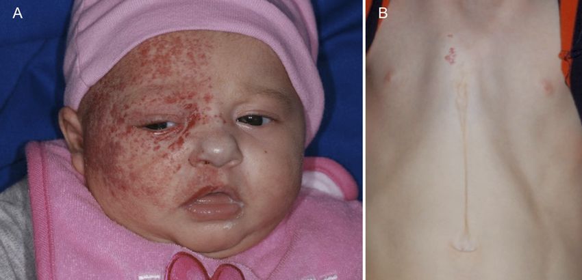
is actively proliferating.96–98 See the          IHs had feeding and oral sensory                     develop noisy breathing or a hoarse
section entitled “Management of                  problems that resulted in failure to                 cry.106
Ulcerated IH” for a discussion in                thrive.105                                           The cutaneous findings associated
greater detail.                                                                                       with underlying airway involvement,
                                                 Airway Involvement and Obstruction                   when present, help to identify those
                                                 Airway IHs can occur in the presence                 patients at greatest risk of airway
Bleeding
                                                 or absence of skin findings.                          IHs. Cutaneous IHs in a “beard”
Although concern for potential                   Symptomatic obstructive airway IHs,                  distribution, defined as involving the
bleeding in IH is common among                   including supra- and subglottic IHs,                 preauricular regions, chin, anterior
caregivers and providers, it occurs              usually present with progressive                     neck, or lower lip (Fig 11), have been
rarely and almost exclusively in                 biphasic inspiratory and expiratory                  associated with airway
ulcerated lesions. The majority of               stridor during the first 6 to 12 weeks                involvement.106,107 Infants with IHs
bleeding that occurs in nonulcerated             of age as the lesion is proliferating.105            within this distribution bilaterally
IHs is minor and easily controllable             Affected infants may also rapidly                    appear to be at an even higher risk of
with pressure. The most common
such scenario is an IH that has
sustained minor surface trauma (ie,
from friction or a fingernail), bled
minimally, stopped bleeding
spontaneously or with minimal
sustained pressure, and subsequently
presents with surface hemorrhagic
crusting.
In a large prospective study of
ulcerated IHs, bleeding occurred in
41% of lesions but was clinically
significant in only 2% of these
cases.96,98 Significant bleeding
requiring blood transfusion or other
intervention is infrequently
reported.98 Rare instances of life-
threatening bleeding have been
observed, including 1 report of
ulceration of a segmental neck IH into
arterial vessels, the bleeding from
which necessitated transfusions,
systemic and topical treatment of the
IH, embolization, and surgical
excision.102

Feeding Impairment
Feeding impairment can occur in
infants with IHs involving either the
perioral region or the airway. Infants
with ulcerated lip IHs may be unable
to latch onto a nipple secondary to
severe pain, which can lead to
impaired feeding.103 Obstructive
airway IHs may complicate
breathing and swallowing, also
leading to impaired feeding.104 In               FIGURE 11
                                                 A, The presence of multiple IHs in the “beard” distribution is associated with a higher likelihood of
a small case series in infants with
                                                 airway involvement (reproduced with permission from J Pediatr. 1997;131(4):643–646 ©Elsevier).106
complicated facial IHs, several with             B and C, Patient with airway involvement requiring tracheotomy is shown with “beard” involvement
ulcerated perioral lesions or airway             at the lip and chin (B) as well as the parotid area and neck (C).

                                    Downloaded from www.aappublications.org/news by guest on October 23, 2021
PEDIATRICS Volume 136, number 4, October 2015                                                                                                   e1069
having associated airway compromise usually improves with anomalies. The best-known such
involvement; in a recent series in treatment of both the heart failure association is PHACE (Online
17 infants with airway IHs, bilateral and the IH. Mendelian Inheritance in Man
involvement of the lower facial Diffuse lesions of the liver may also 606519).114 The disorder is also
segment was present in 13 be associated with severe referred to as PHACES to include
(76%).106,108 Early referral to consumptive hypothyroidism caused potential ventral midline defects,
otolaryngology of infants with severe by excess production of type 3 specifically Sternal cleft and/ or
stridor and a cutaneous IH in the iodothyronine deiodinase. Liver IHs Supraumbilical raphe. Originally
“beard” distribution is advisable, are discussed in greater detail in the described as a “syndrome,” PHACE is
because airway involvement can be subsection entitled “Liver” of “IHs more appropriately termed an
life-threatening if diagnosis and With Special Anatomic Concerns.” association, although there are recent
treatment are delayed. In less data suggesting that chromosomal
symptomatic children, a high region 7q33 may provide a genetic
kilovoltage radiograph of the airway susceptibility to exhibit the PHACE
Highlights of This Section
may be useful in identifying phenotype.115
subglottic IH. Airway IHs are • Segmental IHs are far more
The spectrum of anomalies in PHACE
discussed in greater detail in the likely than focal IHs to result
syndrome and the ipsilateral
subsection under “IHs With Special in a complication, usually
relationship between such anomalies
Anatomic Concerns” entitled ulceration.
and cutaneous IH strongly suggest
“Airway.” • Focal IHs cause complica- a “developmental field defect,”
tions primarily by virtue of whereby an insult at a critical time in
Visual Impairment and Other Ocular their location on or near vi-
Complications embryogenesis gives rise to similar
tal structures.
developmental outcomes.116 The
IHs occurring within the orbit have the • Facial IHs cause complica- precise timing of such an insult in
potential to cause mechanical ptosis, tions more frequently than PHACE syndrome is speculative, but
strabismus, anisometropia, or nonfacial IHs and are several both the anatomic IH patterns and
astigmatism, which can quickly lead to times more likely to receive several of the associated structural
the development of amblyopia.108 some form of therapy. abnormalities point to changes early
Studies have identified specific
• Minor bleeding from an during the first trimester, probably
characteristics of periocular IHs, which
ulcerated IH is common, but within the first 3 to 12 weeks of
place the child at higher risk of
rarely of clinical significance; gestation before or during early
amblyopia. These include periocular
bleeding from a non- vasculogenesis.117 PHACE syndrome
IHs that are larger than 1 cm in
ulcerated IH is rare. is now understood to be
diameter, nasal location of the IH,
associated ptosis, eyelid margin • Patients with an extensive predominantly a congenital
change, or displacement of the IH in the “beard” distribu- vasculopathy. In fact, many of its
tion are more likely to have features can be explained as
globe.109–111 Orbital IHs are discussed
involvement of the airway. downstream events of arteriopathy
in greater detail in the subsection
• High-risk periocular IHs are with resultant ischemia, and it has
entitled “Eye and Orbit” under “IHs
With Special Anatomic Concerns.” those that are that are larger been hypothesized that vascular
than 1 cm in diameter, lo- dysplasia may be a key or even
Congestive Heart Failure and cated near the nose, associ- primary event in the pathogenesis of
Hypothyroidism ated with ptosis or eyelid PHACE syndrome.118
Although rare, high-output congestive margin change, or displacing Consensus criteria were recently
heart failure can occur in infants with the globe. developed for the diagnosis of PHACE
large IHs as a result of arteriovenous • Diffuse IH of the liver may syndrome (Tables 2 and 3).25 Clinical
shunting of a large blood volume be associated with severe examination of the skin and eyes as
through the lesion. This complication consumptive well as detailed imaging of the head,
has been reported in infants with hypothyroidism. neck, and chest are required to make
large cutaneous IHs and RICHs and in the diagnosis. More than 90% of
those with diffuse or multifocal infants with PHACE syndrome exhibit
hepatic IHs.91,92,112,113 Symptomatic more than 1 extracutaneous anomaly,
infants may present with difficulty IH Syndromes and Associations although very few manifest the
feeding, poor growth, heart murmur, A small subset of children with IH will complete spectrum.119 In contrast to
or hepatomegaly. The cardiac exhibit associated congenital nonsyndromic IH, PHACE syndrome
Downloaded from www.aappublications.org/news by guest on October 23, 2021
e1070 FROM THE AMERICAN ACADEMY OF PEDIATRICSTABLE 2 Consensus Algorithm for the Diagnosis of PHACE Syndrome is more common in full-term
PHACE Syndrome Possible PHACE Syndrome singleton infants of normal birth
Facial hemangioma Facial hemangioma Hemangioma of the No hemangioma plus
weight, although females are still
.5 cm in diameter .5 cm in diameter neck or upper 2 major criteria more commonly affected.120
plus 1 major criterion plus 1 minor torso plus 1 major
The hallmark of PHACE syndrome is
or 2 minor criteria criterion criterion or 2
minor criteria a large, segmental, often superficial
Adapted from ref 25.
IH, characteristically located on the
face, scalp, and/or neck (Fig 12).
PHACE syndrome–associated IHs
most commonly affect facial segments
1 and/or 3, which also confers a
particularly high risk of associated
central nervous system
TABLE 3 Consensus Diagnostic Criteria for PHACE Syndrome involvement.84,119
Organ System Major Criteria Minor Criteria
PHACE syndrome is not exceedingly
Cerebrovascular Anomaly of major cerebral arteries Persistent embryonic artery other rare, and probably even more
Dysplasiaa of the large than trigeminal artery
common than Sturge-Weber
cerebral arteriesb Proatlantal intersegmental
Arterial stenosis or occlusion artery (types 1 and 2) syndrome. 121 The segmental IH
with or without moyamoya Primitive hypoglossal artery associated with PHACE is
collaterals Primitive otic artery sometimes confused with the port
Absence or moderate-severe wine stain associated with Sturge-
hypoplasia of the large
Weber syndrome, especially in the
cerebral arteries
Aberrant origin or course of newborn period before significant
the large cerebral arteriesb IH proliferation or in cases of
Persistent trigeminal artery “minimal growth” IH in which there
Saccular aneurysms of any is an absence of significant
cerebral arteries
proliferation. The risk of PHACE
Structural brain Posterior fossa anomaly Enhancing extraaxial lesion with syndrome in an infant presenting
Dandy-Walker complex or features consistent with with a large, segmental IH ($22 cm2)
unilateral/bilateral cerebellar intracranial hemangioma
of the head or neck is approximately
hypoplasia/dysplasia Midline anomalyc
Neuronal migration disorderd one-third.120
Cardiovascular Aortic arch anomaly Ventricular septal defect Cerebrovascular anomalies, present
Coarctation of aorta Right aortic arch (double aortic arch) in more than 90% of patients, are the
Dysplasiaa most common extracutaneous feature
Aneurysm of PHACE syndrome, followed by
Aberrant origin of the subclavian
cardiac anomalies (67%) and
artery with or without a
vascular ring structural brain anomalies (52%).84
The most common arterial
Ocular Posterior segment abnormality Anterior segment abnormality abnormality in PHACE syndrome is
Persistent hyperplastic Microphthalmia
primary vitreous Sclerocornea
dysgenesis of the anterior circulation,
Persistent fetal vasculature Coloboma particularly within the internal
Retinal vascular anomalies Cataracts carotid artery.119 The neuroanatomic
Morning glory disc anomaly and cerebrovascular anomalies
Optic nerve hypoplasia observed in PHACE may lead to
Coloboma
Peripapillary staphyloma
a number of neurologic sequelae,
including motor and speech delays,
Ventral or midline Sternal defect Hypopituitarism seizures, migraine-like headaches,
Sternal cleft Ectopic thyroid
and rarely, arterial ischemic
Supraumbilical raphe
Sternal defects stroke.121 Hearing loss (conductive or
Adapted from ref 25. sensorineural) has also been reported
a Includes kinking, looping, tortuosity, and/or dolichoectasia. in PHACE syndrome, particularly
b Internal carotid artery, middle cerebral artery, anterior cerebral artery, posterior cerebral artery, or vertebrobasilar
when the IH involves the ear and
system
c Callosal agenesis or dysgenesis, septum pellucidum agenesis, pituitary malformation, or pituitary ectopia. periauricular scalp, which can be
d Polymicrogyria, cortical dysplasia, or gray matter heterotopia. related to the presence of ipsilateral
Downloaded from www.aappublications.org/news by guest on October 23, 2021
PEDIATRICS Volume 136, number 4, October 2015 e1071to be segmental and often “minimal
growth” in morphology.85 Such IHs
were often extensive (eg, involving the
entire leg) and showed additional
potential for ulceration and, rarely,
underdevelopment of the affected limb.
Like PHACE syndrome, the cutaneous
IHs and underlying anomalies showed
regional correlation. Myelopathies,
particularly spinal dysraphism, were
the most common extracutaneous
anomaly. Interestingly, arterial
anomalies were noted in a minority of
patients who were specifically studied
FIGURE 12
A, Frontotemporal segmental IH typical of PHACE syndrome. B, Sternal clefting characteristic of for such anomalies, although the true
PHACE syndrome (scar is congenital, not surgical). incidence and long-term risks are
unknown. Imaging is region-specific;
intracranial IH involving auditory risk infants, progressive
MRI is useful in delineating the extent
structures. It has been suggested that cerebrovascular changes may be
of lumbosacral involvement and
children with PHACE syndrome and identified early, and neurosurgical
potential myelopathy, whereas
periauricular IH be evaluated with revascularization procedures can be
additional imaging with MRA may be
both MRI and audiometric testing.122 performed to potentially reduce arterial warranted for infants with extensive
Cardiovascular anomalies are the ischemic stroke–related morbidity and lower limb involvement to assess for
second most common extracutaneous mortality.125 The presence of severe arterial anomalies.
manifestation of PHACE syndrome. The cerebrovascular and/or cardiovascular
aortic coarctation observed differs from arterial anomalies in patients with
classic coarctation in that it occurs in PHACE syndrome may preclude the use
Highlights of This Section
a more proximal location, often of propranolol for treatment of IH in
this population, or require dose • PHACE syndrome includes
involves the arteries feeding the upper
modification. (see section entitled features of Posterior fossa
extremities, and affects longer
“Medical Therapy for IH”). defects, Hemangiomas, cere-
segments, which may preclude
brovascular Arterial anoma-
detection based on a blood pressure LUMBAR syndrome (Lower body IH lies, Cardiovascular
gradient between the upper and lower and other cutaneous defects, Urogenital anomalies including co-
extremities. The aortic anomalies in anomalies and ulceration, Myelopathy, arctation of the aorta, and
PHACE syndrome are often noted to be Bony deformities, Anorectal Eye anomalies.
particularly unusual and severe, often malformations and arterial anomalies,
requiring surgical repair; thus, detailed • The hallmark of PHACE
and Renal anomalies) may be best
imaging of the arch is essential.123 syndrome is a large, seg-
considered the “lower half of the body”
mental IH, characteristically
Even in asymptomatic infants, MRI or variant of PHACE syndrome.85 located on the face, scalp,
magnetic resonance angiography LUMBAR has also been previously and/or neck.
(MRA) of the head and neck is described under the competing
indicated, especially given the known acronyms SACRAL87 (Spinal • The most common extra-
potential for progressive vasculopathy dysraphism, Anogenital anomalies, cutaneous features of PHACE
and resultant ischemic events in a small Cutaneous anomalies, Renal and syndrome are cerebrovascu-
subset of severely affected patients.124 urologic anomalies, associated with lar anomalies, followed by
Arterial ischemic stroke, a rare but Angioma of Lumbosacral localization) cardiac anomalies and
devastating complication, appears to be and PELVIS86 (Perineal hemangioma, structural brain anomalies.
more likely in patients with PHACE External genitalia malformations, • LUMBAR syndrome may be
who exhibit significant narrowing or Lipomyelomeningocele, Vesicorenal best considered the “lower
nonvisualization of large cerebral abnormalities, Imperforate anus, Skin half of the body” variant of
arteries, especially when more than 1 tag). The IHs are usually segmental PHACE syndrome and may
vessel is involved and/or if there are lumbosacral or anogenital lesions. In an be associated with urogeni-
associated cardiovascular morbidities analysis of 24 new patients and tal, anal, skeletal, and spinal
such as coarctation of the aorta.125 a review of 29 published cases, IHs in cord anomalies.
Through serial neuroimaging of high- association with LUMBAR were noted
Downloaded from www.aappublications.org/news by guest on October 23, 2021
e1072 FROM THE AMERICAN ACADEMY OF PEDIATRICSYou can also read