DISCOVERIES 2020 - University of Washington Orthopaedics & Sports Medicine
←
→
Page content transcription
If your browser does not render page correctly, please read the page content below

DISCOVERIES 2020

University of Washington Orthopaedics & Sports Medicine
University of Washington
Department of Orthopaedics
   and Sports Medicine
    Discoveries 2020
Department of Orthopaedics and Sports Medicine University of Washington Seattle, WA 98195 Editor-in-Chief: Howard A. Chansky, MD chansky@uw.edu Assistant Editors: Christopher H. Allan, MD callan@uw.edu Stephen A. Kennedy, MD, FRCSC sajk@uw.edu William D. Lack, MD wdlack@uw.edu Managing Editor: Fred Westerberg fwesterb@uw.edu Front Cover Illustration: Space Needle lit purple at night for Togetheruw campaign: https://uwphotos.smugmug.com/Places-UW-and-WA/Seattle/i-dfrVtcG/A Permission Requests: All inquiries should be directed to the Managing Editor, University of Washington, Department of Orthopaedics and Sports Medicine, 1959 NE Pacific Street, Box 356500, Seattle, WA 98195-6500, or at the email address above.
Contents

                                              1
                                          Foreword

                                              3
                                    From the Chair’s Desk

                                             4
              Rick and Anne Matsen Honorary Professorship for Shoulder Research

                                             6
                                         New Faculty

                                              7
                    Department of Orthopaedics and Sports Medicine Faculty

  Zebrafish: An Emerging Model for Orthopedic 13 Björn Busse, PhD, Jenna L. Galloway, PhD,
                                    Research     Ryan S. Gray, PhD, Matthew P. Harris, PhD,
                                                 and Ronald Y. Kwon, PhD

             Independent Investigative Inquiry 25 William D. Lack, MD

Triangles of Pain; Understanding the Symptoms 26 Winston J. Warme, MD
    that Accompany Sternoclavicular Pathology

            2019 Japanese Traveling Fellows 28 Victoria M. Cannon, BA, Conor P. Kleweno, MD,
                                               and Reza Firoozabadi, MD, MA

      University of Washington Medical Center 29 WIlliam D. Lack, MD and Albert O. Gee, MD
         and Northwest Hospital Orthopaedics

      Seattle Children’s Hospital Orthopaedics 31 Gregory A. Schmale, MD

     Harborview Medical Center Orthopaedics 33 Carlo Bellabarba, MDCM

               VA Puget Sound Orthopaedics 35 Nicholas P. Iannuzzi, MD

     Orthopaedic Surgery Residency Program 36 Christopher Y. Kweon, MD,
                                              Nicholas P. Iannuzzi, MD,
                                              Stephen A. Kennedy, MD, FRCSC,
                                              and Lisa A. Taitsman, MD, MPH
38
Graduating Residents
40
Incoming Residents
42
ACEs and Fellows
44
Research Grants
47
Department Publications 2019-2020
54
Alumni
57
EndowmentsForeword
I
n this time of unprecedented global Washington from Wuhan, China on been accompanied by a very gradual
upheaval, it is my humble privilege to January 15. There were predictions of return to something that nobody would
present to you what I hope is a brief rapid spread but the six-week interval refer to as “normal”. Every employee
respite from another day of worrisome before the first death was reported may must sign an attestation to their health
news and widespread illness. Managing have led to a false sense of security. every day they show up for work
Editor Fred Westerberg, the three co- By this time, dozens of residents at while many continue to work from
editors Drs. Chris Allan, Steve Kennedy the Life Care Center in Kirkland were home. Every surgical patient needs
and Will Lack and all of the contributors sick with COVID-19 and many would to be tested for COVID-19 and all
have put together another stellar not survive. Elderly residents of other employees will be gradually tested for
annual summary of the Department, nursing homes around the country the presence of antibodies that would
Discoveries 2020, while using their were eventually similarly severely indicate previous exposure to COVID.
ingenuity and skill to take advantage afflicted. Subsequently many public Many more outpatient visits are being
of the modern collaborative tools that schools with COVID-positive students handled via telehealth modalities and
permit working closely together while began to voluntarily close as did some many patients continue to be wary of
physically separated. organizations, notably on March 4 the the risks of contracting COVID while
Perhaps even an understatement, Fred Hutchinson Cancer Research outside of their home and particularly
Center instructed all employees
performing “nonessential” work to work
from home – this order has now been
extended through the month of August.
By March 5 many large companies
followed the actions of the “Hutch” and
on March 6, UW announced that in-
person classes would be cancelled for
the rest of the semester. On March 13,
all schools in the state were instructed
to close and on March 23, following
the decrees of several mayors and
requests by the public, Governor Inslee
issues a shelter-in-place order for the
state. Initially to be in place for at least
2 weeks, the stay-at-home order was
extended to May 4 and then again to
May 31.
Despite these early measures and
Michael J. Goldberg, MD
successfully “flattening the curve”, the Kevin L. Smith, MD
“surge” of patients with COVID-19
stressed our infrastructure and front-line
but nothing has been the same for while in a health care setting. The
health care providers who displayed
our Department since the first case of financial consequences of this tumult
incredible fortitude and courage in
coronavirus disease 2019 (COVID-19) are predictions of up to a 500 million-
caring for so many critically ill patients
was detected in the Pacific Northwest dollar annual deficit for UW Medicine,
who were isolated from their loved
on January 20. That is the day that employee furloughs, unpaid voluntary
ones. The emotional toll on our ED
the first patient in the United States, leave and voluntary salary reductions.
and critical care physicians, staff and
in Snohomish County, was identified Aside from the inconceivable
advanced-practice providers cannot
to have severe acute respiratory number of deaths, perhaps what is
be underestimated. While the impact
syndrome (SARS-CoV-2) due to most jarring is the conversion of routine
on the orthopaedic faculty, staff and
infection with COVID-19. This case set face-to-face interactions to mask-to-
trainees was not as direct, we also
off a national cascade of events and mask interactions. Barring development
had to rapidly adapt to the risks of
consequences that perhaps only a few of effective treatment or vaccines
exposure and to the needs of our
epidemiologists and authors of fiction which hopefully will occur within the
patients and health care system. Most
could have envisioned. Of course, next 1-2 years, the medical recovery
impactful from a financial perspective
the consequences for the rest of the from the COVID pandemic will likely
was the governor’s decree on March
planet have been no less disruptive and be lengthy with intermittent surges and
19 that restricted “non-urgent” surgical
deadly, and in some countries quite a tightening of infectious precautions.
procedures.
bit worse. Unfortunately, most experts predict that
The lifting of restrictions on elective
The initial patient had returned to the economic devastation may not be
surgery and social gathering have
Discoveries 2020 1entirely reversed for at least five years. Rick Matsen and Dr. Doug Harryman. Please stay in good health and
“Life” in UW Medicine and the As fate would have it, he never left! Kevin good cheer through these challenging
Department continues, albeit with completed his residency in Orthopaedic times. In the 2021 issue of Discoveries
physical distancing and a large dose Surgery at McLaren Regional Medical I hope to be reporting on the sustained
of “zoom” conferences. We have had Center in Flint, MI before coming to financial health of the Department of
faculty join the Department over the do his fellowship at the University of Orthopaedics and Sports Medicine and
past year as well as several of our Washington School of Medicine. Kevin more importantly, on the progress of our
faculty announce their retirement. Dr. then spent more than a decade in the nation’s battle against COVID-19 and its
David Fitz has joined our team at the Department based at UWMC where societal and economic consequences.
Northwest Campus where he will focus he was beloved by his colleagues, Do not hesitate to reach out to myself,
on adult reconstructive surgery and nurses and the residents. In fact, for his Fred Westerberg, or any of the co-
management of patients with arthritis of excellent clinical and educational skills, editors with questions and suggestions
the hip and knee. For more information the residents awarded him Teacher of for future editions of Discoveries.
on Dr. Fitz please see Dr. Gee’s site the Year. He also ran the residency
update in this issue of Discoveries. interview and selection process for
F o r 1 6 y e a r s D r. M i c h a e l years and made the process both
Goldberg has been a linchpin of Seattle effective and fun.
Children’s Hospital’s skeletal dysplasia After he left the UW as an Associate
program and served as the Director Professor, Dr Smith practiced briefly
of the Skeletal Health Program, a at the now defunct Group Health
Howard A. Chansky, MD
multidisciplinary clinic involving both Cooperative before moving to Northwest
Professor and Chair
orthopaedic surgeons and pediatric Hospital where he practiced for the past
geneticists. While you are probably decade. In the end he came full circle
familiar with Dr. Goldberg’s renowned this past January and re-joined the
clinical and scientific expertise in the Department of Orthopaedics & Sports
realm of skeletal dysplasia, you may Medicine. It was great to have him back
not know that he also dedicated (an in the Department and my good friend
understatement) time every year to “Kev” said that it was a difficult and
the children at Camp Korey, a camp sad decision, but he decided to retire
for children with life altering conditions, from orthopaedic surgery this past
including orthopaedic conditions such April. “Gosh, I’ve had a ball and we’ve
as skeletal dysplasias. accomplished soooo much. It’s been a
Dr. Michael Goldberg has been WONDERFUL ride, personally as well
extremely prolific and I attribute this as professionally, but it’s time for me to
to his seemingly having several move on and pursue new adventures”.
successful careers all tied together Amongst many great qualities, what I
by his commitments to evidence- will always remember about Dr. Smith
based care delivered by compassion is his dedication to his patients and to
and humanistic caregivers. His life his residents and colleagues. He was
as an orthopaedic surgeon has been a stalwart of call coverage and took on
bracketed by a focus on evidence- any patient regardless of complexity or
based care in the earlier phases of social issues. I wish Kevin and his wife
his career and a focus on burnout Diane good health and a wonderful
and compassionate care in the later retirement where they get to spend
phases. No doubt it is this focus that led more time with their two daughters.
the Schwartz Center for Compassionate
Healthcare in Boston to recognize the
wisdom of bringing Dr. Goldberg into
their program. Perhaps most
notably, throughout his career he
has been a passionate advocate for
his patients, children with the most
complex syndromes, and their families.
So, after 16 years at SCH, what
Dr. Goldberg refers to as his second
retirement is about to begin. Please
join me in congratulating Michael and
his wife Fran and wishing them a long
healthy retirement.
Dr. Kevin Smith came to Seattle
in 1995 to spend one year doing his
Shoulder & Elbow Fellowship with Dr.
2 Discoveries 2020From the Chair’s Desk
M
embers of our department from disheartening regularity. We all share can help, listen to their perspective.
disparate backgrounds have the same sense of outrage over these Support the department’s efforts at
understandably expressed deaths and yet expressing outrage - encouraging and pursuing diversity
sadness and outrage at the recent “horrific” and “enough is enough” - is and inclusion. Take your usual excellent
deaths Ahmaud Arbery, Breonna clearly not enough if we are to see real care of the neediest in our community
Taylor and now George Floyd. I share change. to another level. Ask our leaders
their sentiments. The UW Medicine I cannot profess to have in government - our sheriff, mayor,
community has endured several the answers to this long-standing governor, representatives - what they
struggles over the past year - radioactive stain on our community and our nation are doing to address endemic racism
cesium upending laboratories at the and we will all have our own personal and violence and let them know that
R&T building, aspergillus severely approaches. While we won’t see the their words and actions are important
curtailing care of children at SCH, and abolition of racism and violence in our for your continued support. Vote. Let
the coronavirus pandemic leading country in the span of our lives, the your partners, staff and residents
to massive loss of life. However, the status quo isn’t acceptable and we are know that they are welcome in our
senseless deaths of these three and not free from the moral imperative to department. Even small gestures and
other African-Americans have occurred help, to at least reach out and express efforts, consistently applied, can make
at the hands of our fellow citizens or our shame and support, and to listen a difference as we strive for a more
those who enlisted to protect us, and to our black neighbors, colleagues perfect department, community and
they serve as a pointed reminder of our and members of our department. nation.
nation’s history of racism. Perhaps the best next step is listening
The sheer nonchalant brutality closely to their everyday fears - fears
of the killing of Mr. Floyd, televised that are existential and fears that most
for the world to view, has captured our of us live our lives without needing to
attention and I am sure many of us pay any regard to. Fears that do not
are trying to make any sort of sense arise out of paranoia but fears that
of it and explain it to our children or spring from words, deeds and events Howard A. Chansky, MD
to friends outside of the United States. in our daily lives. Fears that are now Professor and Chair
These tragic and senseless deaths greatly magnified and made manifest
unfortunately are not isolated, in fact by recent events.
they are just the latest three that have Many are hurting, disillusioned and
come to light in a long legacy of similarly fearful - this is a time to care for each
horrific deaths. At this point I am not other, our trainees, and particularly
even sure what “horrific” means in the those members of our department of
context of events that occur with such color. Express solidarity, ask how you
Discoveries 2020 3Rick and Anne Matsen
Honorary Professorship for Shoulder Research
I
t is my great pleasure to announce the
new Rick and Anne Matsen Honorary
Professorship for Shoulder
Research. Dr. Jesse Roberts, a grateful
patient of Dr. Matsen’s, funded this
professorship in honor of Dr. and
Mrs. Matsen (pictured to the right). As
we all know, Dr. Matsen is one of the
world’s foremost surgeons and pioneers
of modern shoulder surgery. And Rick
would be the first to tell anyone that he
could not have achieved this without the
support of his wife Anne.
The goal of this professorship
is to support a member of the
Department who can best advance
the field of shoulder care and surgery
through innovative patient-centered
research and secondarily to provide
strategic support to the Department
of Orthopaedics and Sports Medicine.
The committee charged with selecting
the first holder of this professorship has
chosen Dr. Jason Hsu. The committee
made their selection based on Dr.
Hsu’s “growing international academic
reputation, his outstanding surgical
care, his thoughtful clinical and research
mentorship, and his willing service,
inspiring leadership and exemplary
professionalism align directly with the
intent of the donors.”
This wonderful gift and professorship
will advance the goals of the department
by helping to retain critical faculty such
as Dr. Hsu and it will advance the field
of shoulder surgery. Please join me in
congratulating Jason Hsu and Rick and
Anne Matsen.
Howard A. Chansky, MD
Professor and Chair
4 Discoveries 2020Rick and Anne Matsen
Honorary Professorship for Shoulder Research
D
r. Jason Hsu is an Associate Professor in the Department of Orthopaedics and Sports Medicine. Dr. Hsu completed his
medical degree at Northwestern University Feinberg School of Medicine, followed by his residency at the University
of Pennsylvania followed by a fellowship at Washington University. He came to UW Medicine in 2014 and has
become a highly valued educator, surgeon and leader in the Department of Orthopaedics and Sports Medicine. Dr. Hsu’s
clinical interest lies in arthroscopic rotator cuff repair, arthroscopic and open procedures for shoulder instability, primary and
revision shoulder replacement surgery. His research interests have focused on the prevention, diagnosis and treatment of
periprosthetic shoulder infections. Dr. Hsu has distinguished himself as a national leader in shoulder and elbow surgery and
there is no more fitting first holder of the Rick and Anne Matsen Honorary Professorship for Shoulder Research.
Discoveries 2020 5New Faculty
David W. Fitz, MD Christian M. Peterson, DO
Assistant Professor Clinical Assistant Professor
Northwest Hospital Northwest Hospital
Hip and Knee Sports Medicine
dwfitz@uw.edu chrismp@uw.edu
D C
avid Fitz, MD is an Assistant Professor at hristian M. Peterson, DO is board certified by
the University of Washington Department the American Osteopathic Board of Orthopedic
of Orthopaedics and Sports Medicine. He Surgery and the National Board of Osteopathic
specializes in Adult Joint Reconstruction. His areas of Medical Examiners. He attended medical school at
clinical interest are hip and knee arthroplasty, partial Kirksville College of Osteopathic Medicine, completed
knee replacement, post-traumatic reconstruction of the his internship and residency at Doctors Hospital in
hip and knee, as well as geriatric fracture care. He is Columbus, Ohio, and completed fellowships in spine
based at our Hip & Knee Center at Northwest Hospital. surgery at Ohio University and in sports medicine and
Dr. Fitz attended college at Harvard and medical arthroscopy at the Institute for Bone and Joint Disorders
school at Northwestern University, where he also in Phoenix, Arizona.
completed his internship and residency. After graduation, Dr. Peterson belongs to the American Academy
he completed at fellowship at Massachusetts General of Orthopedic Surgeons, the American Osteopathic
Hospital in Boston, MA. Association, the American Osteopathic Association
Dr. Fitz has published original research on of Sports Medicine, the Association of Professional
“Differences in Post-Operative Outcome Between Team Physicians, the Washington Osteopathic Medical
Conversion and Primary Total Hip Arthroplasty”, “The Association and the Washington State Orthopedic
Impact of Metabolic Syndrome on 30-Day Complications Association.
Following Total Joint Arthroplasty”, “Preoperative Opioid Dr. Peterson’s clinical interests include advanced
Use Negatively Affects Patient-reported Outcomes After arthroscopic procedures and sports medicine.
Primary Total Hip Arthroplasty”, “Relationship of the Dr. Peterson is the team physician for Bishop Blanchet
Posterior Condylar Line and the Transepicondylar Axis: High School and Bellevue Community College. He is also
A CT-Based Evaluation”, as well as many other articles a consulting physician for Seattle Pacific University.
in orthopaedics. He joins our department as a Clinical Assistant
He is also active in the orthopaedic community as Professor based at Northwest Hospital.
a member of American Association of Hip and Knee
Surgeons and American Academy of Orthopaedic
Surgeons.
6 Discoveries 2020Department of Orthopaedics and Sports Medicine Faculty
Howard A. Chansky, MD Carlo Bellabarba, MD
Professor and Chair Professor
VA Puget Sound Health Care System Division Chief
University of Washington Medical Center Harborview Medical Center
Northwest Hospital Spine and Trauma
Adult Reconstructive Surgery cbella@uw.edu
chansky@uw.edu
Christopher H. Allan, MD Stephen K. Benirschke, MD
Associate Professor Professor
University of Washington Medical Center Harborview Medical Center
Hand and Wrist Foot and Ankle
callan@uw.edu beniskb@uw.edu
Steven D. Bain, PhD Sarah D. Beshlian, MD
Research Associate Professor Clinical Associate Professor
Harborview Medical Center Northwest Hospital
Research Hand and Wrist
sdbain@uw.edu sarbesh@uw.edu
David P. Barei, MD Todd J. Blumberg, MD
Professor Assistant Professor
Harborview Medical Center Seattle Children’s Hospital
Trauma Pediatric Orthopaedics
barei@uw.edu todd.blumberg@seattlechildrens.org
Jennifer M. Bauer, MD, MS Michael E. Brage, MD
Assistant Professor Associate Professor
Seattle Children’s Hospital Harborview Medical Center
Pediatric Orthopaedics Foot and Ankle
jennifer.bauer@seattlechildrens.org bragem@uw.edu
Daphne M. Beingessner, MD Richard J. Bransford, MD
Professor Professor
Harborview Medical Center Harborview Medical Center
Trauma Spine
daphneb@uw.edu rbransfo@uw.edu
Discoveries 2020 7Department of Orthopaedics and Sports Medicine Faculty
Kenneth Chin, MD Reza Firoozabadi, MD, MA
Clinical Assistant Professor Associate Professor
Northwest Hospital Harborview Medical Center
University of Washington Medical Center Trauma
Foot and Ankle rezaf2@uw.edu
kenchin@uw.edu
Robert S. Clawson, MD David W. Fitz, MD
Clinical Associate Professor Assistant Professor
Northwest Hospital Northwest Hospital
Fractures & Trauma Hip and Knee
robert.clawson@nwhsea.org dwfitz@uw.edu
Mark C. Dales, MD Edith M. Gardiner, PhD
Clinical Associate Professor Research Associate Professor
Seattle Children’s Hospital Harborview Medical Center
Spine Research
mark.dales@seattlechildrens.org edigar@uw.edu
Robert P. Dunbar, MD Albert O. Gee, MD
Associate Professor Associate Professor
Harborview Medical Center Division Chief
Trauma University of Washington Medical Center
dunbar@uw.edu Sports Medicine
ag112@uw.edu
Russell J. Fernandes, MSc, PhD Michael Githens, MD, MS
Research Associate Professor Assistant Professor
University of Washington Medical Center Harborview Medical Center
Research Trauma
rjf@uw.edu mfg28@uw.edu
Navin D. Fernando, MD Ted S. Gross, PhD
Assistant Professor Professor
Northwest Hospital Vice Chair, Research
Hip and Knee Harborview Medical Center
navinf@uw.edu Research
tgross@uw.edu
8 Discoveries 2020Department of Orthopaedics and Sports Medicine Faculty
Mia S. Hagen, MD David M. Hudson, PhD
Assistant Professor Research Assistant Professor
University of Washington Medical Center University of Washington Medical Center
Sports Medicine Research
smia@uw.edu dmhudson@uw.edu
Douglas P. Hanel, MD Nicholas P. Iannuzzi, MD
Professor Assistant Professor
Harborview Medical Center Division Chief
Hand and Wrist VA Puget Sound Health Care System
dhanel@uw.edu General Orthopaedics
iannuzzi@uw.edu
Jared L. Harwood, MD, MBA Stephen A. Kennedy, MD, FRCSC
Assistant Professor Associate Professor
University of Washington Medical Center Northwest Hospital
Tumor Harborview Medical Center
harwoodj@uw.edu Hand and Wrist
sajk@uw.edu
Jonah Hebert-Davies, MD Conor P. Kleweno, MD
Assistant Professor Associate Professor
Harborview Medical Center Harborview Medical Center
Trauma Trauma
jdavies2@uw.edu ckleweno@uw.edu
Jason E. Hsu, MD Walter F. Krengel III, MD
Associate Professor Clinical Professor
University of Washington Medical Center Seattle Children’s Hospital
Shoulder and Elbow Spine
jehsu@uw.edu wally.krengel@seattlechildrens.org
Jerry I. Huang, MD Christopher Y. Kweon, MD
Associate Professor Assistant Professor
University of Washington Medical Center University of Washington Medical Center
Hand and Wrist Sports Medicine
jihuang@uw.edu ckweon@uw.edu
Discoveries 2020 9Department of Orthopaedics and Sports Medicine Faculty
Ronald Y. Kwon, PhD Vincent S. Mosca, MD
Associate Professor Professor
Harborview Medical Center Seattle Children’s Hospital
Research Pediatric Orthopaedics
ronkwon@uw.edu vincent.mosca@seattlechildrens.org
William D. Lack, MD Sean E. Nork, MD
Assistant Professor Professor
Northwest Hospital Harborview Medical Center
General Orthopaedics Trauma
wdlack@uw.edu nork@uw.edu
Seth S. Leopold, MD Viral R. Patel, MD
Professor Acting Instructor
University of Washington Medical Center University of Washington Medical Center
Northwest Hospital Spine
Adult Reconstructive Surgery pviralr@uw.edu
leopold@uw.edu
Antoinette W. Lindberg, MD Christian M. Peterson, DO
Clinical Assistant Professor Clinical Assistant Professor
Seattle Children’s Hospital Northwest Hospital
Orthopaedic Oncology Sports Medicine
antoinette.lindberg@seattlechildrens.org chrismp@uw.edu
Paul A. Manner, MD Bruce J. Sangeorzan, MD
Professor Professor
University of Washington Medical Center Harborview Medical Center
Northwest Hospital Foot and Ankle
Adult Reconstructive Surgery bsangeor@uw.edu
pmanner@uw.edu
Frederick A. Matsen III, MD Michael G. Saper, DO
Professor Assistant Professor
University of Washington Medical Center Seattle Children’s Hospital
Shoulder and Elbow Pediatric Orthopaedics
matsen@uw.edu michael.saper@seattlechildrens.org
10 Discoveries 2020Department of Orthopaedics and Sports Medicine Faculty
Gregory A. Schmale, MD Winston J. Warme, MD
Associate Professor Professor
Acting Division Chief University of Washington Medical Center
Seattle Children’s Hospital Shoulder and Elbow
Pediatric Orthopaedics warmewj@uw.edu
gregory.schmale@seattlechildrens.org
Suzanne E. Steinman, MD Klane K. White, MD, MSc
Clinical Associate Professor Professor
Seattle Children’s Hospital Seattle Children’s Hospital
Pediatric Orthopaedics Pediatric Orthopaedics
suzanne.steinman@seattlechildrens.org klane.white@seattlechildrens.org
Lisa A. Taitsman, MD, MPH Suzanne M. Yandow, MD
Professor Professor
Harborview Medical Center Seattle Children’s Hospital
Trauma Pediatric Orthopaedics
taitsman@uw.edu suzanne.yandow@seattlechildrens.org
Scott Telfer, EngD Haitao Zhou, MD
Research Assistant Professor Assistant Professor
University of Washington Medical Center Harborview Medical Center
Research Spine
telfers@uw.edu hzhou71@uw.edu
Matthew Thompson, MD
Assistant Professor
University of Washington Medical Center
Orthopaedic Oncology
mthomp2@uw.edu
Florence Unno, MD
Acting Assistant Professor
Northwest Hospital
Trauma
unno@uw.edu
Discoveries 2020 11Department of Orthopaedics and Sports Medicine Faculty
Emeritus Faculty Affiliate Faculty
Stanley J. Bigos, MD William R. Ledoux II, PhD
Professor Emeritus Jeremy S. Somerson, MD
Peter R. Cavanagh, PhD Sundar Srinivasan, PhD
Professor Emeritus
David R. Eyre, PhD Senior Fellow
Professor Emeritus Charlotte Gistelinck, PhD
Sigvard T. Hansen, Jr., MD
Professor Emeritus Clinical Instructor
M. Bradford Henley, MD David Gendelberg, MD
Professor Emeritus
Roger V. Larson, MD Acting Instructor
Associate Professor Emeritus Claire Watson, PhD
Douglas G. Smith, MD
Professor Emeritus Teaching Associate
Lynn T. Staheli, MD Hannah Bae, ARNP
Professor Emeritus Alexander L. Bertelsen, PA
Carol C. Teitz, MD EChing V. Bertelsen, PA
Professor Emeritus Edward G. Blahous, DPM
Theodore A. Wagner, MD Lauren A. Colpo, PA
Clinical Professor Emeritus Emily H. Clayton, PA
Mahra Davidson, PA
Joint Faculty Jason S. Erickson, PA
Monique S. Burton, MD Aaron J. Eusterbrock, PA
Randall M. Chesnut, MD Joseph L. Fiorito, PA
Armagan Dagal, MD Peter C.A. Hall, PA
Jeffrey B. Friedrich, MD Jennifer S. Hamilton, PA
Kimberly Harmon, MD Douglas J. Ichikawa, DPM
Mark A. Harrast, MD Sophia Jeannot, ARNP
Stanley A. Herring, MD Cherie H. Johnson, DPM
Thomas Jinguiji, MD Tony D.H. Kim, PA
Brian Krabak, MD Julianne V. Krause, PA
John Lockhart, MD Kaitlen E. Laine, PA
John W. O’Kane, Jr., MD Erik C. Lilja, DPM
John E. Olerud, MD Connie U. Ly, PA
Celeste Quitiquit, MD Sara Mahmood, DPM
Nicholas B. Vedder, MD Jason E. Maris, PA
Fangyi Zhang, MD Katie L. Moore, ARNP
Rockwell G. Moulton, DPM
Adjunct Faculty Janice M. Olivo, PA
Roger E. Bumgarner, PhD Amanda L. Pedersen, PA
Cora C. Breuner, MD Dena R. Pruitt, PA
Charles H. Chesnut III, MD Priya Shah, PA
Randal P. Ching, PhD Jennifer L. Stambaugh, PA
Joseph Cuschieri, MD Michael D. Taylor, PA
Gregory C. Gardner, MD Emily M. Thach, PA
Dennis Kao, MD Kirsten M. Thompson, PA
Erin Miller, MD Stasia Turner, ARNP
Grant E. O’Keefe, MD Wyatt Visca, PA
Susan M. Ott, MD
Michael L. Richardson, MD
Miqin Zhang, PhD
12 Discoveries 2020Zebrafish: An Emerging Model for Orthopedic Research
Björn Busse, PhD1, Jenna L. Galloway, PhD2, Ryan S. Gray, PhD3,
Matthew P. Harris, PhD4, and Ronald Y. Kwon, PhD5
Abstract neuroscience, 3 regeneration, 4 and can have multiple co-orthologs for
Advances in next-generation disease 5) that are difficult in other human genes (e.g., human RUNX2
sequencing have transformed our vertebrate models. Such avenues has two zebrafish co-orthologs, runx2a
ability to identify genetic variants include in vivo imaging of cell dynamics, and runx2b). While this can complicate
associated with clinical disorders of and genetic and chemical screens. testing of gene function due to issues
the musculoskeletal system. However, Moreover, zebrafish can be used such as functional redundancy,
the means to functionally validate and as a pre-screening tool to prioritize such difficulties can be alleviated
analyze the physiological repercussions more labor and cost-intensive studies through simultaneous knockdown or
of genetic variation have lagged behind that require de novo mutant mouse knockout of co-orthologs.1,7 In other
the rate of genetic discovery. The generation. The potential benefits of cases, maintenance of two copies
zebrafish provides an efficient model incorporating zebrafish into a research of a gene in the zebrafish often is
to leverage genetic analysis in an in program must be weighed with balanced by the partitioning function
vivo context. Its utility for orthopedic limitations, including infrastructure costs of a gene, or subfunctionalization. As
research is becoming evident in regard (which vary depending on institution), such, the retention of co-orthologs in
to both candidate gene validation differences in zebrafish and human the zebrafish often permits nuanced
as well as therapeutic discovery in genetics and physiology, and the fact analysis of gene function in genes
tissues such as bone, tendon, muscle, that many experimental approaches that might be lethal in mice. In mouse,
and cartilage. With the development are still in their infancy, and thus remain analysis of many genes often requires
of new genetic and analytical tools to be rigorously validated. Indeed, the combinatorial genetic techniques to
to better assay aspects of skeletal use of zebrafish to understand clinical provide conditional spatial or temporal
tissue morphology, mineralization, disorders of the musculoskeletal system regulation of gene function, whereas in
composition, and biomechanics, has only begun to be established. the zebrafish simple genetic alterations
researchers are emboldened to Here, we introduce the experimental can be studied (e.g. Fgfr18). The often
systematically approach how the advantages of the zebrafish, discuss partitioned function of paralogues in the
skeleton develops and to identify the its genetic and physiological similarities zebrafish also permits loss-of-function
root causes, and potential treatments, and differences to humans, and survey analysis to the model the effect of more
of skeletal disease. © 2019 Orthopaedic recent applications to musculoskeletal nuanced alleles such as regulatory
Research Society. Published by Wiley development and disease. This review shifts in gene function underlying many
Periodicals, Inc. J. Orthop. Res elaborates on a workshop of the common skeletal pathologies. Such
same name at the 2019 Orthopaedic pathologies cannot easily be modeled
Keywords Research Society Meeting, conducted with knockout strategies in the mouse
zebrafish; bone; tendon; muscle; by the authors, the purpose of which and due to the complex anatomical
cartilage was to introduce emerging orthopedic nature of many skeletal disorders,
research in zebrafish to facilitate cross- often cannot be modeled in vitro even
Introduction talk, establish foundations, and develop when allele-specific cell lines are
Small animal models amenable to new models of clinical disorders. constructed. A large number of skeletal
rapid-throughput biology are needed disease models have been identified in
to accelerate the discovery of new Genetics zebrafish, 9-11 and this number is steadily
treatments for clinical disorders of the Genetic similarity to humans increasing.
musculoskeletal system. Complex, A key criterion in the selection
multi-cellular interactions are difficult of an appropriate disease model Techniques
to recapitulate in a dish. While such is its genetic similarity to humans. The ease of forwarding genetics in
processes can be studied in animal Approximately, 71% of human protein- zebrafish gives this model an advantage
models, ready-made mutant lines often coding genes possess at least one for unbiased discovery of mutant
do not exist (e.g., see section “Disease zebrafish ortholog.6 This is comparable phenotypes. N-ethyl-N-nitrosourea
loci”). The zebrafish (Danio rerio) is to mouse, as ~80-90% of human (ENU) mutagenesis screens in zebrafish
a small, tropical freshwater fish that, protein-coding genes possess at least have uncovered a large number of
by virtue of its unique experimental one ortholog in mouse (http://www. variants relevant to fundamental aspects
attributes (e.g., small size, low informatics.jax.org/homology.shtml). of skeletogenesis,12-14 morphological
cost, genetic tractability, and optical As zebrafish arose from a common evolution, 15 and human skeletal
transparency), has opened powerful ancestor that underwent an additional diseases.16-20 Early examples involved
avenues for biomedical research round of whole-genome duplication the identification of a large collection
(including studies of development,1,2 relative to mice and humans, zebrafish of mutants with defects in formation of
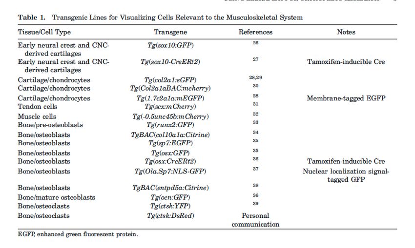
Discoveries 2020 13Table 1: Transgenic Lines for Visualizing Cells Relevant to the Musculoskeletal System. the jaw and branchial arches.12-14 The demonstrates the feasibility of hundreds of embryos can be injected same screen also identified mutations, systematic genome-wide analysis. by a single user in one morning. This specifically affecting the adult form.21 The In addition to forward genetics, allows for efficient creation of induced mutants identified in these early large- zebrafish are also readily amenable mutations or replacements at specific scale screens served as foundation to reverse genetics, that is, testing for genetic loci. Screening phenotypes in for experimental analysis and were phenotypic consequences following the injected G0 founder “crispant” animals proof that not only could specific targeted interference of gene function. can further enable rapid and cost- mechanisms of skeletal development The advent of TALEN-23 and CRISPR- effective assessment of gene function.1 be identified, but that mutation could be based gene editing has substantially In addition to alleviating the time and in genes homologous to human genes expanded the means by which the resources needed to breed alleles to associated with skeletal diseases. Such scientific community can approach homozygosity, G0 screens are also screens often yield specifically defined reverse genetics in zebrafish. For gene amenable to multiplexing strategies, in point mutations which provide nuanced editing using CRISPR, administration which multiple genes are targeted in the changes in gene function that simple of Cas9:gRNA ribonucleoproteins same animal. The ability to detect adult loss-of-function mutations or frameshifts (RNPs) generates double-stranded skeletal phenotypes in G0 zebrafish for cannot. Screens for dominant mutations breaks at defined loci. Errors in the genes associated with recessive forms affecting skeletogenesis, in which non-homologous end joining (NHEJ) of OI (bmp1a and plod2) was recently mutations often lead to dominant- repair mechanism lead to insertions and demonstrated.25 negative properties as well as alleles deletions (indels) at the cut site, often Zebrafish also provide a versatile with increased functions (hyper- or leading to loss of function (e.g., due to system to test gene function through neomorphic alleles), are proving to non-sense-mediated decay triggered by the use of transgenesis. This allows be informative and useful in disease a premature stop codon). Alternatively, for stable or inducible (e.g., heat modeling. For example, recent screens multiple RNPs can be used to induce shock-induced) protein expression, or have identified dominant mutants site-spanning deletions that delete conditional gene targeting (e.g., Cre- closely mirroring collagenopathies promoter regions or entire gene loci, mediated recombination). The Tol2 and osteogenesis imperfecta (OI) which may help reduce activation of transposon system is commonly used (col1a1a/b, col1a2), Adams-Oliver compensatory pathways triggered for introducing transgenes. There exists Syndrome (dll4), and hyperhidrotic by messenger RNA degradation. 24 a large, growing panel of zebrafish ectodermal dysplasia (edar).16 Finally, Moreover, Cas9:gRNAs can be co- fluorescent reporter lines for cell types the Zebrafish Mutation Project,22 which injected with a donor template which, within the musculoskeletal system has phenotyped a large number of following homology-directed repair (Table 1). As zebrafish are also relatively zebrafish mutant alleles and made (HDR), can result in precise gene edits. transparent and develop externally, them available to the community, Because zebrafish develop externally, development can be easily observed 14 Discoveries 2020
in real-time. With these two attributes,     cartilage and bone until later stages,                  important for proper tendon formation,45
use of transgenic reporters for particular   it has the same somitic compartments,                   and cyp26b1 loss of function studies
cell types and proteins have provided        s c l e r o t o m e , s y n d e t o m e , 48 a n d      suggest that retinoic acid is required for
an unmatched ability to visualize the        myotome, fated to become skeletal,                      tendon cell condensation.61,62 Recent
dynamics of skeletal patterning and          tendon, and muscle tissues as in higher                 studies have shown that mechanical
regeneration. These advantages also          vertebrates. Prior to 5 dpf, the axial                  force, through release of TGF-β,
permit the visualization of cell behaviors   musculoskeletal structures primarily are                regulates the formation of tendon cell
in specific genetic contexts to gain         composed of muscle and myosepta, a                      projections, which are thought to be
mechanistic understanding of disease         scleraxis-expressing myotendinous                       involved in extracellular matrix (ECM)
etiology.                                    tissue that links the myomeres.49,50 The                production.31 In the adult, the cranial
    Because of their small size and low      bony elements form through direct/                      tendons have similar ultrastructure to
cost, zebrafish are also amenable to         intramembranous ossification, or via                    mammalian tendons with highly ordered
drug discovery via chemical screens.         a cartilage or cartilage-like template                  type I collagen fibrils observed by
In such screens, large libraries of small    (e.g., via perichondral or endochondral                 transmission electron microscopy.45 In
molecules are tested to identify specific    o s s i f i c a t i o n ) . 51-54 T h e m o d e s o f   addition, they can be readily visualized
compounds that affect gene function or       ossification can differ in zebrafish                    using second harmonic generation
developmental processes. In a typical        and mammals in similar bones. For                       (SHG) imaging (Fig. 1F).
screen, zebrafish embryos/larvae are         example, in mouse, the vertebrae                            Analogous to higher vertebrates,
dispensed into 48- or 96- well plates,       form by endochondral ossification,55                    striated muscle of zebrafish contain
drugs are administered by adding             in zebrafish, vertebrae form by direct                  three main components: contractile
them to the water, and phenotypes            mineralization of the notochord sheath                  proteins, lipids, and connective
are assessed (e.g., via morphological,       (perichordal ossification), without                     tissue.63 Vertebrae are connected by
fluorescent, or behavioral readouts).        passing through a cartilaginous                         intervertebral ligaments. 64 Zebrafish
This strategy can be adapted                 stage.56 In some bones, osteoblasts                     possess both slow- and fast-twitch
to adults. 40,41 The identification of       and osteoclasts act in concert to model                 muscle fibers, which are topographically
dorsomorphin as a selective inhibitor        bone shape into adulthood.57 Although                   separated. 65 Together with cellular
of bone morphogenetic protein (BMP)          uncommon, osteon-like structures                        mineralized bone tissue, muscles,
type I receptors were discovered in a        in zebrafish have been reported for                     tendon, and other soft tissues, the
large zebrafish chemical screen and led      lateral ethmoid bone.54 Notably, these                  zebrafish skeleton facilitates locomotion,
to the development of analogs for the        structures contained solely one lamella                 provides mechanical support, and
treatment of heterotopic ossification.42     and no osteocytes. Indeed, most                         protects internal organs. In Figure 2,
In another screen, phosphodiesterase         skeletal elements in adult zebrafish                    we compare the inter-vertebral space
(PDE) inhibitors were found to alter         skeletons are osteocytic and do not                     in the zebrafish and mouse as a case
phenotypes in a zebrafish model of           show osteons or hemiosteons indicative                  study of how skeletal structures in
Duchenne muscular dystrophy.43 See           of human-like secondary remodeling. In                  each species typically exhibit both
Wiley et al.44 for a recent review of        vertebrae of adult zebrafish, osteocyte                 morphophysiological similarities and
chemical screens in zebrafish.               lacunar orientation shows a preferred                   differences.
                                             orientation (Fig. 1E). 58 While the
Formation and Integration of the             mechanosensing and remodeling                               Conservation of Developmental
Zebrafish Musculoskeletal System             characteristics of osteocytic bone in                   programs
    Development and patterning               zebrafish remain to be fully understood,                    The molecules that govern
    Fully developed, the zebrafish           lacunae in zebrafish indicate smaller                   zebrafish skeletal development are
skeleton comprises several functional        volumes with less numerous canaliculi                   highly conserved with mammals.
groups including the cranial skeleton,       compared with mice and humans.                          Sox9, a transcription factor necessary
axial skeleton, caudal skeleton,                  Tendons are the tissue interface                   for chondrogenesis and skeletal
unpaired fins (dorsal, anal, the caudal      between the muscle and bone. 59                         development,66 has two co-orthologs
fins), paired fins (pectoral and pelvic      Concurrent to skeletal development,                     in the zebrafish, sox9a, and sox9b.
fins), and elasmoid scales (Fig. 1A-         transcripts of scleraxis a (scxa) are                   They are expressed in overlapping
D). As in all vertebrates, the zebrafish     found in the forming tendon cells                       and complementary patterns during
cranial skeleton and its associated          adjacent to the developing cartilage                    development with sox9a in the
connective tissues, tendons and              and muscle by two dpf. These cells                      pharyngeal arches and later restricted
ligaments, arise from the cranial neural     aggregate and differentiate, turning                    to the pre-chondrogenic mesenchyme
crest; the fin skeletal elements arise       on expression of tendon matrix genes,                   that will form the jaw cartilage and
from the lateral plate mesoderm, and         tenomodulin (tnmd), thrombospndin-4                     fin scapulocoracoid, and with sox9b
the myosepta and axial skeleton from         (tsp4b), and type I collagen (col1a1a/b,                in the premigratory neural crest and
somitic paraxial mesoderm. 45-47 The         col1a2).49,50 As in mammals, initiation                 fin endochondral disc. 7 The sox9a
cranial musculoskeletal system forms         of the axial tendon program depends                     expressing chondrocytes also express
rapidly and can function by 5 days           on signals from the muscle. Cranial                     col2a1 and are Alcian Blue positive
post fertilization (dpf). The pectoral       and fin tendons form in the absence of                  before three dpf. These skeletal
fin cartilage and muscles are also           muscle, but require muscle for tendon                   elements will undergo perichondral
developing at this time. Although            maintenance.45,60 FGF and transforming                  or endochondral ossification and later
the axial skeleton does not form             growth factor-β (TGF-β) are also                        become Alizarin red-positive cranial

                                                         Discoveries 2020                                                                  15
Figure 1: Imaging of tissue structure, composition, and quality. (A) Contact X-Ray of a juvenile zebrafish. The vertical line shows the histological plane for the
image in (D). (B) Microcomputed tomography (2µm isotropic voxel size) of an adult zebrafish skull. (C) Quantitative backscattered scanning electron imaging
(qBEI) in the spine of an adult zebrafish. Bone growth occurs at the vertebral endplates. (D) H&E stained section of the zebrafish trunk. Muscle fiber density
and cross-sectional muscle fiber area are readily assessed. (E) High-resolution imaging of a vertebral body via X-Ray Microscopy highlighting the osteocyte
lacunar network. The osteocyte-lacunar orientation may reflect orientation of collagen fibers, and loading patterns in zebrafish vertebrae. The lacunar orientation
follows a specific pattern, i.e. longitudinal orientation in the center of the vertebrae and circumferential orientation near the endplate regions. (F) An adult
tendon attached to the maxilla in zebrafish is imaged using in vivo Second Harmonic Generation (SHG) imaging, an indicator of type I collagen organization
and density. The right panel shows dual imaging of tendon in concert with osteoblasts (green: osteocalcin+ cells expressing the ocn:GFP transgene). [Color
figure can be viewed at wileyonlinelibrary.com]
bones. In perichondral ossification, The skeleton serves as a key organ mammalian skeleton is acting as a site
perichondral cells become runx2a/b, which mediates systemic signaling of hematopoiesis, as well as fat storage,
osterix (sp7/osx), and collagen 10 affecting the physiology. Although many in the marrow cavities. Zebrafish
positive osteoblasts and initiate of these non-structural functions of the possess bone marrow spaces,53 which
o s s i f i c a t i o n 67. S i m i l a r t o o t h e r skeleton are just being identified, it is evident in endochondral bones, which
vertebrates, indian hedgehog co- is clear that many have conservation are filled with fatty tissue.54 However,
orthologs (ihha/b) are expressed by between humans and zebrafish. unlike in humans, this is never colonized
chondrocytes and are thought to signal One key function of the skeleton is by hematopoietic stem cells (HSC).
to patched, Hh receptors (ptc1/2) in to facilitate mineral homeostasis. Thus, zebrafish bone marrow spaces
the perichondrium and mediate bone The skeleton participates in part by lack hematopoietic tissue.53 A number
formation.68,69 Other cranial elements, regulating phosphate homeostasis in the of zebrafish bones possess adipocytes
such as the maxilla undergo direct kidney through bone-kidney crosstalk. within their marrow spaces,54 however,
intramembranous ossification via osx- There is evidence that Fgf23, which it is unknown whether this adiposity
expressing osteoblasts.54,70 For many of in humans and mice, is synthesized responds to the metabolic demands,
these genes and cell types, reporter and in osteocytes and regulates kidney as it does in mice.72 Finally, the skeleton
lineage-tracing transgenic zebrafish phosphate reabsorption, also regulates can regulate the metabolic processes
lines have been generated, which, phosphate homeostasis in zebrafish.71 independent of mineral metabolism. For
along with the optical access provided In mammals, the skeleton also serves instance, the bone-derived hormone
by zebrafish, allow unprecedented as a calcium and phosphorus reservoir. osteocalcin has been implicated in
ability to visualize skeletogenesis (Fig. Because calcium regulation can occur glucose homeostasis, cognition, and
3). through the gills in fish, compared with male fertility.73 Whether the zebrafish
humans, the physiological role of the skeleton functions as an endocrine
Physiology During Development skeleton in calcium homeostasis in organ through osteocalcin secretion
and in Homeostasis fish may differ.53 Another function of the requires further investigation.
16 Discoveries 2020Aging specific for neutrophils (mpx:GFPi114Tg)83 been established to force exercise and
Compared with early development, or macrophages (mpeg:eGFP).84 There stimulate natural modes of skeletal
processes such as homeostasis and are also several methods to functionally loading in zebrafish. In this way, the
aging have not been studied in depth deplete immune cell populations complex interplay of cellular, structural,
in the zebrafish. As certain debilitating (reviewed in Keightley et al.80), which and compositional bone characteristics
conditions arise in the skeleton as a can permit temporal control over cell can be assessed using multiscale
function of age, such as osteopenia type-specific cell ablation to assess approaches in zebrafish to study the
and osteoporosis, the ability of the the role of immune cell populations effects of genetic and environmental
zebrafish to model components of at different stages of the regenerative interactions on the skeletal system
these processes could be important. process, as has been performed for tail in vivo. During early development in
Zebrafish typically have a lifespan fin regeneration.85 zebrafish, swim training alters timing
of approximately 2-3 years (though Zebrafish have a significant capacity of skeletogenesis.92 In adult zebrafish,
5 years or more is possible),74 and for epimorphic regeneration.3,14 One swim training increases vertebral bone
exhibit growth throughout life. There is example is the caudal fin, which formation and alters quality.58 Moreover,
evidence that zebrafish skeletal function regenerates following amputation.86 this type of forced exercise also induces
declines with age. For instance, tendon Similar to salamander limb muscle adaptations in adult zebrafish.93
mechanical properties diminish with regeneration, fin regeneration involves This paradigm opens up avenues for
age.75 Moreover, alterations of vertebral a heterogeneous pool of progenitors genetic and small molecule screens to
bone and disc are observed in aged called the blastema, which is comprised, identify signaling pathways critical for
zebrafish.76 The bone dependence on at least in part, of mature cells at the musculoskeletal adaptation to loading
estrogen has been modeled in another amputation stump that dedifferentiated, and exercise.
small teleost, medaka, and thus the including osteoblasts. 36 A variety of
basic properties of the etiology are pathways known to be important Phenotyping
likely present in zebrafish.77 With more for skeletogenesis in mammals are MicroCT
analysis of late developmental stages, recapitulated during fin redevelopment, The three dimensional (3D)-high-
it is likely more insight will emerge from as reviewed in Watson et al. 86 An resolution micro-computed tomography
the zebrafish into how the skeletal intact musculoskeletal system is has become established as a powerful
system ages and its consequences. required for normal regeneration as method to assess bone morphology
zebrafish subjected to injection of and microstructure in zebrafish.18,58,94,95
Regeneration and Repair botulinum toxin, which inhibits synaptic Using a 5 μm voxel size, bone structure
Zebrafish have not been used as release at cholinergic nerves, exhibit indices as vertebral bone volume,
a common model for understanding impaired regeneration.87 This model thickness, and eccentricity can be
human fracture repair. This is in part due has also revealed the existence of characterized.18,58 Neural arch area,
to lack of accessible long bones, as well mesenchymal progenitor populations which reflects modeling arising from
as its high regenerative capacity, which within specific regions that robustly osteoblast and osteoclast activity, can
may utilize different repair mechanisms respond to injury and generate new also be captured.94 Because of their
than in mammals. Previous studies osx+ osteoblasts.88,89 Dedifferentiation small size, whole body, high-resolution
have examined the repair properties of mature osteoblasts also occurs scans are readily acquired.95 Software
of damaged membranous bones of during repair of zebrafish fin fractures for semi-automated segmentation
the skull roof78 as well as mandible.79 and skull injuries.78 While osteoblast enables in-depth phenotyping at a large
Although these are not directly dedifferentiation is more limited in number of skeletal sites. By quantifying
comparable with analysis of long bone mammals, fin repair after fracture hundreds of measures this was shown to
fractures studied in mouse and most exhibits some similarities to mammalian increase the sensitivity in discriminating
commonly seen in patients, there were long bone fracture, including formation mutant populations.95 Moreover, the
some similarities in terms of the genes of a remodeling callus,90 and recruitment osteocyte lacunar network in the
and cell types involved. For example, of osteoclasts.91 Recently, it was shown vertebral tissue can be imaged at high
runx2+ cells in the periosteum were that neutrophils dynamically colonize resolution with lab-based nano-CTs and
likely involved in new bone formation the fracture site. When infected with 3D X-ray Microscopy (3DXRM). The
and proper formation of the cartilage Staphylococcus aureus, neutrophils orientation of the osteocyte lacunae
callus relied upon Indian hedgehog a were retained in the fracture site and in relation to the long and short axis of
(ihha).79 In addition to examination of repair was reduced.91 Further studies the vertebral bodies, sphericity, mean
intrinsic regenerative mechanisms, the examining the utility of the fin fracture lacunar volume and lacunar density can
transparency of the zebrafish permits model to study the aspects of fracture be quantified.18,58 Finally, synchrotron-
the analysis of extrinsic cell populations biology are warranted. based X-ray microCT, when combined
in the healing process. Studies have with tissue-contrast stains, can yield
shown that the immune system plays Musculoskeletal Loading whole-organism images suitable for
an important role in mediating tissue While the zebrafish skeleton has a cell-level quantitative histological
regeneration. 80,81 Visualization of reduced role in resisting gravitational phenotyping in zebrafish.96
immune infiltration after injury can loads relative to humans, there is
be accomplished through the use of evidence that the zebrafish skeleton Histomorphometry
transgenic reporter lines that either label can respond to exercise, as well as In zebrafish, histologic sections
all leukocytes (cd45:DsRed)82 or are disuse. Swim training routines have stained using von Kossa/van
Discoveries 2020 17Figure 2: Comparison of the intervertebral disc (IVD) in mouse and zebrafish. (A-A’) and (B-B’): Midline section of a Safranin-O/Fast green staining of an
intervertebral disc region in mouse (6-months) (A-A’) and zebrafish (1-year) (B-B’). (C) and (D): Cartoon schematic of insets for mouse (C) and zebrafish (D). In
mouse, the IVD is composed of a proteoglycan-rich lamellar fibrocartilaginous cartilage called the annulus fibrosus (AF) which surrounds the nucleus pulposus
(NP) joins adjacent bony vertebrae at the level of the cartilaginous end plate (CEP). Zebrafish IVD retains notochord-derived vacuolated cells embedded in a
fibrocartilaginous matrix, however, there is no NP-like structure observed in zebrafish. An analogous structure to the outer AF layer in is observed as a small
acellular intervertebral ligament (IVL). Histologically, the NP in mouse appears to be composed of: an outer tissue layer which stains for Safranin-O (orange in
(C)); an inner cell layer (dotted red line in (C)); and an inner tissue layer that does not stain well for Safranin-O (yellow in (C)). In contrast, the zebrafish IVD has
only weak Safranin-O staining (magenta in (B’, D)) in an interior region adjacent to the intervertebral ligament (IVL) (B’, blue in (D)). Twist positive osteoblast
progenitor cells ((Tw+)ObP) are observed adjacent to the IVL. AF, annulus fibrosis; CEP, cartilaginous endplate; GP, growth plate; IVL, intervertebral ligament;
NP, nucleus pulposus; (Tw+)ObP, twist positive osteoblast progenitors; vert, vertebrae. [Color figure can be viewed at wileyonlinelibrary.com]
Gieson, Goldner’s modified Masson- the mean calcium content in the OI is a disease of the collagen
trichrome, and toluidine blue enable mineralized bone tissue, as well as matrix, which results in brittle bones
static bone histomorphometry, and the homogeneity of mineralization18,58. and skeletal deformities. In humans,
is performed in accordance with Vibrational spectroscopy methods (e.g. , collagen type I is a heterotrimer
standardized nomenclature set Fourier transform infrared spectroscopy composed by two alpha chains, α1(I)
forth by the ASBMR nomenclature (FTIR) and Raman spectroscopy) and α2(I), which trimerize in a 2:1
committee for practitioners of bone have also been adapted to zebrafish ratio, respectively, to form a fibril with a
histomorphometry.97 Calcein labeling bone. 18,98 Parameters such as the triple-helix structure. In zebrafish, the
or double labeling with calcien and mineral-to-matrix-ratio, carbonate- collagen type I triple helix is composed
Alizarin Red S can be performed and to-phosphate ratio, cross-link-ratio of three α chains, α1(I), α2(I), and
double labels can be evaluated for (collagen maturity), and crystallinity α3(I), which are encoded for by the
dynamic bone histomorphometry.18,58 (purity, size of mineral crystals) of genes col1a1a, col1a2, and col1a1b,
Such an approach was used to quantify the bone were shown to provide respectively.99 Most human patients with
increases in mineral apposition rate information about the molecular and OI are attributed to mutations in type I
(MAR), mineralizing surface per bone compositional bone characteristics.18 collagens, with the majority of mutations
surface (MS/BS), and bone formation Finally, nanoindentation of vertebrae disrupting the conserved Gly-X-Y motifs
rate (BFR) at the vertebral endplates can be performed in zebrafish to responsible for fibrillar assembly of the
in zebrafish subjected to swimming assess local mechanical and material collagen heterotrimers.100 In zebrafish,
exercise.58 properties such as Young’s modulus several dominant mutants have been
(elastic modulus), hardness, and identified carrying heterozygous glycine
Assessment of Bone Composition, fracture toughness.18 The biomechanical substitution in the α1 chain of collagen
Mineral Density Distribution, and properties of zebrafish cranial tendons type I, and which exhibit severe,
Mechanical Properties can also be measured. A maxillary pathological features of classical OI.
Recently, quantitative backscattered tendon was found to have stress-strain This was demonstrated in the chihuahua
electron imaging (qBEI) has been nonlinearity and a linear modulus mutant, which exhibited changes in
established as an effective means to similar to mammalian tendon data.75 vertebral tissue composition.18 A large
measure the bone mineral density panel of zebrafish mutants of col1a1
distribution in zebrafish. 18,58 Gray Disease Applications genes with qualitative and quantitative
value histograms were used to assess Collagenopathies defects in collagen type I have been
18 Discoveries 2020You can also read