DURIAN SHELL HUSK EXTRACT ASSISTED SYNTHESIS OF COPPER OXIDES NANOPARTICLES FOR THE PHOTODEGRADATION OF PARACETAMOL - UKM
←
→
Page content transcription
If your browser does not render page correctly, please read the page content below
Malaysian Journal of Analytical Sciences, Vol 23 No 5 (2019): 818 - 827
 DOI: https://doi.org/10.17576/mjas-2019-2305-07

 MALAYSIAN JOURNAL OF ANALYTICAL SCIENCES ISSN
 1394 - 2506
 Published by The Malaysian Analytical Sciences Society

 DURIAN SHELL HUSK EXTRACT ASSISTED SYNTHESIS OF COPPER
 OXIDES NANOPARTICLES FOR THE PHOTODEGRADATION OF
 PARACETAMOL
 (Ekstrak Sekam Kulit Durian Membantu Sintesis Nanozarah Tembaga Oksida Untuk
 Fotodegradasi Paracetamol)
Ahmad Masudi1, Nurfatehah Wahyuny Che Jusoh1,2*, Zunika Bakri1, Aishah Abdul Jalil2,3, Roshafima Rasit Ali 1,2,
 Nur Farhana Jaafar4
 1
 Department of Chemical Process Engineering, Malaysia-Japan International Institute of Technology (MJIIT),
 Universiti Teknologi Malaysia Kuala Lumpur, Jalan Sultan Yahya Petra, 54100 Kuala Lumpur, Malaysia
 2
 Center of Hydrogen Energy, Institute of Future Energy
 3
 School of Chemical and Energy Engineering, Faculty of Engineering
 Universiti Teknologi Malaysia, 81310 UTM Johor Bahru, Johor, Malaysia
 4
 School of Chemical Sciences,
 Universiti Sains Malaysia, 11800 USM Penang, Malaysia

 *Corresponding author: nurfatehah@utm.my

 Received: 13 February 2019; Accepted: 20 September 2019

 Abstract
A series of copper oxide (CuO) nanoparticles catalysts were prepared via electrochemical method. The addition of different
concentrations of Durian Shell Husk (DSH) extract in the electrolyte system was evaluated for degradation of paracetamol
under visible light irradiation. The catalysts were characterized by using X-ray diffraction (XRD), Fourier Transform Infrared
(FTIR) and Scanning Electron Microscopy (SEM). The characterization data showed that the different sizes of CuO were
obtained when the amount of DSH extract added into the electrolyte system were varied. The crystallite size of CuO prepared
with DSH was decreased from 40 nm to 29 nm, which showed the role of DSH extract as a capping agent. The particle sizes
reduction caused a significant increment towards paracetamol degradation. The initial degradation rate was increased from
2.67×10⁻² to 7.28×10⁻² mg/L.min. From this study, the optimum condition was noticed for CuO that was prepared by using
0.06 mg/L of DSH extract at a paracetamol initial concentration of 10 mg/L (pH 9) by using 0.1 g/L catalyst. Finally, the result
could contribute to the production of copper oxide with ideal sizes using abundant agriculture waste extract for the removal of
paracetamol in wastewater.

Keywords: electrochemical, copper oxide, durian shell husk, paracetamol

 Abstrak
Satu siri pemangkin nanozarah tembaga oksida (CuO) telah disediakan melalui kaedah elektrokimia. Penambahan pelbagai
kepekatan ekstrak sekam kulit durian (DSH) dalam sistem elektrolit telah dinilai untuk degradasi paracetamol di bawah sinar
cahaya boleh lihat. Pemangkin telah dicirikan menggunakan pembelauan sinar-X (XRD), spektroskopi inframerah transformasi
Fourier (FTIR) and mikroskopi imbasan elektron (SEM). Data pencirian menunjukkan bahawa pelbagai saiz CuO diperoleh
apabila jumlah ekstrak DSH yang ditambah di dalam sistem elektrolit telah diubah-ubah. Saiz kristal CuO yang disediakan
menggunakan DSH menurun dari 40 nm ke 29 nm, yang mana menunjukkan peranan ekstrak DSH sebagai ejen pelekatan.
Pengurangan saiz zarah ini menyebabkan kenaikan ketara terhadap degradasi paracetamol. Kadar degradasi awal meningkat
daripada 2.67×10⁻² kepada 7.28×10⁻² mg/L.min. Daripada kajian ini, keadaan optimum telah dikenalpasti untuk CuO yang
disediakan menggunakan 0.06 mg/L ekstrak DSH pada kepekatan awal paracetamol 10 mg/L (pH 9) dengan menggunakan

 818

Ahmad et al: DURIAN SHELL HUSK EXTRACT ASSISTED SYNTHESIS OF COPPER OXIDES
 NANOPARTICLES FOR THE PHOTODEGRADATION OF PARACETAMOL

0.1 g/L pemangkin. Akhirnya, hasil kajian ini dapat menyumbang kepada penghasilan tembaga oksida dengan saiz yang ideal
menggunakan ekstrak sisa buangan pertanian untuk penyingkiran paracetamol dalam air sisa.

Kata kunci: elektrokimia, tembaga oksida, sekam kulit durian, paracetamol

 Introduction
The pharmaceutical market in Malaysia increases 9.5% annually, which is double than average of the Asia Pacific
region. The market is still predicted to grow from $ 2.3 billion in 2015 to $ 3.6 billion in 2020. The increasing
demand of drugs will be followed by large municipal and discharge waste which is hazardous to the environment.
Paracetamol (PCT) is the largest analgesic and antipyretic drug without prescription worldwide. PCT was detected
0.033-0.071 μg/L at water surface in Korea [1], 0.22-6.8 μg/L at treated water in Spain [2] and 0.01-0.07 μg/L in
surface and sewage water in Malaysia [3]. Therefore, it was proven that the conventional technology still needs
some advancements.

Advanced Oxidation Process (AOPs) is one of promising method to degrade emerging pollutants. This technology
is based on massive reaction between organic radicals with pollutants. The radical compounds were produced from
heterogeneous catalyst (for example, TiO2 and ZnO) which activates after exposure to external light such as
ultraviolet, visible or solar light. The common photocatalysts are TiO2 and ZnO [4]. However, these catalysts
mainly absorp UV light and consume extensive energy [5, 6]. CuO is p-type semiconductor which has the potential
to become a photocatalyst with low band gap energy in the range of 1.2 eV -1.8 eV and active under visible or
fluorescent lamp [7, 8]. There are many approaches reported for synthesis of CuO, such as precipitation [9], sol-gel
[10] and solution combustion method [11]. However, these methods require an extremely controlled set-up
including high temperature or a long reaction time to obtain the CuO. Therefore, it is necessary to find a simple and
rapid method for synthesis. Electrochemical method seems to be a potential method due to its effectiveness, facile
experimental set-up and can also be operated at ambient temperature and pressure [12]. However, from the previous
study, an expensive ionic liquid are usually used as a synthesis media in electrochemical method [13].

Hence, it is crucial to find a low cost material as an alternative media. To date, the synthesis of CuO nanoparticles
by using plant extract has received great attention due to the expanding need in environmentally friendly
technologies in material synthesis. Plant extracts are basically enriched in polysaccharide compound that acts as a
stabilizing agent in the synthesis of CuO [14]. However, the synthesis of CuO nanoparticles using plant extract
through electrochemical method is still rare since most of the studies used precipitation and sol gel method.
Therefore, this study aims to synthesize CuO nanoparticles via electrochemical method by using durian shell husk
(DSH) extract as a media. The DSH extract contains polysaccharides which act as capping agent and stabilizer
during the synthesis. As compared to ionic liquid, the DSH extract also has other advantages, such as cost effective
and environmentally friendly. The synthesized CuO nanoparticles were then analysed by X-ray diffraction (XRD),
Fourier Transform Infrared (FTIR) and Scanning Electron Microscopy (SEM). Next, the photocatalytic activity of
the CuO nanoparticles was studied towards the degradation of paracetamol (PCT).

 Materials and Methods
Preparation of copper oxides assisted by DSH
An open electrolysis cell consisted of copper plate electrode (2x2 cm2) and platinum plate electrode (2x2 cm2) were
placed in parallel each other. 10 mL of solution containing different amounts of DSH extract (0 mg/L, 0.02 mg/L,
0.06 mg/L and 0.1 mg/L) were added to the solution at a constant stir (150 rpm). The solution also comprised of 0.1
M of tetraethyl ammonium perchlorate (TEAP) as the supporting electrolyte. The electrolysis was conducted at 0°C
and ambient pressure with constant current density (120 mA/cm2) . After electrolysis, the mixture was dried
overnight at 60 °C and calcined at 550 °C for 2 hours. The catalyst was donated as CuOx (x= 0, 0.02, 0.06 and 0.1
mg/L of DSH extract).

Characterization
Catalyst crystallinity structure was evaluated by using X-ray diffraction (XRD, D8 Advance Bruker X-ray
diffractometer). Meanwhile the chemical functional groups in catalyst were identified by Fourier Transformation

819

Malaysian Journal of Analytical Sciences, Vol 23 No 5 (2019): 818 - 827
 DOI: https://doi.org/10.17576/mjas-2019-2305-07

Infrared (FTIR, Perkin Elmer Spectrum GX FTIR spectrometer). Finally, the catalyst morphologies were observed
with Scanning Electron Microscopy (SEM, JEOL JSM-6300).

Photodegradation test
PCT degradation was tested under visible light irradiation (36 watts). Before exposure to the visible light, a certain
amount of catalyst was added into 100 mL of PCT solution in dark condition and was continuously stirred. This
procedure was conducted to achieve equilibrium between the PCT solution and catalyst. The solution was exposed
to the visible light radiation for another 1 hour. Then, 1.5 mL of solution was taken periodically for every 10
minutes and degradation of PCT was analyzed by using a UV-Vis spectrometer (Shimadzu UV-2600) at maximum
wavelength of 245 nm.

 Results and Discussion
Catalyst characterization
Crystallinity and purity of copper oxides (CuO) nanoparticles were confirmed with X-ray diffraction (XRD). Based
on the XRD pattern as presented in Figure 1, the diffraction peaks appeared at 33°, 35°, 39°, 49°, 53°, 57°, 62°, 68°
and 75°. These peaks correspond to monoclinic CuO and match with JCPDS no.5-661 [15]. The crystallite sizes (D)
of CuO0, CuO0.02, CuO0.06 and CuO0.1 from Debye Scherrer equation were 40.74, 40.73, 29.6370 and 29.6371 nm
respectively. From this result, its was obvious that the various DSH concentrations affected the CuO particle size.
As reported by Song et al, the particle size of the synthesized gold nanoparticles was decreased at a high
concentration of Magnolia kobus plant extract [16]. At low concentration, the gold nanoparticles were hexagonal or
triangular in shape, but changed to spherical at high concentration. In this present study, at low concentration of
DSH (0.02 mg/L), the amount of DSH might not be enough to cover all the Cu 2+ ions, and thus could not act as a
good capping agent. However, the crystallinity of CuO nanoparticles increased while using 0.06 mg/L, followed by
significant reduction in particle size. Meanwhile, the particle size was almost the same at a higher concentration of
DSH (0.1 mg/L). The concentration of plant extract needed to be optimized since its did not only affect the particle
size, but also the morphology catalyst as reported in literature [17].

 -111 111
 220
 (d) 110 -202 020 202-113 022 -312203
 Intensity (a.u.)

 (c)

 (b)

 (a)
 20 30 40 50 60 70 80

 2-theta ( )

 Figure 1. XRD patterns of (a) CuO0 (b) CuO0.02 (c) CuO0.06 (d) CuO0.1

The functional groups of raw DSH and series of CuO were determined by using FTIR spectroscopy to verify the
formation mechanism of the catalyst. FTIR spectra of DSH in Figure2(a) shows four major spectra at 3323 cm -1,
1732 cm-1, 1610 cm-1 and 1033 cm-1. The peak at 3323 cm-1 corresponded to stretching of –OH group of
macromolecular association. Meanwhile, the peaks at 1732 cm-1 and 1610 cm-1 were contributed from the carbonyl
group and –OH stretching from H2O. The band around 1033 cm-1 referred to the C-O band of polysaccharide group.
In CuO catalyst series, there was no observed DSH peak due to the removal of DSH after calcination at 550 °C. In

 820
Ahmad et al: DURIAN SHELL HUSK EXTRACT ASSISTED SYNTHESIS OF COPPER OXIDES
 NANOPARTICLES FOR THE PHOTODEGRADATION OF PARACETAMOL

these catalysts, a new intense peak appeared at 560 cm-1, which corresponded to the Cu-O stretching bond, had
proven the existence of CuO nanoparticles [18].

 (e)

 % Transmission (d)

 (c)

 (b)
 (carbonyl)
 (polysaccharide)
 1610 1732 3323 (-hydroxyl) (a)
 1033

 400 800 1200 1600 2000 2400 2800 3200 3600 4000
 Wavelength (cm-1 )

 Figure 2. FTIR result for (a) DSH (b)CuO0 (c) CuO0.02 (d) CuO0.06 (e) CuO0.1

The CuO morphology was spherical shaped as presented by the SEM result in Figure 3. The SEM result had
convinced that CuO0.06 is much smaller than CuO0 which proved that DSH is good stabilizer. A study by Naika et
al. also obtained the spherical shape of CuO using Gloriosa superba L plant extract which dominantly contained the
alkaloid compound [19].

 (a) (b)

 Figure 3. SEM result for (a) CuO0 (b) CuO0.06

Photodegradation test
The series of synthesized CuO nanoparticles were tested for paracetamol degradation. Several parameters were
studied to obtain the optimum condition.

Effect of pH
Initial pH solution affected the interaction between pollutants and catalyst (Figure 4). The point of zero charge
(PZC) of CuO and PCT were at 7.9 and 9.2, respectively [20-22]. At lower pH than pHPZC, the molecules became
positively charged and became negative charged at higher pH than pHPZC. In pH 5 and 7, both CuO and PCT were
in positively charged, which would lead to repulsion with each other. Meanwhile, the degradation rate increased at
pH 9 due to the attractive interaction between the negatively charged CuO and positively charged PCT. However,
the reaction rate decreased again as both CuO and PCT were negatively charged.

821
Malaysian Journal of Analytical Sciences, Vol 23 No 5 (2019): 818 - 827
 DOI: https://doi.org/10.17576/mjas-2019-2305-07

 0.035

 Rate of reaction (mg/L.min)
 0.03
 0.025
 0.02
 0.015
 0.01
 0.005
 0
 5 7 9 11
 pH

 Figure 4. The effect of initial pH for PCT degradation

Effect of catalyst dosage
The rate of photocatalytic degradation was also influenced by catalyst dosage. Figure 5 display the effect of catalyst
dosage from 0.1 to 1 g/L for PCT degradation. The rate of reaction was slightly increased at catalyst dosage of 0.1
to 0.5 g/L. Then, the rate of reaction increased drastically by using catalyst dosage of 1 g/L. The increase in
degradation rate with increasing catalyst dosage was due to the increase in the number of active sites and active
radicals in the solution. A similar result was also observed in another copper-based catalyst to degrade the PCT [23].
The degradation efficiency increased from 38.6% to 90% when the catalyst dosage increased from 0.1 to 0.3 g/L.
This phenomenon occurs due to increase in active sites when the catalyst dosage was increased.

 0.07
 Rate of reaction (mg/L.min)

 0.06
 0.05
 0.04
 0.03
 0.02
 0.01
 0.00
 0.1 0.3 0.5 1
 Catalyst dosage (g/L)

 Figure 5. The effect of catalyst dosage

Effect of DSH concentration
DSH is one of potential stabilizers in nanoparticle formation. As reported, the concentration of plant extract had
differentiated the particle size of nanoparticle [24]. This study was conducted at different concentrations of DSH
extract which were 0, 0.02, 0.06 and 0.1 mg/L. Figure 6 shows the different catalytic activities of the catalysts for
PCT degradation. It was observed that the highest reaction rate was obtained with CuO prepared by using 0.06
mg/L of DSH extract. The introduction of 0.02 mg/L DSH showed only little improvement on its catalytic activity
as compared to without DSH extract. At low concentration, DSH may not covered all the CuO nuclei, which then

 822
Ahmad et al: DURIAN SHELL HUSK EXTRACT ASSISTED SYNTHESIS OF COPPER OXIDES
 NANOPARTICLES FOR THE PHOTODEGRADATION OF PARACETAMOL

caused the large particle size was confirm with XRD. The particle size of CuO decreased significantly after the
addition of 0.06 mg/L DSH and it was also highly crystalline indicating a complete covering of CuO nuclei.
However, the particle size increased again after the insertion of 0.1 mg/L DSH. This phenomenon may be assigned
to the agglomeration of DSH extract which hindered CuO nuclei stabilization.

 0.07
 Rate of reaction (mg/L.min)
 0.06
 0.05
 0.04
 0.03
 0.02
 0.01
 0.00
 CuO
 CuO0 CuO
 CuO0.02 CuO
 CuO0.06 CuO
 CuO0.1
 0 0.02 0.06 0.1

 Catalyst

 Figure 6. The effect of DSH concentration

Effect of reaction temperature
Figure 7 demonstrates the effect of reaction temperature for PCT degradation. In this study, the effect of
temperature was carried out at 30, 40 and 50 °C. It was observed that the degradation rate at 30 °C was much higher
than at 40 and 50 °C. The highest degradation rate was 6.99 x 10⁻² mg/L.min. Therefore, the degradation of PCT
was considered as an exothermic reaction with unnecessary high temperature condition is needed. This gives an
advantage to this reaction as the other copper catalyst is commonly an endothermic reaction which requires an
extensive energy [25]. Then, by using the degradation constant and temperature, activation energy (Ea, J/mol) can
be estimated by using Arrhenius equation as presented in equation (1),

 − 1
 ln kapp = ( ) + ln A (1)

This equation consisted of R as the gas constant (8.314 J.K/mol) where kapp and A are as apparent rate constant and
frequency factor, respectively. This plot exhibited high R2 (0.9995) with activation energy of 101 kJ/mol, as
presented in inset Figure 7. This activation energy was lower than other copper catalyst as reported in literature [25].
With lower activation energy, the reaction time was faster, and the energy consumption can be reduced.

 0.08
 0.0031 0.0032 0.0033
 0.07
 Rate of reaction (mg/L.min)

 0
 2
 -2 R = 0.9995
 ln (kapp )

 0.06
 Ea = 101 kJ/mol
 0.05 0.0699 -4
 0.04 -6
 -8
 0.03 0.0189 1/T (1/K)
 0.02
 0.0055
 0.01
 0.00
 30 40 50
 Temperature ( C)

 Figure 7. Effect of reaction temperature

823
Malaysian Journal of Analytical Sciences, Vol 23 No 5 (2019): 818 - 827
 DOI: https://doi.org/10.17576/mjas-2019-2305-07

Effect of initial concentration
The Initial concentration of pollutant is essential in a degradation studies, especially for industrial application. This
study showed that reaction rate increased from 0.0407 to 0.122 mg/(L.min), when increasing the PCT concentration
from 5 to 20 mg/L. The reaction rate decreased to 0.0170 mg/(L.min) at higher initial concentration of 30 mg/L.
From this result, its was obvious that CuO nanoparticles were efficient until 20 mg/L of PCT as presented in Table
1. At higher concentration than 20 mg/L, the turbidity of solution hindered the light penetration, and then decreased
degradation rate significantly.

 Table 1. Effect of initial concentration of PCT

 Concentration of PCT Rate of Reaction
 (mg/L) (mg/L.min)
 5 0.04
 10 0.08
 20 0.12
 30 0.02

Propose mechanism for CuO formation and PCT degradation
The possible mechanism for CuO formation was proposed according to the characterization results and literature as
shown in Figure 9. The electrolysis at anode and cathode involved the following reactions (2-3):

 Anode : Cu → Cu2+ + 2e- (2)
 Cathode : 2H2O + 2e- → 2H2 + 2OH- (3)

The copper plate was oxidized to generate copper cations (Cu2+) and released electrons at the anode. As reported,
DSH is rich with polysaccharide compound. The presence of polysaccharide in electrolyte formed a complex
compound with Cu2+ to form Cu[polysaccharide]2+. This Cu[polysaccharide]2+ then played an important role in the
formation of CuO nuclei. The polysaccharide reacted as a capping agent as well as a stabilizer which controlled the
growth of CuO nuclei. Therefore, as observed in XRD, the particles size of synthesized CuO in the presence of DSH
extract was much smaller than in the absence of DSH extract. Since the electrochemical synthesis was conducted in
open air, part of oxygen could be dissolved and formed a highly crystalline CuO [26]. Meanwhile at the cathode, the
generated electrons reacted with the hydroxyl group from electrolyte to form hydrogen gas on the electrode surface.

 2e-

 CuO nuclei

 Cu2+

 Cu
 [polysaccharide]2+
 CuO Crystal
 Cu Pt

 DZH extract

 Figure 9. Propose mechanism for CuO nanoparticles formation

 824
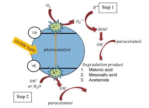
Ahmad et al: DURIAN SHELL HUSK EXTRACT ASSISTED SYNTHESIS OF COPPER OXIDES
 NANOPARTICLES FOR THE PHOTODEGRADATION OF PARACETAMOL

PCT degradation was initiated by the reaction between free electron from Cu oxidation and oxygen to produce O 2∙-.
Then, the O2- will react with hydrogen ion (H +) from water to produce ∙OH with HOO∙ as the intermediate reaction.
Meanwhile, the holes in VB were positive enough to oxidize water or OH - on the copper surface to produce
hydroxyl radicals (∙OH). Therefore, ∙OH would react with PCT and produce degradation product. The degradation
product mainly consists of carbocyclic acid which further decreases the pH value after reaction [27]. The proposed
degradation is presented in Figure 10.

 Figure 10. Propose PCT degradation

 Conclusion
Pure copper oxide (CuO) nanoparticles were successfully prepared by using electrochemical method. CuO was
spherical in shape and the particles sized was highly affected by the durian shell husk (DSH) concentration. The
particle size decreased drastically from 40 nm to 29 nm in the presence of optimum DSH content. The
photodegradation rate of paracetamol (PCT) by using CuO in the presence of DSH increased by more than twice as
compared to in the absence of DSH. The optimum condition for PCT degradation was obtained by using 1 g/L of
CuO0.06 in pH 9 at 30 °C to degrade 20 mg/L of PCT with the highest degradation rate of 0.1222 mg/L.min. These
findings suggested that DSH extract was remarkably important for an efficient nanoparticle’s synthesis and PCT
degradation. For future prospect, the combination of CuO with a catalyst support or other metal oxides will be a
great contribution to enhance the photocatalytic activity and stability of the catalyst.

 Acknowledgement
The authors would like to express appreciation for the financial support by the Fundamental Research Grant Scheme
from Ministry of Higher Eduacation (Grant No. 5F031), Research University Grant (Grant No. 19H37) and MJIIT
Incentive Scheme (Ahmad Masudi) form MJIIT UTM.

 References
1. Gómez, M. J., Martínez Bueno, M. J., Lacorte, S., Fernández-Alba, A. R. and Agüera. A. (2007). Pilot survey
 monitoring pharmaceuticals and related compounds in a sewage treatment plant located on the Mediterranean
 coast. Chemosphere, 66(6): 993-1002.
2. Kim, S. D., Cho, J., Kim, I. S., Vanderford, B. J. and Snyder. S. A. (2007) Occurrence and removal of
 pharmaceuticals and endocrine disruptors in South Korean surface, drinking, and waste waters. Water Research,
 41(5):1013-1021.

825
Malaysian Journal of Analytical Sciences, Vol 23 No 5 (2019): 818 - 827
 DOI: https://doi.org/10.17576/mjas-2019-2305-07

 3. Al-Odaini, N. A., Zakaria, M. P., Yaziz, M. I. and Surif. S. (2010). Multi-residue analytical method for human
 pharmaceuticals and synthetic hormones in river water and sewage effluents by solid-phase extraction and
 liquid chromatography–tandem mass spectrometry. Journal of Chromatography A, 1217(44): 6791-6806.
 4. Sidik, D. A. B., Hairom, N. H. H., Zainuri, N. Z., Desa, A. L., Misdan, N., Yusof, N., Ong, C. B., Mohammad,
 A. W. and Aripen. N. S. M. (2018). Photocatalytic degradation of industrial dye wastewater using zinc oxide-
 polyvinylpyrrolidone nanoparticles. Malaysian Journal of Analytical Sciences, 22(4): 693-701.
 5. Vaiano, V., Sacco, O. and Matarangolo, M. (2018). Photocatalytic degradation of paracetamol under UV
 irradiation using TiO2-graphite composites. Catalysis Today, 315: 230-236.
 6. Jusoh, N.W.C., Jalil, A. A., Triwahyono, S., Karim, A. H., Salleh, N. F., Annuar, N. H. R., Jaafar, N. F.,
 Firmansyah, M. L., Mukti, R. R. and Ali, M.W. (2015). Structural rearrangement of mesostructured silica
 nanoparticles incorporated with ZnO catalyst and its photoactivity: Effect of alkaline aqueous electrolyte
 concentration. Applied Surface Science, 330: 10-19.
 7. Mazumder, N. A. and Rano. R. (2018). Synthesis and characterization of fly ash modified copper oxide
 (FA/CuO) for photocatalytic degradation of methyl orange dye. Materials Today: Proceeding, 5: 2281-2286.
 8. An, J. and Zhou. Q. (2012). Degradation of some typical pharmaceuticals and personal care products with
 copper-plating iron doped Cu2O under visible light irradiation. Journal of Environmental Sciences, 24(5): 827-
 833.
 9. Phiwdang, K., Suphankij, S., Mekprasart, W. and Pecharapa. W. (2013). Synthesis of CuO nanoparticles by
 precipitation method using different precursors. Energy Procedia, 34: 740-745.
10. Kayani, Z. N., Ali, Y., Kiran, F., Batool, I., Butt, M. Z., Umer, M., Riaz, S. and Naseem. S. (2015). Fabrication
 of copper oxide nanoparticles by sol-gel route. Materials Today: Proceedings, 2(10): 5446-5449.
11. Dong, C., Xing, X., Chen, N., Liu, X. and Wang, Y. (2016). Biomorphic synthesis of hollow CuO fibers for
 low-ppm-level n -propanol detection via a facile solution combustion method. Sensors and Actuators B:
 Chemical, 230: 1-8.
12. Sapawe, N., Jalil, A. A., Triwahyono, S., Sah, R. N. R. A., Jusoh, N. W. C., Hairom, N. H. H. and Efendi, J.
 (2013). Electrochemical strategy for grown ZnO nanoparticles deposited onto HY zeolite with enhanced
 photodecolorization of methylene blue: Effect of the formation of SiOZn bonds. Applied Catalysis A: General,
 456: 144-158.
13. Jusoh, R., Jalil, A. A., Triwahyono, S., Idris, A., Haron, S., Sapawe, N., Jaafar, N. F. and Jusoh, N. W. C.
 (2014). Synthesis of reverse micelle α-FeOOH nanoparticles in ionic liquid as an only electrolyte: Inhibition of
 electron–hole pair recombination for efficient photoactivity. Applied Catalysis A: General, 469: 33-44.
14. Chutrakulwong, F. and Thamaphat, K. (2014). Durian peeling extract mediated green synthesis of silver
 nanoparticles. Advanced Materials Research, 875-877: 18-22.
15. Raghav, R., Aggarwal, P. and Srivastava. S. (2015). Tailoring oxides of copper-Cu2O and CuO nanoparticles
 and evaluation of organic dyes degradation. AIP conference proceeding, 1724: 020078-1-020078-5.
16. Song, J.Y., Jang, H. K. and Kim. B. S. (2009). Biological synthesis of gold nanoparticles using Magnolia kobus
 and Diopyros kaki leaf extracts. Process Biochemistry, 44(10): 1133-1138.
17. Mittal, A. K., Chisti, Y. and Banerjee, U. C. (2013). Synthesis of metallic nanoparticles using plant extracts.
 Biotechnology Advances, 31: 346-356.
18. Srivastava, R., Anu Prathap, M. U. and Kore. R. (2011). Morphologically controlled synthesis of copper oxides
 and their catalytic applications in the synthesis of propargylamine and oxidative degradation of methylene blue.
 Colloids and Surfaces A: Physicochemical and Engineering Aspects, 392(1): 271-282.
19. Suresh, D., Kumar, D., Nagabhushana, H., Nagaraju, G. Manjunath, K., Naika, H. R. and Lingaraju, K. (2014).
 Green synthesis of CuO nanoparticles using Gloriosa superba L. extract and their antibacterial activity. Journal
 of Taibah University for Science, 9(1): 7-12.
20. Guedes, M., Ferreira, J. M. F. and Ferro. A. C. (2009). A study on the aqueous dispersion mechanism of CuO
 powders using Tiron. Journal of Colloid and Interface Science, 330(1): 119-124.
21. Mestre, A.S., Bexiga, A. S., Proença, M., Andrade, M., Pinto, M. L., Matos, I., Fonseca, I. M. and Carvalho, A.
 P. (2011). Activated carbons from sisal waste by chemical activation with K 2CO3: Kinetics of paracetamol and
 ibuprofen removal from aqueous solution. Bioresource Technology, 102(17): 8253-8260.
22. Bernal, V., Erto, A., Giraldo, L. and Moreno-Piraján, C. J. (2017). Effect of Solution pH on the Adsorption of
 paracetamol on chemically modified activated carbons. Molecules, 22(7): 1032.

 826
Ahmad et al: DURIAN SHELL HUSK EXTRACT ASSISTED SYNTHESIS OF COPPER OXIDES
 NANOPARTICLES FOR THE PHOTODEGRADATION OF PARACETAMOL

23. Zhang, Y., Zhang, Q. and Hong. J. (2017). Sulfate radical degradation of acetaminophen by novel iron–copper
 bimetallic oxidation catalyzed by persulfate: Mechanism and degradation pathways. Applied Surface Science,
 422: 443-451.
24. Dubey, S.P., M. Lahtinen, and M. Sillanpää. (2010). Green synthesis and characterizations of silver and gold
 nanoparticles using leaf extract of Rosa rugosa. Colloids and Surfaces A: Physicochemical and Engineering
 Aspects, 364(1-3): 34-41.
25. Shen, Y., Zhang, Z.and Xiao, K. (2015). Evaluation of cobalt oxide, copper oxide and their solid solutions as
 heterogeneous catalysts for Fenton-degradation of dye pollutants. RSC Advances, 5(111): 91846-91854.
26. Jaafar, N. F., Jalil, A. A., Triwahyono, S., Efendi, J., Mukti, R. R., Jusoh, R., Jusoh, N. W. C., Karim, A. H.,
 Salleh, N. F. M. and Suendo, V. (2015). Direct in situ activation of Ag 0 nanoparticles in synthesis of Ag/TiO2
 and its photoactivity. Applied Surface Science, 338: 75-84.
27. Moctezuma, E., Leyva, E., Aguilar, C. A., Luna, R. A. and Montalvo, C. (2012). Photocatalytic degradation of
 paracetamol: Intermediates and total reaction mechanism. Journal of Hazardous Materials, 243: 130-138.

827
You can also read


