Effects of Antibiotics on the Dynamic Balance of Bacteria and Fungi in the Gut of the German Cockroach - Coby Schal
←
→
Page content transcription
If your browser does not render page correctly, please read the page content below
Journal of Economic Entomology, XX(XX), 2020, 1–13
                                                                                                                 doi: 10.1093/jee/toaa205
Biological and Microbial Control                                                                                                Research

                                                                                                                                                                Downloaded from https://academic.oup.com/jee/advance-article/doi/10.1093/jee/toaa205/5910672 by D H Hill Library - Acquis Dept S user on 29 October 2020
Effects of Antibiotics on the Dynamic Balance of Bacteria
and Fungi in the Gut of the German Cockroach
Yaru Li,1 Coby Schal,2 Xiaoyuan Pan,1 Yanhong Huang,3 and Fan Zhang1,4
1
  Key Laboratory of Animal Resistance Biology of Shandong Province, College of Life Science, Shandong Normal University, 88 East
Wenhua Road, Jinan 250014, People of Republic of China, 2Department of Entomology and Plant Pathology, North Carolina State
University, Raleigh, NC, 3State Key Laboratory of Biobased Material and Green Papermaking, Shandong Food Ferment Industry
Research and Design Institute, Qilu University of Technology (Shandong Academy of Sciences), Jinan 250013, 41 Jiefang Road,
People’s Republic of China, and 4Corresponding author, e-mail: zhangfan0531@163.com

Subject Editor: Chow-Yang Lee

Received 17 August 2020

Abstract
The German cockroach, Blattella germanica (L.) (Blattaria: Blattidae) harbored diverse microorganisms in the
digestive tract, including bacteria, fungi, viruses, archaea, and protozoa. This diverse community maintains a
relatively stable balance. Some bacteria have been confirmed to play crucial roles in the insect’s physiology,
biochemistry, and behavior. Antibiotics can effectively eliminate bacteria and disrupt the balance of gut micro-
biota, but the time-course of this process, the structure of the new microbial community, and the dynamics of
re-assemblage of a bacterial community after antibiotic treatment have not been investigated. In the present
study, antibiotic (levofloxacin and gentamicin) ingestion reduced bacterial diversity and abundance in the cock-
roach gut. Within 14 d of discontinuing antibiotic treatment, the number of culturable gut bacteria returned
to its original level. However, the composition of the new bacterial community with greater abundance of
antibiotic-resistant Enterococcus and Dysgonomonas was significantly different from the original community.
Network analysis showed that antibiotic treatment made the interaction between bacteria and fungi closer
and stronger in the cockroach gut during the recovery of gut microorganisms. The study on the composition
change, recovery rules, and interaction dynamics between gut bacteria and fungi after antibiotic treatment are
helpful to explore gut microbes’ colonization and interaction with insects, which contributes to the selection of
stable core gut bacteria as biological carriers of paratransgenesis for controlling Blattella germanica.

Key words: Blattella germanica, antibiotic, gut bacteria, fungi, interaction, niche

The German cockroach, Blattella germanica (L.), is a common in-                         amino acids, and nitrogen metabolism, as well as in defense against
door sanitary pest and can transmit a variety of pathogenic micro-                      pathogen colonization and reinforcement of the host immune system
organisms and parasites (Graczyk et al. 2005, Salehzadeh et al. 2007,                   (Dillon and Dillon 2004, Sabree et al. 2009, Ben-Yosef et al. 2010,
Vazirianzadeh et al. 2014, Zhang and Yang 2019). Because of its                         Feldhaar 2011, Weiss and Aksoy 2011, Douglas 2015, Zhang and
rapid development, high fecundity, adaptability to the human envir-                     Zhang 2018). Fungi are also abundant in the intestinal tract of in-
onment and pervasive resistance to chemical pesticides, the German                      sects, where they help the host synthesize amino acids, proteins, and
cockroach has become an important target for pest control in indoor                     degrade cellulose. For example, yeast-like symbionts in anobiid bee-
environments (Zhang et al. 2014, Zhang et al. 2018b, Yang et al.                        tles can synthesize proteins and provide essential amino acids for
2019, Pan and Zhang 2020).                                                              the host insect and participate in the synthesis of sterols (Noda and
    Many insects host a large number of microorganisms in the gut,                      Koizumi 2003). Gloeophyllum sepiarium can secrete over 20 en-
including bacteria, fungi, viruses, archaea, and protozoa (Gijzen                       zymes such as amylase, cellulase, hemicellulase, and lignase to help
et al. 1991, Moya et al. 2009, López-Sánchez et al. 2009, Hongoh                        termites degrade cellulose and lignocellulose (Noda and Koizumi
2010, Zhang and Zhang 2018). Bacteria are the most diverse and                          2003). And some fungi can also degrade harmful substances such as
abundant taxa, and the gut lumen provides suitable conditions for                       nicotine (Eberhardt 1995, Frankenburg and Vaitekunas 1995, Wang
the formation of bacterial biofilm (Parsek and Singh 2003, Kim et al.                   et al. 2003). A dynamic balance is achieved in the gut between bac-
2014, Zhang et al. 2020b). Gut bacteria have diverse functions in di-                   teria and fungi through biofilm formation, quorum sensing signaling
gestion and absorption of nutrients, including synthesis of vitamins,                   molecules, and antimicrobials secreted by fungi and bacteria (Kim

© The Author(s) 2020. Published by Oxford University Press on behalf of Entomological Society of America.                                                  1
All rights reserved. For permissions, please e-mail: journals.permissions@oup.com.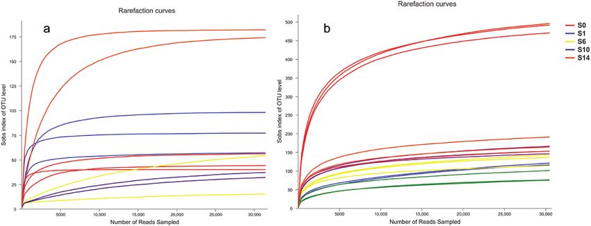
2                                                                                 Journal of Economic Entomology, 2020, Vol. XX, No. XX

et al. 2014, Xiang and Huang 2017). This balance is affected by            after discontinuing the antibiotics (see below): food was cleared
many external factors, such as food, temperature and especially ex-        from the digestive tract of cockroaches by starving them for 24 h;
ogenous antibiotics (Pérez-Cobas et al. 2015).                             their surfaces were disinfected with 75% ethanol for 90 s, and then
    Several strategies are used to remove gut bacteria in insects          thoroughly washed with sterile water to remove the disinfectant.
(Ohtaka 1991, Prosser and Douglas 1991, Tegtmeier et al. 2015,
Rosas et al. 2018), such as sterilizing the egg surfaces and rearing       Gut Microbial Cultures

                                                                                                                                                        Downloaded from https://academic.oup.com/jee/advance-article/doi/10.1093/jee/toaa205/5910672 by D H Hill Library - Acquis Dept S user on 29 October 2020
the hatchlings under aseptic conditions (Tegtmeier et al. 2015). This      The digestive tracts of a total of 120 cockroaches were collected: a
method is not widely used because of the strict aseptic culture con-       control (S0), and 2 d (S2), 4 d (S4), 6 d (S6), 8 d (S8), 10 d (S10), 12 d
ditions for the larvae. In contrast, the gut bacteria are quickly and      (S12), and 14 d (S14) after stopping antibiotic treatment. Each group
easily removed by antibiotics, thus antibiotics are commonly used          consisted of five cockroach guts and replicated three times (8 groups
(Chen and Purcell 1997, Raymond et al. 2009, Sharon et al. 2010,           × 5 cockroaches per group × 3 replications = 120 cockroaches). Gut
Llop et al. 2018, Rosas et al. 2018, Zhang et al. 2018a). Antibiotics      Samples were homogenized in 1 ml of sterile water and ground uni-
are most often used with the method of ingestion, and when used for        formly under sterile conditions using a vitreous bar with a tapered
eliminating bacteria, they usually have many side-effects on insects,      tip (Jinan Zhongchu Hengtong Biotechnology Co., Ltd., Jinan,
including stress, changes in the activity of metabolic enzymes and         China). All samples were vortex-mixed for 1 min and centrifuged at
immune responses (Woo et al. 1999), suppression of growth, devel-          98 g for 5 min. A series of dilutions were carried out and multiple di-
opment and reproduction, and higher mortality (Chen and Purcell            lution gradients were selected for the plate culture. The supernatant
1997, Raymond et al. 2009, Sharon et al. 2010, Llop et al. 2018).          (200 µl) was collected and streaked on Potato Dextrose Agar (PDA)
Moreover, antibacterial agents can selectively eliminate bacteria,         medium using an applicator. The nystatin was added into the cul-
facilitating the proliferation of fungi and other gut microbes.            ture medium to inhibit the growth of mycete and yeast. The number
    Many studies have investigated the composition and community           of cultured bacterial colony was recorded at 48 h. Bacterial and
structure of gut bacteria in insects. Few investigations, however, have    fungal colonies were distinguished based on differences in the size
examined symbiotic gut fungi and their interactions with bacteria,         and morphology of colonies and only the bacterial colonies were
and the resulting dynamic balance represented in species-specific          counted (Renata et al. 2013).
microbial communities. In this article, we analyzed the changes in
community composition, recovery rules of gut bacteria and fungi,
                                                                           High-Throughput Sequencing and Analysis of Gut
and the bacterial-fungal interaction during the process of gut bac-
                                                                           Microbiota
terial recovery of German cockroaches after antibiotic treatment,
                                                                           The guts of a total of 360 cockroaches were collected a control (S0)
which is very useful for studying the microorganism colonization
                                                                           and 1 d (S1), 2 d (S2), 6 d (S6), 10 d (S10), and 14 d (S14) after stop-
and interaction with insects. Recently, new biological control tech-
                                                                           ping antibiotic treatment. Each of these six groups consisted of 20
nologies based on the interaction between insects and symbionts
                                                                           guts and replicated three times (360 cockroaches).
have gradually developed, such as reintroducing genetically modified
                                                                               DNA was extracted using the K2306 Karroten Microbial
symbionts to secrete toxic proteins in the hosts (Daily and Johanna
                                                                           Genomic DNA extraction kit (Novoprotein Scientific Inc., Shanghai,
2012). The symbiont Pantoea agglomerans in the mosquito midgut
                                                                           China). The primers for amplification of the 16S rRNA gene were
was genetically modified to express anti-malaria proteins to disturb
                                                                           338F-806R (5′-ACTCCTACGGGAGGCAGCAG-3′ and 5′-GGACT
malaria development, thereby greatly decreased the plasmodium
                                                                           ACHVGGGTWTCTAAT-3′). The 16S rRNA PCR was performed in
carrying rate of mosquitoes (Wang et al. 2012). Our study could pro-
                                                                           a total volume of 20 µl containing 4 µl of 5 × FastPfu buffer, 2 µl of
vide a stable biological carrier for paratransgenesis through the selec-
                                                                           dNTPs (2.5 mM), 0.8 µl of Forward Primer (5 µM), 0.8 µl of Reverse
tion of stable gut bacteria for the biological control of B. germanica.
                                                                           Primer (5 µM), 0.4 µl of FastPfu Polymerase, 0.2 µl of BSA, 10 ng of
                                                                           DNA template and deionized ultrapure water (to 20 µl). The primers
Materials and Methods                                                      for ITS sequencing were ITS1F-ITS2R (5′-CTTGGTCATTTAGAG
                                                                           GAAGTAA-3′ and 5′-GCTGCGTTCTTCATCGATGC-3′). The ITS
Insects Collection                                                         PCR was performed in a total volume of 20 µl containing 2 µl of 10 ×
German cockroaches were supplied by the Key Laboratory of                  Buffer, 2 µl of dNTPs (2.5 mM), 0.8 µl of Forward Primer(5 µM),
Animal Resistance Biology of Shandong Province and maintained              0.8 µl of Reverse Primer(5 µM), 0.2 µl of Taq Polymerase, 0.2 µl
in a growth chamber. Culture chambers were adjusted to 27 ± 1°C,           of BSA, 10 ng of template DNA and deionized ultrapure water (to
60 ± 5% relative humidity (RH), and a photoperiod of 12:12 (L:D)           20 µl). The PCR conditions were as follows: 95°C for 3 min, 95°C
h. The insects fed on rat pellets (Shandong Experimental Animal            for 30 s, 55°C for 30 s and a final elongation step at 72°C for 10 min
Center, Jinan, China). All experimental insects were adult males.          of 27 cycles.
                                                                               Raw fastq files were demultiplexed, quality-filtered by
Preparation of Gut Samples                                                 Trimmomatic and merged by FLASH (http://ccb.jhu.edu/software/
In previous studies of our lab, a variety of antibiotics have been         FLASH) according to the following criteria: the reads data were
studied to remove the gut bacteria of B. germanica, among which the        truncated at any site with an average quality score of less than
combined use of gentamicin and levofloxacin had the best removal           20 within 50 bp sliding windows, allowing two nucleotides to be
effect (Shi et al. 2017). In the experimental group, adult male cock-      mismatched and removing ambiguous bases in primer-matching,
roaches were provided with sterile water fortified with levofloxacin       splicing sequences overlap longer than 10 bp and removing unspliced
(62 µg/ml) and gentamicin (62 µg/ml) and sterile rat pellets for 4         sequences. Operational taxonomic units (OTUs) were clustered with
d in sterile conditions (B. germanica was cultivated in a sterilized       97% similarity cutoff using UPARSE (version 7.1 http://drive5.
beaker, and the opening of the beaker was sealed with three steril-        com/uparse/) and chimeric sequences were identified and removed
ized layers of gauze). The control cockroaches were provided with          using UCHIME. The community richness indices (Chao1 and Ace)
sterile water. In preparation for gut dissection at specific intervals     and diversity indices (Shannon and Simpson) were estimated using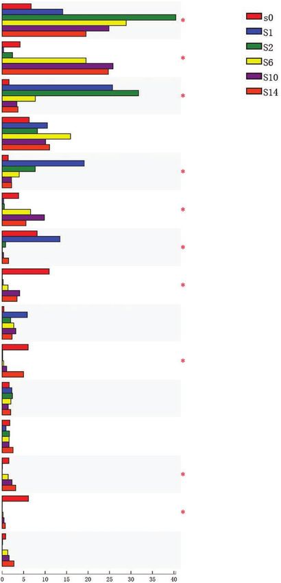
Journal of Economic Entomology, 2020, Vol. XX, No. XX                                                                                                                                     3

MOTHUR (http://www.mothur.org). We used the UniFrac server for

                                                                          Lg value of cultureable bacteria that
                                                                                                                  5.00
community comparisons.                                                                                            4.50                                                          **
                                                                                                                  4.00                                              ***

                                                                                 can be cultured (unit)
                                                                                                                                                           ***
                                                                                                                  3.50                             ***
Network Analysis                                                                                                  3.00
In order to study the relationship among the gut flora of B. germanica                                            2.50                    ***
                                                                                                                                  ***

                                                                                                                                                                                              Downloaded from https://academic.oup.com/jee/advance-article/doi/10.1093/jee/toaa205/5910672 by D H Hill Library - Acquis Dept S user on 29 October 2020
after stopping the antibiotic treatment, the potential relationship be-                                           2.00
                                                                                                                  1.50
tween the bacterial and fungal taxa was described by network ana-
                                                                                                                  1.00
lysis using the CoNet plug-in in Cytoscape (Shannon et al. 2003,                                                  0.50
Soffer et al. 2015). In order to reduce the complexity of calculation                                             0.00
and ensure the accuracy of taxonomic information, network analysis                                                        0       2        4        6       8       10          12   14
was conducted at the genus level of these two microbiomes. To ex-                                                        The number of days to stop feeding antibiotics(days)
plore all the pairwise associations and correlation scores (Spearman
                                                                           Fig. 1. Number of culturable bacteria (log-transformed) from the guts of
correlation, Pearson correlation, Kullback-Leibler dissimilarity,
                                                                           German cockroach adult males on different days after discontinuing the
Bray-Curtis dissimilarity, and mutual information) were calculated
                                                                           availability of antibiotics in their drinking water. 0 indicates males provide
(Faust and Raes 2012, Faust et al. 2012). The Brown method inte-           sterile water, the other numbers indicate the number of days to stop antibiotic
grated the P values of the five methods, only significant correlations     treatment. All treatments were compared to the 0 treatment, with significant
were retained (P < 0.05) (Soffer et al. 2015). After the Brown merging     differences indicated as follows: ** (0.001 < P ≤ 0.01), *** (P ≤ 0.001). Each bar
P values, the Benjamini–Hochberg multiple tests were performed as          represents the mean of five guts and ±SEM is shown.
a correlation (Hu et al. 2017). We import the obtained correlation
into the Gephi platform and use the Frucherma Reingold algorithm           Microbial Community Organization Before and After
for visualization (Bastian et al. 2009). Though the plug-in Network        Antibiotic Treatment
Analyzer in Cytoscape to calculate the network topology param-             Bacteria
eters, the degree, betweenness centrality, and closeness centrality of     After pyrosequencing, 949,839 raw sequences and 2,935 OTUs were
each node in the network (Assenov et al. 2008). The clustering coeffi-     obtained from the 18 samples (three replicates of each: a control,
cient and network density were selected to reflect changes in gut mi-      and 1, 2, 6, 10, and 14 d after stopping antibiotic treatment).
crobial community combination (Barberan et al. 2012), and degree,              The rarefaction curves tended to asymptote, showing saturation
betweenness centrality, and closeness centrality were used to explore      of the sequencing data, substantial abundance of bacterial reads, and
the key hub of the network (Sporns et al. 2007). Network analysis          sufficient depth for analysis of the diversity of the bacterial commu-
was used to explore the relationship between bacteria and fungi in         nity (Fig. 2). The rarefaction curves also showed variation in OTU
gut after antibiotic treatment.                                            density over time after withdrawal of the antibiotics. OTU density of
                                                                           the cockroach gut was highest for the treated cockroaches (S0) and
Statistical Analysis                                                       lowest 1 (S1) and 2 (S2) day after the 4-d antibiotic treatment. These
                                                                           curves also indicated that the bacterial richness in the gut lumen of
The statistical analysis was performed using SPSS version 19.0
                                                                           the German cockroach increased with time since discontinuing the
for Windows (IBM, Armonk, NY). The independent sample T-test
                                                                           antibiotic treatment. Nevertheless, the bacterial richness remained
was performed to assess the significant difference in the number of
                                                                           low compared with the controls even in 14th day after stopping the
culturable bacteria between each treatment group and control group.
                                                                           antibiotic treatment (Fig. 2).
Results with P < 0.05 between groups were considered statistically
                                                                               Nineteen phyla and 122 genera of bacteria were detected.
significant. Using a one-way ANOVA to examine the significant dif-
                                                                           Sequences that could not be classified were assigned ‘no rank’. One-
ference of richness and diversity indices of 18 bacterial samples or 15
                                                                           way ANOVA comparing the six treatment groups indicated that
fungal samples. P < 0.05 is considered a significant difference. The
                                                                           the number of OTUs and the bacterial diversity indices were sig-
Kruskal–Wallis H test with Benjamini–Hochberg false discovery rate
                                                                           nificantly different among the groups (P < 0.05) but the bacterial
(FDR) correction was used to evaluate the relative abundance differ-
                                                                           richness indices were not significantly different (P > 0.05) (Table 1).
ences of bacteria and fungi among multiple groups implemented in
                                                                               Using Bray-Curtis distance based on pyrosequencing data, we
the R software.
                                                                           conducted PCA, PCoA, and NMDS analyses. The PCA score map
                                                                           indicated that S0, S10, and S14 were grouped on the left of the graph
Availability of Data and Materials                                         and separated along PC1, which explained 46.35% of the total vari-
All nucleotide sequences from this study were uploaded to the NCBI         ation, from S1 and S2 (Fig. 3). Treatment group S0 separated from
SRA (Sequence Read Archive) database, and the accession numbers            S2, S6, S10, and S14 along PC2, which explained 35.19% of the
are PRJNA594114 and PRJNA594155.                                           total variation (Fig. 3). Overall, the gut bacterial community soon
                                                                           after the discontinuation of antibiotics (on the 1st and 2nd day) sep-
                                                                           arated from the remaining three groups (on the 6th, 10th, and 14th
Results                                                                    day), which were more similar to each other, and S0 separated from
Culturable Bacteria After Antibiotic Treatment                             the latter groups (Fig. 3). This pattern was generally confirmed with
The 4 d of continuous antibiotic treatment was effective at reducing       PCoA and NMDS analyses (Supp Fig. S1 [online only]).
the number of culturable bacteria in the gut of German cockroach               The relative abundance of 16S rRNA sequences, grouped at
males from Fig. 1. After stopping the antibiotic treatment, how-           the phylum level, are shown in Fig. 4. Firmicutes, Bacteroidetes,
ever, the number of culturable gut bacteria increased rapidly and          Fusobacteria, Actinobacteria, and Planctomycetes were most rep-
recovered to the pretreatment level on 14 d (Fig. 1). The result of the    resented, with Firmicutes being the most abundant in the control
gut count is not the exact number of gut bacteria, it only shows the       cockroaches (41.4%), followed by Bacteroidetes (35.0%). The
number of gut bacteria that can be cultured in vitro.                      most abundant of the gut microbiota on the 1st and 2nd day after
4 Journal of Economic Entomology, 2020, Vol. XX, No. XX
Downloaded from https://academic.oup.com/jee/advance-article/doi/10.1093/jee/toaa205/5910672 by D H Hill Library - Acquis Dept S user on 29 October 2020
Fig. 2. Rarefaction analysis of the different bacterial (a) and fungal (b) samples, including a control (S0), and 1 (S1), 2 (S2), 6 (S6), 10 (S10), and 14 (S14) d after
stopping antibiotic treatment. Sobs represent the observed number of species.
discontinuing the antibiotics were the Firmicutes (65.0 and 60.9%, relative abundances of the fungi at the phylum and genus levels
respectively), but in the last three groups (the 6th, 10th, and 14th are shown in Fig. 6. The 15 most dominant genera are shown in
day), the Bacteroidetes were most abundant (40.1, 45.3, and 45.9%) Fig. 7. The most abundant genus of fungi was Candida, which
and Fusobacteria were second in abundance (30.2, 26.5, and 22.3%) increased from 41.4% in control cockroaches up to 96.7% of all
was the second-highest (Fig. 4, Supp Table S1 [online only]). sequences 6 d after stopping the antibiotic treatment; it declined
The relative abundance of the major bacterial genera is shown to 19.3% 8 d later (Fig. 6, Supp Table S3 [online only]). Of the
in Fig. 4. Bacteria in the gut lumen were most diverse in the control 15 fungus genera, only three genera showed statistically signifi-
cockroaches that were not exposed to antibiotics, as also represented cant changes with respect to antibiotic treatment (unclassified_k_
in Table 1. Antibiotic treatment for 4 d dramatically reduced the Fungi, Mortierella, and unclassified_f_norank_o_Pleosporales; P
bacterial diversity, but the diversity of the bacterial community grad- < 0.05).
ually increased with time after stopping the antibiotic treatment;
nevertheless, even after 14 d, bacterial diversity did not return to Dynamic Network Between Fungi and Bacteria
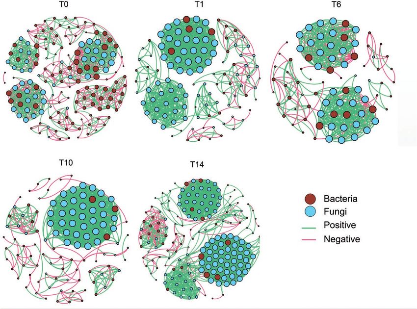
the level of the control cockroaches (Fig. 4). This indicates that anti- Five networks represent the interactions among microbes in control
biotics caused an irreversible change in the gut bacterial community, cockroaches and during the recovery of the microbial community in
for at least 14 d. the gut of German cockroaches after antibiotic treatment (Fig. 8).
The relative abundance of the 15 most dominant bacterial genera These networks differentiate based on the number of bacterial and
was subjected to statistical analysis among the six treatment groups fungal nodes and the number of edges of microbial interactions.
(Fig. 5). For 10 genera, there were statistically significant differ- Compared to the control group, the clustering coefficient and net-
ences (P < 0.05) among the six groups. The most common pattern work density of the antibiotic treatment groups were respectively
was for some genera that were represented at low relative abun- increased by 0.039, 0.024, 0.020, 0.039 and 0.055, 0.078, 0.072,
dance in the control cockroaches (e.g., Candidatus_Soleaferrea, 0.063, which indicated that gut microbiome associations were more
Anaerotruncus, Enterococcus, Desulfovibrio) to undergo dra- tightened with the antibiotic treatment (Table 3). The fungi in the
matic but transient increases or declines in abundance, but return networks of the T6 was reduced by 50 nodes; the T10 and the T14
to their original levels after 14 d on antibiotics-free water. Some were increased by 19 and 64 nodes than the previous group. The
genera, however, remained at lower abundance after 14 d (e.g., bacteria in the networks of the T6, the T10, and the T14 were in-
Tyzzerella_3, Parabacteroides, Alistipes), whereas others attained creased by 13, 9, and 4 nodes than the previous group. It also dis-
higher relative abundances after 14 d without antibiotics (e.g., played that the edges linking bacteria to fungi in the networks of the
Fusobacterium, Dysgonomonas) (Fig. 5). T6 and T10 were reduced by 13 and 7 edges, the T14 was increased
by 294 edges, the edges linking bacteria to bacteria in the networks
Fungi of the T6, the T10, and the T14 were increased by 38, 1 and 31
After pyrosequencing, 698,568 raw sequences and 1,031 OTUs were edges, the edges linking fungi to fungi in the networks of the T6 was
obtained from the 15 fungal samples. A total of eight phyla and 180 reduced by 355 edges and the T10 and T14 were increased by 371
genera of fungi were detected. Sequences that could not be classified and 932 edges than the previous group (Table 3).
were assigned ‘no rank’. Using one-way ANOVA for comparison, the The network analysis demonstrated that antibiotics interfered
number of OTUs and the fungus richness indices were significantly with the balance between bacteria and fungi and caused dysbiosis:
different (P < 0.05) (Table 2). The fungus samples on the 2nd day the percentage of fungi–fungi and fungi–bacteria interactions in-
were unqualified and were not sequenced. creased, while bacteria–bacteria interactions were reduced. In terms
The rarefaction curves tended to asymptote, suggesting that of overall relationships, antibiotic treatment leads to a closer rela-
the depth of the sequencing data was sufficient (Fig. 2). The tionship between gut microflora.Journal of Economic Entomology, 2020, Vol. XX, No. XX 5
Table 1. Richness and diversity indices of 18 bacterial samples representing three replicates each of six groups
Alpha diversity
ID Coverage Threshold Number of OTUs ACE Chao Shannon Simpson
S0-1 0.999245 0.03 385 394 398 4.37 0.0333
Downloaded from https://academic.oup.com/jee/advance-article/doi/10.1093/jee/toaa205/5910672 by D H Hill Library - Acquis Dept S user on 29 October 2020
S0-2 0.999344 0.03 392 401 403 4.59 0.0279
S0-3 0.999114 0.03 395 408 410 4.59 0.0227
S1-1* 0.999475 0.03 68 80 80 1.57 0.3292
S1-2 0.998753 0.03 116 207 160 2.37 0.1548
S1-3 0.999081 0.03 104 136 125 2.22 0.1449
S2-1* 0.999442 0.03 74 91 91 1.37 0.4586
S2-2 0.999475 0.03 92 106 103 1.62 0.3281
S2-3 0.999574 0.03 66 78 79 1.74 0.2514
S6-1 0.999344 0.03 129 143 143 2.49 0.1514
S6-2 0.999213 0.03 130 153 153 2.64 0.1350
S6-3 0.999410 0.03 107 121 145 2.44 0.1599
S10-1 0.999344 0.03 138 152 165 2.72 0.1361
S10-2 0.999344 0.03 145 160 156 2.75 0.1288
S10-3 0.999278 0.03 149 167 168 2.95 0.1091
S14-1 0.999475 0.03 169 178 177 3.30 0.0718
S14-2 0.999377 0.03 143 157 159 3.21 0.0774
S14-3 0.999541 0.03 133 143 140 3.17 0.0828
P value 0.018 0.063 0.097 0.015 0.004
OTUs were defined at the 97% similarity level (the threshold is 0.03). The 18 samples represent three replicates each of six treatment groups that include a con-
trol (S0), and 1 (S1), 2 (S2), 6 (S6), 10 (S10), and 14 (S14) d after stopping the antibiotics treatment. Sample S1-1 (*) and S2-1 (*) were discarded as an outlier in
community composition relative to the other two replicates, and it was also excluded from all subsequent analysis.
Discussion and become dominant in the gut temporarily, such as Candidatus,
Soleaferrea, and Anaerotruncus (Wertz and Breznak 2007, Ryu et al.
Antibiotic treatment is a common method to obtain aseptic insects
2008, Hassan et al. 2011, Yogurtcu and Tuncer 2013, Huang et al.
(Woo et al. 1999, Yong-Ming et al. 2006, Ben-Yosef et al. 2008,
2016, Abdolmaleki et al. 2019, He et al. 2020). The abundance of
Rosas et al. 2018). The combination treatment of levofloxacin and
Enterococcus, Dysgonomonas, and Desulfovibrio were significantly
gentamicin on German cockroaches for 4 d was effective in re-
increased in the recovery period and most of them showed resist-
moving gut bacteria. Both cultivable bacteria and high-throughput
ance to different kinds of antibiotics. Enterococcus is well known
sequencing showed a very low level of bacterial flora. The number
for resistance to gentamicin, streptomycin, chloramphenicol, tetra-
of the cultivable gut bacteria had basically recovered to the original
cycline, and streptomycin; Desulfovibrio are resistant to penicillin
level on the 14th day after stopping antibiotic treatment, but there
and rifampicin; Dysgonomonas are resistant to various beta-
were significantly different in the composition of the gut microbial
lactams, erythromycin, aminoglycosides, and fluoroquinolones, but
community, which was consistent with the studies of the rifampicin
was susceptible to clindamycin, minocycline, and chloramphenicol
treatment on German cockroaches. Rifampin treatment experi-
(Dzierzewicz et al. 2001, Hironaga et al. 2008). To better adapt to
ment showed that bacterial diversity and richness were not com-
the gut environment, Enterococcus can produce tyramine, which is
pletely recovered to the original level after stopping antibiotic for
related to tyrosine metabolism, to enhance the adhesion between
10 d (Rosas et al. 2018). In addition, the composition of the gut
bacteria and guts (Ladero et al. 2013, Pérez-Cobas et al. 2013).
microbiota of cockroaches was different after different antibiotic
Dysgonomonas, Fusobacterium, etc., can rapidly proliferate and oc-
treatments. Our study showed that after the combined utilization of
cupy a certain proportion, which is related to the basic ecological
gentamicin and levofloxacin, the relative abundance of Candidatus,
niche they have realized (Mikaelyan et al. 2016). Interestingly, al-
Soleaferrea, Fusobacterium, Bacteroides, and Anaerotruncus in
though Parabacteroides is resistant to tetracycline and β-lactam
the gut of cockroaches was increased. After the treatment of ri-
antibiotics, its abundance decreased because these bacteria that pos-
fampicin, the abundance of Fusobacterium and Desulfovibrio was
sessed the gene ermF and beta-lactamases (BLAs) were more sensi-
relatively high (Rosas et al. 2018). After the use of doxycycline, the
tive to the aminoglycoside antibiotic gentamicin (Boente et al. 2010,
abundance of Alphaproteobacteria in the gut of cockroaches was
Brook et al. 2013). In addition, we hypothesized that gut bacteria
relatively high (Pietri et al. 2018). After the use of vancomycin,
can not completely recover after treatment with antibiotics, even
the abundance of Enterobacteriaceae, Yersiniaceae, Budviciaceae,
some taxa that are sensitive to antibiotics may remain in the guts or
and Enterobacterales in the gut of cockroaches was relatively high
feces at very low abundance and can colonize their gut niche once
(Domínguez-Santos et al. 2020). Different antibiotics had different
again after antibiotic pressure was removed. In fact, similar results
effects on the removal of gut microbes of German cockroaches, but
had also been showed in Zootermopsis angusticollis, which revealed
the gut microbes of German cockroaches could recover to the ori-
a permanent reduction in bacterial diversity after rifampicin treat-
ginal level after stopping antibiotic treatment (Rosas et al. 2018,
ment (Rosengaus et al. 2011).
Domínguez-Santos et al. 2020).
The interaction between bacteria and fungi is mainly antag-
When gut bacteria were removed by antibiotics, those bacteria
onistic and achieves a dynamic balance. A variety of bacteria in
that are not susceptible to antibiotic and fast-growing bacteria
the gut of B. germanica can produce antimicrobial substances that
may utilize the limited oxygen and other resources to grow rapidly
have a broad spectrum for fungi or bacteria, for example, Bacillus6 Journal of Economic Entomology, 2020, Vol. XX, No. XX
PCA on OTU level
5000 s0
61 63 S1
4000 101
102
Downloaded from https://academic.oup.com/jee/advance-article/doi/10.1093/jee/toaa205/5910672 by D H Hill Library - Acquis Dept S user on 29 October 2020
62 S2
3000 S6
103
S10
2000 22 S14
143
142
1000 141
23
0
PC2(35.19%)
-1000
-2000
-3000
-4000
S_3
S_1
-5000 S_2 S13
-6000
S12
-7000
-8000
-6000 -4000 -2000 0 2000 4000 6000 8000 10000 12000
PC1(46.35%)
Fig. 3. Principal components analysis showing the similarity of the 16 bacterial communities based on the Bray-Curtis distance. Principal components (PCs)
1 and 2 explained 46.35 and 35.19% of the variance, respectively. The 16 bacterial samples, representing three replicates each of six treatment groups that
included a control (S0), and 1 (S1), 2 (S2), 6 (S6), 10 (S10), and 14 (S14) d after stopping antibiotic treatment. One replicate in the S1 and the S2 treatment group
was excluded from analysis as an outlier relative to the other two replicates in the same treatment group. In S0, one replicate is obscured by another replicate.
a b
Community barplot analysis
Community barplot analysis
1 Firmicutes
Bacteroidetes 1 Fusobacterium
Fusobacteria
Proteobacteria Dysgonomonas
Actinobacteria Candidatus_Soleaferrea
Planctomycetes Bacteroides
others Anaerotruncus
0.8
0.8 Enterococcus
Tyzzerella_3
Percent of community abundance on Phylum level
Percent of community abundance on Genus level
Parabacteroides
unclassified_o__Micrococcales
Desulfovibrio
0.6
unclassified_f__Ruminococcaceae
0.6
norank_f__Ruminococcaceae
norank_f__Porphyromonadaceae
Alistipes
Sebaldella
0.4 Christensenellaceae_R-7_group
0.4
Lactobacillus
Lachnoclostridium
unclassified_f__Lachnospiraceae
Weissella
Blattabacterium
0.2 0.2 Rs-D38_termite_group
Tannerella
unclassified_f__Porphyromonadaceae
Paludibacter
norank_o__Rs-K70_termite_group
0 0 norank_c__vadinHA49
s0 S1 S2 S6 S10 S14 s0 S1 S2 S6 S10 S14 unclassified_f__Enterobacteriaceae
Samples Samples norank_f__Rhodospirillaceae
others
Fig. 4. Bacterial composition of the different communities at the Phylum (a) the Genus (b) level. The relative read abundances of different bacterial genera within
the different communities are shown. Taxa with an abundanceJournal of Economic Entomology, 2020, Vol. XX, No. XX 7
Kruskal-Wallis H test bar plot
Fusobacterium 0.0122
Downloaded from https://academic.oup.com/jee/advance-article/doi/10.1093/jee/toaa205/5910672 by D H Hill Library - Acquis Dept S user on 29 October 2020
Dysgonomonas 0.02204
Candidatus_Soleaferrea 0.01599
Bacteroides 0.1163
Anaerotruncus 0.02554
Enterococcus 0.01718
Tyzzerella_3 0.03185
Pvalue
Parabacteroides 0.0274
unclassified_o_Micrococcales 0.06277
Desulfovibrio 0.01589
unclassified_f_Ruminococcaceae 0.7226
norank_f_Ruminococcaceae 0.3027
norank_f_Porphyromonadaceae 0.01908
Alistipes 0.02039
Sebaldella 0.05473
Mean proportions(ˁ)
Fig. 5. Differences at the Genus level of bacterial relative abundance across experimental groups after antibiotic treatment was discontinued. The bar length for
each genus indicates its average relative abundance in each sample group, and the different colors indicate the six groups (the combined three replicates of
each of the six treatment groups included a control (S0), and 1 (S1), 2 (S2), 6 (S6), 10 (S10), and 14 (S14) d after stopping the antibiotic treatment). The P value is
shown, and * indicates that the six groups are significantly different from each other (P < 0.05).8 Journal of Economic Entomology, 2020, Vol. XX, No. XX
Table 2. Richness and diversity indices of 15 fungal samples representing three replicates each of five groups
Alpha diversity
ID Coverage Threshold Number of OTUs ACE Chao Shannon Simpson
Downloaded from https://academic.oup.com/jee/advance-article/doi/10.1093/jee/toaa205/5910672 by D H Hill Library - Acquis Dept S user on 29 October 2020
S0-1 1.000000 0.03 55 67 65 0.26 0.9175
S0-2 1.000000 0.03 42 21 19 0.20 0.9315
S0-3 0.999936 0.03 43 39 39 0.98 0.4994
S1-1 1.000000 0.03 77 42 38 0.89 0.4702
S1-2 1.000000 0.03 96 76 76 1.24 0.4695
S1-3 0.999904 0.03 57 43 42 0.90 0.4692
S6-1 0.999458 0.03 55 178 178 1.72 0.3003
S6-2 0.999841 0.03 14 181 181 2.44 0.2585
S6-3* 0.999872 0.03 37 62 62 0.49 0.8178
S10-1 0.999681 0.03 33 55 55 1.99 0.2058
S10-2* 1.000000 0.03 76 42 42 2.12 0.2370
S10-3 0.999681 0.03 36 43 44 1.58 0.3515
S14-1 0.999681 0.03 175 77 77 2.93 0.1267
S14-2 0.999936 0.03 181 96 96 1.39 0.4890
S14-3* 0.999522 0.03 54 59 60 1.51 0.4276
P value 0.008 0.009 0.009 0.084 0.010
OTUs were defined at the 97% similarity level (the threshold is 0.03). The 15 samples represent three replicates each of five treatment groups that include a
control (S0), and 1 (S1), 6 (S6), 10 (S10), and 14 (S14) d after stopping the antibiotics treatment. Three samples indicated with * (S6-3, S10-2, and S14-3) were
discarded as outliers in community composition relative to the other two replicates in their respective groups, and they were also excluded from all subsequent
analysis.
a b Community barplot analysis
1 Candida
Community barplot analysis unclassified_k__Fungi
Percent of community abundance on Genus level
1 Aspergillus
Ascomycota
unclassified_k__Fungi Penicillium
0.8
Percent of community abundance on Phylum level
Basidiomycota Mortierella
Zygomycota Fusarium
0.8
others unclassified_f__Verrucariaceae
unclassified_f__norank_o__Pleosporales
0.6
unclassified_p__Ascomycota
0.6 Sporobolomyces
Trichomonascus
unclassified_f__Pleosporaceae
0.4
Geminibasidium
0.4
others
0.2
0.2
0 0
S0 S1 S6 S10 S14 S0 S1 S6 S10 S14
Samples Samples
Fig. 6. Composition of the different fungal communities at the Phylum (a) the Genus (b) level. The relative read abundances of different fungus genera within
the different communities are shown. Taxa with an abundanceJournal of Economic Entomology, 2020, Vol. XX, No. XX 9
Downloaded from https://academic.oup.com/jee/advance-article/doi/10.1093/jee/toaa205/5910672 by D H Hill Library - Acquis Dept S user on 29 October 2020
Fig. 7. Differences at the Genus level of fungal relative abundance across experimental groups after antibiotic treatment was discontinued.The bar length for each genus
indicates its average relative abundance in each sample group, and the different colors indicate the six groups (including a control (S0), and 1 (S1), 6 (S6), 10 (S10), and
14 (S14) d after stopping the antibiotic treatment). The P value is shown, and * indicates that the five groups are significantly different from each other (P < 0.05).
network density indicated that bacteria and fungi in the cockroach Conclusions
gut established a closer relationship after antibiotic treatment. We In summary, antibiotic treatment significantly reduced the di-
concluded that it was the consequence of multi-factors by anti- versity and abundance of gut bacteria, changed the composition
biotics to tighten the core microbes and lose the low-abundance of of the gut microbiota, and made the relationship of gut micro-
microbes that temporarily host in the gut. organisms closer, which is useful for studying the gut microbes’10 Journal of Economic Entomology, 2020, Vol. XX, No. XX
Downloaded from https://academic.oup.com/jee/advance-article/doi/10.1093/jee/toaa205/5910672 by D H Hill Library - Acquis Dept S user on 29 October 2020
Fig. 8. The networks visualize antibiotic treatment effects on the co-occurrence pattern between bacteria and fungi in the intestines of German cockroach. The
node size is proportional to the abundance of taxa, and the nodes filled in red are bacterial taxa and in blue are fungi taxa. The edges are colored according to
interaction types; positive correlations are labeled with green and negative correlations are colored in red. The five groups that include a control (T0), and 1 (T1),
6 (T6), 10 (T10), and 14 (T14) d after stopping the antibiotic treatment.
Table 3. Topological indices of each network in Fig. 8 carriers of paratransgenesis for the biological control of
T0 T1 T6 T10 T14 B. germanica.
Clustering coefficient 0.804 0.843 0.828 0.824 0.844
Network density 0.072 0.127 0.150 0.144 0.135 Supplementary Data
Number of nodes 156 114 81 109 177
Number of edges 869 815 485 850 2,107 Supplementary data are available at Journal of Economic
Number of fungal nodes 47 82 36 55 119 Entomology online.
Number of bacterial nodes 109 32 45 54 58 Fig. S1 Sample sorting analysis. Study on bacterial community
Number of edges linking 332 183 170 163 457 composition of 16 samples by PCoA analysis based on Bray-Curtis
bacteria to fungi distance. Principal components (PCs) 1 and 2 explained 45.7% and
Number of edges linking 212 596 241 612 1,544 33.72% of the variance, respectively. NMDs showing the differ-
fungi to fungi
ence of bacterial communities according to Bray-Curtis distance.
Number of edges linking 325 36 74 75 106
These samples, including a control (S0), and 1 (S1), 2 (S2), 6 (S6),
bacteria to bacteria
10 (S10) and 14 (S14) days after stopping antibiotic treatment.
T0 indicates feeding sterile water, the other numbers indicate the number of
days to stop antibiotic treatment.
Acknowledgments
colonization and interaction with host insects. Meanwhile, our This study was supported by the National Natural Science Foundation
study could also lay the foundation for the exploitation of new of China (81572027) and Natural Science Foundation of Shandong
control strategies based on core gut flora. It can help find po- Province (ZR2012CQ040). The Project Supported by the Foundation
tential core microbes from cockroach guts as stable biological of State Key Laboratory of Biobased Material and Green PapermakingJournal of Economic Entomology, 2020, Vol. XX, No. XX 11
(ZZ20190405), Qilu University of Technology, Shandong Academy of Ferrandon, D., J. L. Imler, C. Hetru, and J. A. Hoffmann. 2007. The Drosophila
Sciences. systemic immune response: sensing and signalling during bacterial and
fungal infections. Nat. Rev. Immunol. 7: 862–874.
Förster, T. M., S. Mogavero, A. Dräger, K. Graf, M. Polke, I. D. Jacobsen,
References Cited and B. Hube. 2016. Enemies and brothers in arms: Candida albicans and
gram-positive bacteria. Cell. Microbiol. 18: 1709–1715.
Abdolmaleki, Z., Z. Mashak, and F. Safarpoor Dehkordi. 2019. Phenotypic
Downloaded from https://academic.oup.com/jee/advance-article/doi/10.1093/jee/toaa205/5910672 by D H Hill Library - Acquis Dept S user on 29 October 2020
Frankenburg, W. G., and A. A. Vaitekunas. 1955. Chemical studies on nicotine
and genotypic characterization of antibiotic resistance in the methicillin-
degradation by microorganisms derived from the surface of tobacco seeds.
resistant Staphylococcus aureus strains isolated from hospital cock-
Arch. Biochem. Biophys. 58: 509–512.
roaches. Antimicrob. Resist. Infect. Control. 8: 54.
Gijzen, H. J., C. A. Broers, M. Barughare, and C. K. Stumm. 1991. Methanogenic
Akira, S., S. Uematsu, and O. Takeuchi. 2006. Pathogen recognition and in-
bacteria as endosymbionts of the ciliate Nyctotherus ovalis in the
nate immunity. Cell. 124: 783–801.
cockroach hindgut. Appl. Environ. Microbiol. 57: 1630–1634.
Allaker, R. P., and C. W. Douglas. 2009. Novel anti-microbial therapies for
Graczyk, T. K., R. Knight, and L. Tamang. 2005. Mechanical transmission of
dental plaque-related diseases. Int. J. Antimicrob. Agents 33: 8–13.
human protozoan parasites by insects. Clin. Microbiol. Rev. 18: 128–132.
Assenov, Y., F. Ramírez, S. E. Schelhorn, T. Lengauer, and M. Albrecht. 2008.
Hassan, M., Y. Javadzadeh, F. Lotfipour, and R. Badomchi. 2011.
Computing topological parameters of biological networks. Bioinformatics.
Determination of comparative minimum inhibitory concentration (MIC)
24: 282–284.
of bacteriocins produced by Enterococci for selected isolates of multi-
Barberán, A. S. T. B., E. O. Casamayor, and N. Fierer. 2012. Using network
antibiotic resistant Enterococcus spp. Adv. Pharm. Bull. 1: 75–79.
analysis to explore co-occurrence patterns in soil microbial communities.
He, P., D. Z. Mang, H. Wang, M. M. Wang, Y. F. Ma, J. Wang, G. L. Chen,
ISME J. 6: 343–351.
F. Zhang, and M. He. 2020. Molecular characterization and functional
Bastian, M., S. Heymann, and M. Jacomy. 2009. Gephi: an open source
analysis of a novel candidate of cuticle carboxylesterase in Spodoptera
software for exploring and manipulating networks, pp. 8. 361–362. In
exigua degradating sex pheromones and plant volatile esters. Pestic.
Proceedings of the Third International Conference on Weblogs and Social
Biochem. Physiol. 163: 227–234.
Media, ICWSM, 17–20 May 2009. San Jose, California, USA.
Hironaga, M., K. Yamane, M. Inaba, Y. Haga, and Y. Arakawa. 2008.
Ben-Yosef, M., E. Jurkevitch, and B. Yuval. 2008. Effect of bacteria on nu-
Characterization and antimicrobial susceptibility of Dysgonomonas
tritional status and reproductive success of the Mediterranean fruit fly
capnocytophagoides isolated from human blood sample. Jpn. J. Infect.
Ceratitis capitata. Physiol. Entomol. 33: 145–154.
Dis. 61: 212–213.
Ben-Yosef, M., Y. Aharon, E. Jurkevitch, and B. Yuval. 2010. Give us the tools
Hongoh, Y. 2010. Diversity and genomes of uncultured microbial symbionts
and we will do the job: symbiotic bacteria affect olive fly fitness in a diet-
in the termite gut. Biosci. Biotechnol. Biochem. 74: 1145–1151.
dependent fashion. Proc. Biol. Sci. 277: 1545–1552.
Hu, H. W., J. T. Wang, J. Li, X. Z. Shi, Y. B. Ma, D. Chen, and J. Z. He. 2017.
Boente, R. F., L. Q. Ferreira, L. S. Falcão, K. R. Miranda, P. L. Guimarães,
Long-term nickel contamination increases the occurrence of antibiotic re-
J. Santos-Filho, J. M. Vieira, D. E. Barroso, J. P. Emond, E. O. Ferreira,
sistance genes in agricultural soils. Environ. Sci. Technol. 51: 790–800.
et al. 2010. Detection of resistance genes and susceptibility patterns in
Huang, Y. H., X. J. Wang, F. Zhang, X. B. Huo, R. S. Fu, J. J. Liu, W. B. Sun,
Bacteroides and Parabacteroides strains. Anaerobe 16: 190–194.
D. M. Kang, and X. Jing. 2013. The identification of a bacterial strain
Bosco-Drayon, V., M. Poidevin, I. G. Boneca, K. Narbonne-Reveau, J. Royet,
BGI-1 isolated from the intestinal flora of Blattella germanica, and its anti-
and B. Charroux. 2012. Peptidoglycan sensing by the receptor PGRP-LE
entomopathogenic fungi activity. J. Econ. Entomol. 106: 43–49.
in the Drosophila gut induces immune responses to infectious bacteria and
Huang, Y., X. Hou, S. Liu, and J. Ni. 2016. Correspondence analysis of bio-
tolerance to microbiota. Cell Host Microbe. 12: 153–165.
refractory compounds degradation and microbiological community distri-
Brook, I., H. M. Wexler, and E. J. Goldstein. 2013. Antianaerobic
bution in anaerobic filter for coking wastewater treatment. Chem. Eng. J.
antimicrobials: spectrum and susceptibility testing. Clin. Microbiol. Rev.
304: 864–872.
26: 526–546.
Kester, M., K. E. Vrana, and K. D. Karpa. 2011. Integrated view: pharma-
Buchon, N., N. A. Broderick, M. Poidevin, S. Pradervand, and B. Lemaitre.
cology, 2nd ed. Elsevier, Philadelphia, PA.
2009. Drosophila intestinal response to bacterial infection: activation of
Kim, J. K., J. Y. Kwon, S. K. Kim, S. H. Han, Y. J. Won, J. H. Lee, C. H. Kim,
host defense and stem cell proliferation. Cell Host Microbe 5: 200–211.
T. Fukatsu, and B. L. Lee. 2014. Purine biosynthesis, biofilm forma-
Chen, D. Q., and A. H. Purcell. 1997. Occurrence and transmission of faculta-
tion, and persistence of an insect-microbe gut symbiosis. Appl. Environ.
tive endosymbionts in aphids. Curr. Microbiol. 34: 220–225.
Microbiol. 80: 4374–4382.
Daily, J. P. 2012. Malaria vaccine trials–beyond efficacy end points. N. Engl.
Ladero, V., D. M. Linares, B. Del Rio, M. Fernandez, M. C. Martin, and
J. Med. 367: 2349–2351.
M. A. Alvarez. 2013. Draft genome sequence of the tyramine producer
Dillon, R. J., and V. M. Dillon. 2004. The gut bacteria of insects: nonpathogenic
Enterococcus durans strain IPLA 655. Genome Announc. 1: e00265-13.
interactions. Annu. Rev. Entomol. 49: 71–92.
Lemaitre, B., and J. Hoffmann. 2007. The host defense of Drosophila
Domínguez-Santos, R., A. E. Pérez-Cobas, A. Artacho, J. A. Castro, I. Talón,
melanogaster. Annu. Rev. Immunol. 25: 697–743.
A. Moya, C. García-Ferris, and A. Latorre. 2020. Unraveling assemblage,
Llop, P., A. Latorre, and A. Moya. 2018. Experimental epidemiology of
functions and stability of the Gut Microbiota of Blattella germanica by
antibiotic resistance: looking for an appropriate animal model system.
antibiotic treatment. Front. Microbiol. 11:487.
Microbiol. Spectr. 6: MTBP-0007-2016. doi:10.1128/microbiolspec.
Douglas, A. E. 2015. Multiorganismal insects: diversity and function of resi-
MTBP-0007-2016.
dent microorganisms. Annu. Rev. Entomol. 60: 17–34.
López-Sánchez, M. J., A. Neef, J. Peretó, R. Patiño-Navarrete, M. Pignatelli,
Dzierzewicz, Z., B. Cwalina, M. Jaworska-Kik, L. Weglarz, and T. Wilczok.
A. Latorre, and A. Moya. 2009. Evolutionary convergence and nitrogen
2001. Susceptibility to antibiotics and biochemical properties of
metabolism in Blattabacterium strain Bge, primary endosymbiont of the
Desulfovibrio desulfuricans strains. Acta Pol. Pharm. 58: 439–445.
cockroach Blattella germanica. PLoS Genet. 5: e1000721.
Eberhardt, H. J. 1995. The biological degradation of nicotine by nicotinophilic
Mikaelyan, A., C. L. Thompson, M. J. Hofer, and A. Brune. 2016.
microorganisms. Beitr. Tabakforsch. Int. 16: 119–129.
Deterministic assembly of complex bacterial communities in guts of germ-
Faust, K., and J. Raes. 2012. Microbial interactions: from networks to models.
free cockroaches. Appl. Environ. Microbiol. 82: 1256–1263.
Nat. Rev. Microbiol. 10: 538–550.
Moya, A., R. Gil, A. Latorre, J. Peretó, M. Pilar Garcillán-Barcia, and
Faust, K., J. F. Sathirapongsasuti, J. Izard, N. Segata, D. Gevers, J. Raes, and
F. de la Cruz. 2009. Toward minimal bacterial cells: evolution vs. design.
C. Huttenhower. 2012. Microbial co-occurrence relationships in the
FEMS Microbiol. Rev. 33: 225–235.
human microbiome. Plos Comput. Biol. 8: e1002606.
Neyen, C., and B. Lemaitre. 2016. Sensing Gram-negative bacteria: a phylo-
Feldhaar, H. 2011. Bacterial symbionts as mediators of ecologically important
genetic perspective. Curr. Opin. Immunol. 38: 8–17.
traits of insect hosts. Ecol. Entomol. 36: 533–543.12 Journal of Economic Entomology, 2020, Vol. XX, No. XX
Noda, H., and Y. Koizumi. 2003. Sterol biosynthesis by symbiotes: cyto- Shi, W. L., F. Yang, X. X. Sun, J. Lu, D. Q. Zhao, and F. Zhang. 2017.
chrome P450 sterol C-22 desaturase genes from yeastlike symbiotes of rice Effect of multiple antibiotics on intestinal bacteria removal of re-
planthoppers and anobiid beetles. Insect Biochem. Mol. Biol. 33: 649–658. sistant Blattella germanica. J. Inner Mongolia Univ. (Nat Sci). 48:
Ohtaka, C. 1991. Effects of heat treatment on the symbiotic system of an 291–296.
aphid mycetocyte. Symbiosis. 11: 19–30. Soffer, N., J. Zaneveld, and R. Vega Thurber. 2015. Phage-bacteria net-
Pan, X. Y., and F. Zhang. 2020. Advances in biological control of the German work analysis and its implication for the understanding of coral disease.
Downloaded from https://academic.oup.com/jee/advance-article/doi/10.1093/jee/toaa205/5910672 by D H Hill Library - Acquis Dept S user on 29 October 2020
cockroach, Blattella germanica (L.). Biol. Control 142: 104104. Environ. Microbiol. 17: 1203–1218.
Parsek, M. R., and P. K. Singh. 2003. Bacterial biofilms: an emerging link to Sporns, O., C. J. Honey, and R. Kötter. 2007. Identification and classification
disease pathogenesis. Annu. Rev. Microbiol. 57: 677–701. of hubs in brain networks. PLoS One 2: e1049.
Peleg, A. Y., E. Tampakakis, B. B. Fuchs, G. M. Eliopoulos, R. C. Moellering, Stiles, M. E., and W. H. Holzapfel. 1997. Lactic acid bacteria of foods and
Jr, and E. Mylonakis. 2008. Prokaryote-eukaryote interactions identi- their current taxonomy. Int. J. Food Microbiol. 36: 1–29.
fied by using Caenorhabditis elegans. Proc. Natl. Acad. Sci. U. S. A. 105: Tang, L., J. Lian, X. J. Lu, Y. R. Dai, X. S. Tan, and C. G. Zheng. 2005.
14585–14590. Preliminary study on antifungal effect of intestinal bacteria of Blattella
Pérez-Cobas, A. E., M. J. Gosalbes, A. Friedrichs, H. Knecht, A. Artacho, germanica. Natural Enemies of Insects. 27:140–144.
K. Eismann, W. Otto, D. Rojo, R. Bargiela, M. von Bergen, et al. 2013. Tegtmeier, D., C. L. Thompson, C. Schauer, and A. Brune. 2015. Oxygen af-
Gut microbiota disturbance during antibiotic therapy: a multi-omic ap- fects colonization and metabolic activities of gut bacteria in a gnotobiotic
proach. Gut. 62: 1591–1601. cockroach model. Applied Environ. Microbiol. 82: 03130-15.
Pérez-Cobas, A. E., E. Maiques, A. Angelova, P. Carrasco, A. Moya, and Vallabhapurapu, S., and M. Karin. 2009. Regulation and function of
A. Latorre. 2015. Diet shapes the gut microbiota of the omnivorous cock- NF-kappaB transcription factors in the immune system. Annu. Rev.
roach Blattella germanica. FEMS Microbiol. Ecol. 91: fiv022. Immunol. 27: 693–733.
Peters, B. M., M. A. Jabra-Rizk, M. A. Scheper, J. G. Leid, J. W. Costerton, Vazirianzadeh, B., R. Dehghani, M. Mehdinejad, M. Sharififard, and
and M. E. Shirtliff. 2010. Microbial interactions and differential protein N. Nasirabadi. 2014. The first report of drug resistant bacteria isolated
expression in Staphylococcus aureus -Candida albicans dual-species bio- from the brown-banded cockroach, Supella longipalpa, in Ahvaz, South-
films. FEMS Immunol. Med. Microbiol. 59: 493–503. western Iran. J. Arthropod. Borne. Dis. 8: 53–59.
Pietri, J. E., C. Tiffany, and D. Liang. 2018. Disruption of the microbiota af- Wang, G., Y. K. Ma, and Y. Q. Wang. 2003. Analysis of the advantages and
fects physiological and evolutionary aspects of insecticide resistance in the beneficial flora of naturally fermented tobacco. Yunnan Tobacco. 74:
German cockroach, an important urban pest. PLoS One 13: e0207985. 27–30.
Prosser, W. A., and A. E. Douglas. 1991. The aposymbiotic aphid: an ana- Wang, S., A. K. Ghosh, N. Bongio, K. A. Stebbings, D. J. Lampe, and M. Jacobs-
lysis of chlortetracycline-treated pea aphid, Acyrthosiphon pisum. J. Insect Lorena. 2012. Fighting malaria with engineered symbiotic bacteria from
Physiol. 37: 713–719. vector mosquitoes. Proc. Natl. Acad. Sci. U. S. A. 109: 12734–12739.
Raymond, B., P. R. Johnston, D. J. Wright, R. J. Ellis, N. Crickmore, and Weiss, B., and S. Aksoy. 2011. Microbiome influences on insect host vector
M. B. Bonsall. 2009. A mid-gut microbiota is not required for the patho- competence. Trends Parasitol. 27: 514–522.
genicity of Bacillus thuringiensis to diamondback moth larvae. Environ. Wertz, J. T., and J. A. Breznak. 2007. Stenoxybacter acetivorans gen. nov.,
Microbiol. 11: 2556–2563. sp. nov., an acetate-oxidizing obligate microaerophile among diverse
Renata, R. V., B. A. P. Guarnieri, S. J. C. G. Da, and C. A. S. Regina. 2013. O2-consuming bacteria from termite guts. Appl. Environ. Microbiol. 73:
Characteristics of Saccharomyces cerevisiae yeasts exhibiting rough col- 6819–6828.
onies and pseudohyphal morphology with respect to alcoholic fermenta- Woo, P. C., H. W. Tsoi, L. P. Wong, H. C. Leung, and K. Y. Yuen. 1999.
tion. Braz. J. Microbiol. 44: 1121–1131. Antibiotics modulate vaccine-induced humoral immune response. Clin.
Rosas T., C. García-Ferris, R. Domínguez-Santos, P. Llop, A. Latorre, and Diagn. Lab. Immunol. 6: 832–837.
A. Moya. 2018. Rifampicin treatment of Blattella germanica evidences a Xiang, B. Q., and Y. C. Huang. 2017. Research progress on the interaction
fecal transmission route of their gut microbiota. FEMS Microbiol. Ecol. between Candida albicans and common bacteria. Int. J. Biomed. Eng. 40:
94. doi:10.1093/femsec/fiy002. 384–388.
Rosengaus, R. B., C. N. Zecher, K. F. Schultheis, R. M. Brucker and Yang, C. L., H. Y. Zhu, and F. Zhang. 2019. Comparative proteomics analysis
S. R. Bordenstein. 2011. Disruption of the termite gut microbiota and between the short-term stress and long-term adaptation of the Blattella
its prolonged consequences for fitness. Appl. Environ. Microbiol. 77: germanica (Blattodea: Blattellidae) in response to Beta-Cypermethrin. J.
4303–4312. Econ. Entomol. 112: 1396–1402.
Royet, J., and R. Dziarski. 2007. Peptidoglycan recognition proteins: pleio- Yogurtcu, N. N., and Y. Tuncer. 2013. Antibiotic susceptibility patterns of
tropic sensors and effectors of antimicrobial defences. Nat. Rev. Microbiol. Enterococcus strains isolated from Turkish Tulum cheese. I Int. J. Dairy
5: 264–277. Technol. 66: 236–242.
Ryu, J. H., S. H. Kim, H. Y. Lee, J. Y. Bai, Y. D. Nam, J. W. Bae, D. G. Lee, Yong-Ming, R., J. Xu, and S. S. Liu. 2006. Effects of antibiotics on fitness of
S. C. Shin, E. M. Ha, and W. J. Lee. 2008. Innate immune homeostasis by the B biotype and a non-B biotype of the whitefly Bemisia tabaci. Entomol.
the homeobox gene caudal and commensal-gut mutualism in Drosophila. Exp. Appl. 121: 159–166.
Science. 319: 777–782. Zhang, F. 2012. Identification and antifungal study of the intestinal bac-
Ryu, J. H., E. M. Ha, and W. J. Lee. 2010. Innate immunity and gut–microbe terium BGI-17 of Blattella germanica. Chin. J. Vector Biol. Control. 23:
mutualism in Drosophila. Dev. Comp. Immunol. 34: 0–376. 39–41.
Sabree, Z. L., S. Kambhampati, and N. A. Moran. 2009. Nitrogen recycling Zhang, F., and R. Yang. 2019. Life history and functional capacity of the
and nutritional provisioning by Blattabacterium, the cockroach endosym- microbiome are altered in beta-cypermethrin-resistant cockroaches. Int.
biont. Proc. Natl. Acad. Sci. U. S. A. 106: 19521–19526. J. Parasitol. 49: 715–723.
Salehzadeh, A., P. Tavacol, and H. Mahjub. 2007. Bacterial, fungal and para- Zhang, X. C., and F. Zhang. 2018. The potential control strategies based on
sitic contamination of cockroaches in public hospitals of Hamadan, Iran. the interaction between indoor cockroaches and their symbionts in China.
J. Vector Borne Dis. 44: 105–110. Adv. Insect. Physiol. 55: 55–122.
Shannon, P., A. Markiel, O. Ozier, N. S. Baliga, J. T. Wang, D. Ramage, Zhang, F., Y. H. Huang, S. Z. Liu, L. Zhang, B. T. Li, X. X. Zhao, Y. Fu,
N. Amin, B. Schwikowski, and T. Ideker. 2003. Cytoscape: a software J. J. Liu, and X. X. Zhang. 2013. Pseudomonas reactans, a bacterial
environment for integrated models of biomolecular interaction networks. strain isolated from the intestinal flora of Blattella germanica with anti-
Genome Res. 13: 2498–2504. Beauveria bassiana activity. Environ. Entomol. 42: 453–459.
Sharon, G., D. Segal, J. M. Ringo, A. Hefetz, I. Zilber-Rosenberg, and Zhang, F., X. J. Wang, Y. H. Huang, Z. G. Zhao, S. S. Zhang, X. S. Gong,
E. Rosenberg. 2010. Commensal bacteria play a role in mating prefer- L. Xie, D. M. Kang, and X. Jing. 2014. Differential expression of hemo-
ence of Drosophila melanogaster. Proc. Natl. Acad. Sci. U. S. A. 107: lymph proteins between susceptible and insecticide-resistant Blattella
20051–20056. germanica (Blattodea: Blattellidae). Environ. Entomol. 43: 1117–1123.You can also read