Egyptian Journal of Veterinary Sciences
←
→
Page content transcription
If your browser does not render page correctly, please read the page content below

41                                                                Egypt. J. Vet. Sci. Vol. 52, No.3, pp. 361-372 (2021)

                Egyptian Journal of Veterinary Sciences
                                         https://ejvs.journals.ekb.eg/

Histomorphological Study of The Pons and Medulla Oblongata of
African Striped Group Squirrel (Xerus erythropus)
Ajeigbe Sheriff Olawale1, Tauheed Abubakar Muazu2 and James Oliver Nzalak1
1
  Department of Veterinary Anatomy, Ahmadu Bello University Zaria, Kaduna State,
Nigeria.
2
  Department of Veterinary Anatomy, Bayero University Kano, Kano State, Nigeria.

                        T    HE study was undertaken to investigate the histological and functional relationships be-
                             tween the pons and Medulla Oblongata of African Striped Ground Squirrel (Xerus eryth-
                        ropus). Twenty (20) adult African striped ground squirrels were divided in to two groups: 10
                        males and 10 females. The ground squirrels were obtained from the surrounding villages of
                        Zaria Local Government, Kaduna State Nigeria. Each Squirrel was euthanized using ketamine
                        hydrochloride at dose 80mg/kg BW followed by gentle perfusion with neutral formal saline.
                        A pair of scissors, chisel and scalpel blade was used to gently extract the brain (craniotomy).
                        The extracted brain was fixed in bouins solution for 24hours and processed histologically. The
                        pons and medulla are composed of conspicuous nucleus abducens, nucleus facialis and nucleus
                        trigeminal. The nucleus facialis was found dorsal to the paramedian pontine reticular formation
                        as group of large multipolar neurons. A conspicuous nucleus abducens was found as clusters of
                        medium neurons located lateral to the trigeminal nucleus. In myelencephalon, the cuneate fas-
                        cicles, intermediate sulcus and lateral sulcus were not evident on dorsal surface but there were
                        grossly visible pyramids and olivary prominence on the ventral surface. Similarly, the nucleus
                        olivary were prominent, nucleus cochlearis and spinal nucleus trigemini were well developed,
                        nucleus hypoglossi, raphe obscurus, lateral reticular nucleus, gigantocellular reticular nucleus
                        were all very distinct. In conclusion, the presence of distinct nuclei in the pons and medulla
                        oblongata gives squirrels fine voluntary skills with good motor coordination and balance and
                        good visual acuity for improved diurnal adaptation.

                        Keywords: African striped ground squirrel, Medulla Oblongata, Pons.

Introduction                                                   juvenile has soft grayish tan fur while adults have
                                                               buffy grey to sandy brown fur and light tan or
Squirrels are mammals that belong to order roden-
                                                               buff around the head and underside. Their ears are
tia. They are members of the family sciuridae and
                                                               small and closely appressed to the head; eyes are
subfamily Xerinae, genus Xerus (African ground
                                                               black, large and place high on the head enabling
squirrel), species Xerus erythropus consisting of
                                                               the animal to detect approaching danger from al-
small or medium size rodents [1]. They are indig-
                                                               most any direction. Their tail has hairs much lon-
enous to America, Africa and Eurasia [2]. Ground
                                                               ger than those on the body, which fan out to the
squirrel has an overall uniform appearance
                                                               sides, and are multi-colored along their length,
and their fur color varies with ages and season.
                                                               presenting a grizzled appearance [3]. They have
Striped ground squirrels are diurnal herbivores,
                                                               versatile claws for grasping, climbing and dig-
active during the day and spend almost their en-
                                                               ging. Their weight varies during an annual cycle
tire lives on the ground, although they are capable
                                                               but typical weight of adult is about (0.5-1) kg [4].
of climbing into bushes to reach their food. The
                                                               The squirrels live in open woodlands, grassy areas

 Corresponding author: Ajeigbe Sheriff Olawale, E.mail: Sherifdvm@gmail.com, Tel.: 08161690114
 )Received 09/05/2021; accepted 22/07/2021(
 DOI. 10.21608/ejvs.2021.75614.1232
 ©2021 National Information and Documentation Centre (NIDOC)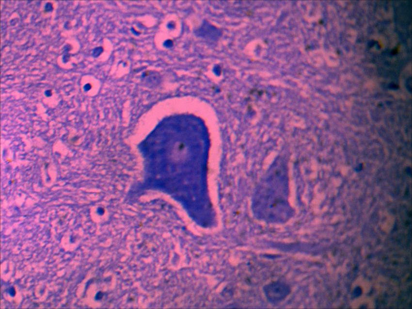
362                                        AJEIGBE SHERIFF OLAWALE et al.

like cemeteries, pastures, parks, or rocky country.           Zaria and its environs. They were acclimatized
In Nigeria (especially south Nigeria), the squir-             for one month in standard laboratory cages in
rel is one of the most commonly consumed small                the animal pen of the Department of Veterinary
mammal species (bush meat) by rural dwellers as               Anatomy, Ahmadu Bello University, Zaria,
a supplementary protein diet[1,5]. They are also              Nigeria. The animals were given access to food
used by Nigerian farmers in cultural ceremonies,              and water ad libitum throughout the experimental
for medicinal purposes (i.e. components of a anti-            period. They were physically examined during
poison drug and as ingredients in the prevention              the pre-experimental period and only apparently
of convulsion in children and also used to en-                healthy ones were utilized.
hanced fertility in men) [5, 6].
                                                              Morphometric parameters
    The pons is a broad, horseshoe shaped                         The body weights of each squirrel were
portion of the brain consisting of large bundles              obtained using a weighing balance model JJ1000,
of nerve fibers. The pons connects the two halves             USA with a capacity of 1000g and sensitivity
of the cerebellum and can be broadly divided                  of 0.01g. The mean length, width and depth of
into two parts; the basilar part, located ventrally           the pons and medulla oblongata were obtained
and the pontine tegmentum, located dorsally [7].              with a vernier caliper (MG6001DC, General
A number of cranial nerve nuclei are present in               Tools and Instruments Company, New York;
the pons which includes the pontine and motor                 sensitivity: 0.01mm). Gross pictures were taken
nucleus of the trigeminal nerve (CN V), located               using canon digital camera power shot (SX170
in the mid-pons while abducens nucleus (CN VI),               IS) with 64-megapixel sensor (focal length: 28-
facial nerve nucleus (VII) and vestibulocochlear              448mm, 7.5cm (3.0”) TFT. Histological pictures
nuclei (vestibular nuclei and cochlear nuclei)                were taken using light microscope (Amscope,
(VIII) are located at the lower end in the pons               T120B) and a digital microscope camera (DCM
[8].. The pons also contains nuclei that relay                510 megapixel, ScopePhoto® China) at ͯ40, ͯ100,
signals from the forebrain to the cerebellum,                 ͯ250, ͯ400.
along with nuclei that deal primarily with sleep,
respiration, bladder control and posture [9].                 Brain extraction
The medulla oblongata is the conical part of the                  Each squirrel was euthanized using ketamine
brain that extends from the pons to the medulla               anesthetic at (80mg/kg bw) [4]. Each brain was
spinalis [10]. It is broad above where it joins the           perfused (intra-cardiac route) with 10% phosphate
level of the foramen magnum [11]. The medulla                 buffered formalin. Each skull was exposed after
oblongata is divided into two parts: an opened                skinning and stripping off all the facial muscles
part (closed to the pons) and a closed part (closed           within 30 minutes of euthanasia. Craniotomy
to the spinal cord), the most rostral part of the             was carried out through the calvaria to exposed
medulla oblongata is the corpus trapezoideum                  the dura matter, which was later cut with curved
[10]. Although, several studies have been                     pointed scissors. The falx cerebri and tentorium
conducted on the pons and medulla oblongata                   cerebelli were pulled from the longitudinal and
of other rodents, little or no research has been              transverse fissures by gentle traction. The cerebral
carried out on the pons and medulla oblongata                 vein was transected and at this stage, the brain
of squirrels, thus this study will provide baseline           was still in the cranium fixed in 10% phosphate
data for detailed neuroanatomical descriptions                buffered formalin for two days to enhance easy
of the pons and medulla oblongata of ground                   extraction as described by Ramaswamy [4].
squirrels. This will in turn provide further                  Harvest of Pons and Medulla oblongata
laboratory research opportunities using ground                    The pons and medulla oblongata were
squirrels as models and also provide additional               isolated from the rest of the brain by gently
information required for successful breeding as               pulling apart the two cerebral hemispheres at
ground squirrels are a good source of protein for             the occipital lobe to expose the corpus callosum.
domestic farmers in Nigeria.                                  The entire corpus callosum together with septum
Materials and Methods                                         pellucidum and the body and rostral commissure
                                                              of the fornix were severed in the midline and
Experimental animals and management                           this separates the cerebrum from the brain-
   Twenty (20) African striped ground squirrels               stem and cerebellum. Then the floculli of the
(10 males and 10 females) were used for this study.           cerebellum was raised manually to expose the
The animals were captured live from the wild in               cerebellar peduncle which was severed starting
Egypt. J. Vet. Sci. Vol. 52, No. 3 (2021)
HISTOMORPHOLOGICAL STUDY OF THE PONS AND MEDULLA OBLONGATA OF AFRICAN …                                   363

with the laterally located brachium restiformis,             blocks were sectioned at 5-6 µm thickness of
followed by the middle brachium pontis and then              slice using a rotary microtome. Histological
the brachium conjuctivum. The brain-stem was                 sections were stained with hematoxylin and eosin
free from the cranial nerves by simple trimming              [12]. Cresyl fast violet stain was used for Nissl
using a scalpel blade, an incision was made at the           substance and nerve nuclei evaluation [13].
transverse fissure between the pons and caudal
colliculi to isolate the pons and medulla oblongata          Results
from the midbrain while incision made at the                 Histological features of Pons and medulla
pontomedullary junction separates the medulla                oblongata of the squirrel brain
oblongata from the pons.                                         Nucleus facial: The nucleus was seen dorsal
Histology                                                    to paramedian pontine reticular formation and
   The pons and medulla oblongata of squirrel                caudal to trigeminal nerve as group of large mul-
were fixed at 10% neutral buffered formalin,                 tipolar nuclei (Plate V). It is a relatively large
dehydrated in a graded series of alcohol, cleared            nucleus with large amount of cytoplasm and the
in xylene, then embedded in paraffin wax. The                nissyl substance was clumpy and densely stain, its
                                                             nucleolus is central (Plate XII).

Plate V: Coronal section of the pons. A: Nucleus abducens, B: nucleus trigemini, C: Nucleus facial D: paramedian
       pontine reticular formation, E: trigemino-thalamic tract. (cresyl violet stains, x40).

Plate XII: Coronal section of the pons showing neurons of motor nucleus of facial nerve (red) and neuron of motor
         nucleus of abducent nerve (black). Cresyl violet stain x400.
                                                                           Egypt. J. Vet. Sci. Vol. 52, No. 3 (2021)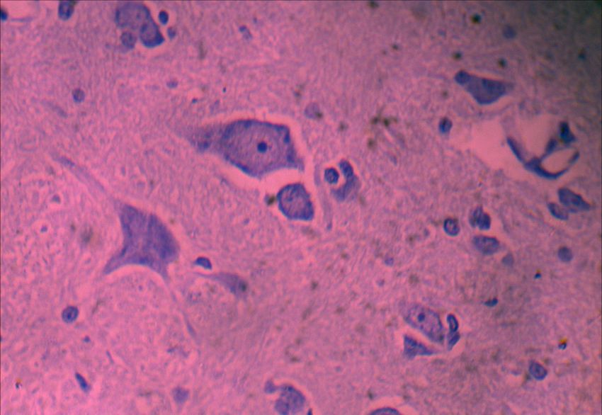
364                                        AJEIGBE SHERIFF OLAWALE et al.

Nucleus abducens: It was a conspicuous nucleus                and uniformly organized neurons forming a
found as a cluster of medium size neurons and                 bean shape (Plate I) and (Plate V). The nucleus
located lateral to the trigeminal nucleus (Plate V).          is a mixture of the medium and large nuclei. It
The nucleus was a mixture of small and medium                 was bounded caudally by spinal trigeminal
size neurons with a large cytoplasmic nuclear ratio,          tract and tractus pyramidalis, dorsolaterally by
densely stain with central nucleolus (Plate VI).              nucleus hypoglossi, medially and ventromedially
                                                              by nucleus ambiguus and reticular nucleus of
Nucleus and tract of trigeminal nerve: The                    medulla ventral respectively (Plate I).
nucleus trigemini on coronal view are evenly

Plate VI: Coronal section of medulla oblongata showing gigantocellular reticular nucleus. Note its large and varied
          cytoplasmic nucleus ratio. Cresyl violet stain x250.

Plate I: Coronal section of medulla oblongata at 0.8mm rostral to the obex. A: nucleus spinalis and.trigemini, B:
          nucleus hypoglossi, C: linear nucleus of medulla, D: nucleus ambiguus, E: nucleus cochlearis, F: reticular
          nucleus of medulla dorsal, G: reticular nucleus of medulla ventral, H: tractus pyramidalis. (cresyl violet
          stain, x40).

Egypt. J. Vet. Sci. Vol. 52, No. 3 (2021)
HISTOMORPHOLOGICAL STUDY OF THE PONS AND MEDULLA OBLONGATA OF AFRICAN …                                  365

Vestibular nucleus: These were small size                   found ventral to nucleus tractus solitarius with a
nucleus located on the ventrolateral surface of             pyramidal shape. It had a 1:1 nucleus cytoplasmic
the pontomedullary junction. The nucleus was                ratio and slight eccentric nucleolus and coarse
found medial to the cerebellar peduncle and also            nissyl substances (Plate X).
ventral to pontocerebellar tract and was of spindle
(fusiform) shape (Plate IV).                                Nucleus olivaris: This nucleus was well stained
                                                            on the ventral coronal section of myelencephalon
Nucleus ambiguus: This nucleus was most                     and appeared as a large and multipolar neuron
prominent at about 0.8mm rostral to the obex                with a centrally located nucleolus and large
where it appeared as a circular group of few (15-           cytoplasm. The nucleus varied between conical
20) oval nuclei. Its neurons were compacted, large          and oval shape with a clumpy nissyl substances
and were found medial to nucleus spinal trigemini           (Plate VIII).
(Plate I). However, more rostrally the nucleus was

Plate IV: Coronal section of pontomedullary junction. J: vestibular nucleus, K: pontocerebellar tract, I: middle
           cerebellar peduncle, L: pontocerebellar fibers (Cresyl violet stain x250).

Plate X: Coronal section of medulla oblongata at 0.9mm rostral to the obex showing neurons of nucleus ambiguus.
         Note the 1:1 nucleus cytoplasmic ratio and slightly eccentric nucleolus. Cresyl violet stain x250.

                                                                          Egypt. J. Vet. Sci. Vol. 52, No. 3 (2021)
366                                        AJEIGBE SHERIFF OLAWALE et al.

Plate VIII: Corona section of medulla oblongata showing neurons of the nucleus olivaris. Note the large and
           multipolar neuron in a variety of oval and conical shape. Cresyl violet stain x400.

Nucleus     cochlearis:    This    was     well               Nucleus solitary tract: It was a spheroidal nucleus
demonstrated in histological section of                       found at 0.9mm rostral to the obex on the open
myelencephalon that was 0.8mm rostral to the                  dorsomedial medulla oblongata. It was faint and
obex. It was composed of large elliptical group               lied medial to the dorsal motor nucleus of the
of nuclei with eccentric nucleoli (Plate IX).                 vagus nerve (Plate II).
It was located on the dorsolateral portion of                 Nucleus hypoglossi: This nucleus was found
the medulla oblongata, dorsal to the reticular                dorsal to nucleus ambiguus and dorsomedial to
nucleus of the medulla and lateral to linear                  the nucleus spinalis trigemini. It was found as
nucleus of the medulla (Plate I). Its neurons                 cluster of discoid or spheroidal and multipolar
are mixture of pseudounipolar and unipolar                    neurons (Plate II). The neuron has a mixture of
with long dendrites (Plate IX).                               small and large nuclei with a small cytoplasm and
                                                              centrally located nucleolus (Plate XI).

Plate IX: Corona section of medulla oblongata showing neurons of the nucleus cochlearis. Note the large
          pseudounipolar and unipolar neuron with elliptical shape. Cresyl violet stain x250.

Egypt. J. Vet. Sci. Vol. 52, No. 3 (2021)
HISTOMORPHOLOGICAL STUDY OF THE PONS AND MEDULLA OBLONGATA OF AFRICAN …                                    367

Plate II: Coronal section of medulla oblongata at 0.9mm rostral to the obex. A: dorsal motor nucleus of vagus,
          B: nucleus of the tractus solitarius, C: tractus solitarius, D: nucleus cuneatus, E: nucleus ambiguous,
          F: nucleus hyoglossi, G: nucleus of roller, H: lateral reticular nucleus, I: paragigantocellular reticular
          nucleus. (Cresyl violet stain, x40).

Plate XI: Coronal section of medulla oblongata at 0.8mm caudal to the obex, showing neurons of nucleus hypoglossi.
          Note the mixture of small and large nuclei and centrally located nucleolus. Cresyl violet stain X250.

Nucleus raphe obscures, nucleus cuneatus and                  very distinct nuclear cell group of the lateral re-
lateral reticular nucleus: These nuclei were well             ticular nucleus which was easily recognized with
represented in histologic section that was 0.8mm              the nissyl stained section. The parvocellular was
caudal to the obex. The nucleus raphe obscurus                a cell rich region containing small fusiform and
migrate medially to occupy a midline position be-             multipolar neurons (Plate III). The lateral reticular
tween paramedian reticular nucleus. The cuneate               nucleus in this section are characterized by large
nucleus was large and consist of scattered neu-               nucleus with eccentric nucleolus and a very small
rons. The most striking feature of the section is the         cytoplasmic nuclear ratio (Plate XIII).

                                                                             Egypt. J. Vet. Sci. Vol. 52, No. 3 (2021)
368                                        AJEIGBE SHERIFF OLAWALE et al.

Plate III: Coronal section of medulla oblongata at 0.8mm caudal to the obex. A: nucleus gracilis, B: nucleus
           cuneatus, C: Raphe obscurus nucleus, D, E: paramedian reticular nucleus, F: pyramidal decussation,
           G: inferior olivary, H: medial longitudinal fasciculus, I: nucleus hypoglossi, J: lateral reticular nucleus,
           K: lateral reticular nucleus parvocellular. (cresyl violet stain, x40).

Plate XIII: Coronal section of medulla oblongata at 0.8mm caudal to the obex showing neuron of nucleus reticu-
           laris lateralis, note the spheroidal and oval shape. Cresyl violet stain x400.

Gigantocellular reticular nucleus: This has                     Nucleus of corpus trapezoideum: It was an
a coarse nissyl granules and large centrally                    elongated nucleus that was well represented
located nucleus with central nucleolus. It                      on the dorsal surface of the closed medulla at
consists of two populations of neurons which                    high magnification (Plate VII). It has coarse and
are largely multipolar and intensely stained                    densely stained nissyl substances, large cytoplasm
with large nucleus (Plate VI).                                  and centrally located nucleolus.

Egypt. J. Vet. Sci. Vol. 52, No. 3 (2021)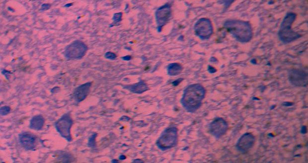
HISTOMORPHOLOGICAL STUDY OF THE PONS AND MEDULLA OBLONGATA OF AFRICAN …                                   369

Plate VI: Coronal section of medulla oblongata at 0.8mm caudal to the obex showing gigantocellular reticular
           nucleus. Note its large and varied cytoplasmic nucleus ratio. Cresyl violet stain x250.

Plate VII: Corona section of medulla oblongata showing neurons of nucleus dorsalis corporis trapezoidei. Note its
           elongated shape, large cytoplasm and coarse nissyl substance. Cresyl violet stain x400.

                                                                           Egypt. J. Vet. Sci. Vol. 52, No. 3 (2021)
370                                        AJEIGBE SHERIFF OLAWALE et al.

Discussion                                                    structures of the head and it is the motor nerve for
                                                              mastication and contain proprioceptive fibers.
    The pons and medulla oblongata are the caudal
                                                              The lateral reticular nucleus was conspicuously
extend of the brain-stem. The pons was convex and
                                                              located caudal to the obex almost at the level
smooth ventrally and triangular dorsally while the
                                                              of spinomedullary junction suggesting that
medulla oblongata was open rostrally and closed
                                                              the squirrel has fine motor function of posture,
caudally. The presence of conspicuous olivary
                                                              scratching and grasping with their limbs, this is
prominence, vestibular nucleus, pyramidal tract
                                                              in agreement with the findings of Bror and Carl-
and the histological characteristics of the olivary
                                                              fredrik [22] who reported a conspicuous lateral
nucleus in this study shows that squirrels have
                                                              reticular nucleus in cats which he said is a major
fine voluntary skill, good motor coordination and
                                                              precerebellar centre of mossy fiber information to
balance. This is similar to what was reported in
                                                              the cerebellum from the spinal cord that is district
grasscutter, African giant rats and wistar rat by
                                                              from direct spinocerebellar paths and provide
Jones et al.[14]. Also, this finding is contrary to
                                                              the cerebellum with segregated information
the report of Sricharoenvej et al.[15]. in flying fox
                                                              from several spinal system controlling posture,
who reported poorly developed olivary nucleus
                                                              reaching, grasping, location and scratching. The
and attributed it to their poor motor coordination.
                                                              nuclei of reticular formation (raphe obscures,
Squirrels have well-developed pyramidal tracts,
                                                              gigantocellular reticular nucleus, pontine
which is contrary to the findings of Majewska-
                                                              reticular nucleu and parvocellular reticular
michaska [16] who reported small pyramidal
                                                              nucleus) observed in this study is suggestive of
tract in guinea pigs. It can be presumable that
                                                              good motor function, maintain good behavioral
squirrels have good motor coordination as in
                                                              arousal and consciousness (sleep, alert, rapid eye
African giant rats and grasscutter if not better,
                                                              movement), this is in agreement with the findings
such as symmetrical limb movement, climbing,
                                                              of Mcnamara and Nunn [23] who reported that
fast runners and manus manipulation during
                                                              the nucleus of reticular formation plays vital
grasping and sitting with hind limbs. The nucleus
                                                              role in maintaining behavioural arousal and
solitary tract was found rostral to the obex, this
                                                              consciousness in mammals. Reticular formation
is in contrary with the findings of Ibe [17] who
                                                              nucleus was observed with some projections
reported that solitary tract nucleus of African
                                                              to the thalamus and cerebral cortex similar to
giant rat is located in the obex. The hypoglossal
                                                              what was reported by Thorpy and Yager [24],
nucleus was found rostral to the obex at the level
                                                              these projections also have projections exert
of the 4th ventricle, this is in agreement with the
                                                              some control on consciousness like alertness and
findings of Ajayi et al.[18]. who reported that
                                                              sleep. injury to this reticular formation nuclei
the hypoglossal nucleus of grasscutter is located
                                                              could result in irreversible coma[24]. The raphe
rostral to the obex at the level of the 4th ventricle.
                                                              nucleus observed in this study is suggestive of
Defects in hypoglossal nucleus may result in
                                                              good visual acuity, this is in agreement with the
uncoordinated vocalization, swallowing and
                                                              findings of Lewy et al.[25] who reported that
chewing. The hypoglossal nucleus is devoid of
                                                              raphe nucleus plays a vital role in rapid and non-
numerous subdivisions but contains multipolar
                                                              rapid eye movement, and also in sleep/wake
neurons that are mixture of both large and small,
                                                              cycle hormone secretion in rodents. The presence
this is in agreement with the findings of Arora and
                                                              of conspicuous nucleus abducens is suggestive
Prakash [19] who reported that the hypoglossal
                                                              of a good visual acuity, this is in agreement with
nucleus of albino rats is devoid of numerous
                                                              the findings of Alexander [8] who reported that
subdivisions. The trigeminal nucleus was very
                                                              the main function of abducens is to generate
conspicuous on the lateral aspect of the medulla
                                                              coordinated movement of both eyes in the
oblongata, this is in agreement with the findings
                                                              same direction, however, abducens nucleus is
of Dean et al. [20] who reported that trigeminal
                                                              composed of motor and interneurons. The motor
nucleus is very conspicuous on the lateral aspect
                                                              neuron drives the contraction of ipsilateral rectus
of the medulla oblongata of mammals. The
                                                              muscle and contraction of this muscles rotate the
trigeminal nucleus has three subdivisions with
                                                              eyes outward (abduction) while the interneurons
no distinct bother lines, this is in agreement with
                                                              relay signals to the contralateral occulomotor
the findings of Young et al. [21] who reported
                                                              nucleus where motor neurons drives the
that trigeminal nucleus has three subdivisions in
                                                              contraction of ipsilateral medial rectus muscles
mammals. Trigeminal nerve supply sensations
                                                              which rotates the eyes inward (adduction).
to the face, mucous membrane and other
Egypt. J. Vet. Sci. Vol. 52, No. 3 (2021)
HISTOMORPHOLOGICAL STUDY OF THE PONS AND MEDULLA OBLONGATA OF AFRICAN … 371
Conclusion 8. Alexander, D.L. Veterinary Neuro-Anatomy and
Clinical Neurology (2nd ed.), pp.16-19, 98-102
The presence of distinct nuclei in the pons (1983).
and medulla oblongata of African striped ground
squirrels gives them fine voluntary skills with 9. Saladin, K. S. Anatomy and Physiology: the unity
good motor coordination and balance and a good of form and function. Dubuque, IA: McGraw-Hill
visual acuity for improved diurnal adaptation. (2007).
Funding statement 10. Al-shehri, H.M Cranial nerve nuclei in the me-
This research did not receive any specific grant dulla oblongata. Retrieved from scribe at http://
from funding agencies in the public, commercial, www.scribd.com/doc/512776/medulla oblongata
or not-for-profit sectors. nucleus-final (2007).
Conflict of interest: 11. Inderbir, S. Essentials of Anatomy. Jaypee broth-
No conflict of interest ers’ medical publishers (p) limited. New delhi,
6,219-224 (2003).
Ethical Statement
The experimental procedures were approved 12. Kiernan, J.A. Histochemistry of staining methods
by the Institutional Animal Ethics Committee with for normal and degenerating myelin in the central
an approval number of ABU/CAUC/2016/038. and peripheral nervous systems. J. Histotech.,
The present investigation was carried out at Fac- 30(2),87-91 (2007).
ulty of Veterinary Medicine, Ahmadu Bello Uni-
13. Drury, R. A. B. Carlton’s histological tech-
versity Zaria, Nigeria.
nique. Ann. Inter. Med., 67,233-237 (1967).
Authors contributions
14. Jones, N., Stolz, T., Batinic, C. and Caston, J. Ef-
All authors contributed substantially to the
fects of lesion of the inferior olivary complex in
design, acquisition, and analysis of the study.
learning of equilibrium behavior in the young rat
Writing and revising for intellectual consumption
during ontogenesis: Total lesion of the inferior ol-
were also collectively done.
ive by 3-acetylpyridine. Brain Res., 697,216-224
References (1995).
1. Adeola, M.O. and Decker, E. Wildlife utilization 15. Sricharoenvej, S., Niyomchan, A., Lanlua, P., Pi-
in rural Nigeria. Proceedings of the International yawinijwong, S. and Roongruangchai, J. Micro-
symposium and conference on wildlife manage- vasculature of the medulla oblongata in the lyle,
ment in Sub-Saharan Africa, Harare, Zimbabwe, s flying fox (Pteropus lylei). Anat. Hist. Embryol.,
pp. 512-521 (1987). 37,401-407 (2008).
2. Whatton, F. Squirrel of the world. JHU press Bal- 16. Majewska-michaska, E. Vascularisation of the
timore, UK, p. 8 (2012). brain in guinea pigs III. Vascular architecture of
the medulla oblongata, pons and cerebellum. Folia
3. Wilson, D.E. and Reeder, D.M. Mammal species Morphol., 56,41-44, (1997).
of the world. A taxonomic and Geographic Refer-
ence (3rd ed.), JHU press Baltimore, UK (2005). 17. Ibe, C.S. Anatomic study of the mesencephalic
tectum and myelencephalon in the African giant
4. Ramaswamy, S. Removal of the brain. A new rat (cricetomys gambianus, water house-1840).
procedure, Italian J. Anat. Embryol., 82, 105-110 Vet. Res., 97-99 (2010).
(1978).
18. Ajayi, I.E., Ojo, S.A., Onyeanusi,B.I., George,
5. Ajayi, S.S. Utilization of forest wildlife in West B.D., Ayo, J.O., Salami, S.O. and Ibe, C.S. Gross
Africa. FAO, Rome, p.79 (1979). Observations and Morphometry of the Medulla
6. Adeola, M.O. Importance of wild animals and Oblongata of the African Grasscutter (Thryono-
their parts in the culture, religious festivals, and mys swinderianus). JVA, 4(1),5-8(2011).
traditional medicine of Nigeria. Environ. Conser., 19. Arora, L. and Prakash, R. Animal studies: Cyto-
19,125-134 (1992). morphometry of hypoglossal nucleus in rats. In-
7. Krinke, G. J. Neuropathologic screening in ro- dian Journal for the practicing Doctor, 3,11-12
dents and other species. J. Am Coll Toxicol., 8, (2006).
141-146 (1989).
Egypt. J. Vet. Sci. Vol. 52, No. 3 (2021)372 AJEIGBE SHERIFF OLAWALE et al.
20. Dean, C., Geiger, L.K., Spertel, B.M., Ohtake, P.J.
and Forster, H.V. An anatomic atlas of the me-
dulla oblongata of adult goat. J. Appl. Physiol.,
87(3),1220-1229 (1999).
21. Young, B., Lowe, J.S., Stevens, A. and Health,
J.W. Wheaters functional histology. A text and
color atlas: (5th ed.), Chur. livingstone, Elsevier,
122p. (2006).
22. Bror, A. and Carl-fredrik, E. Lateral reticu-
lar nucleus. A precerebellar centre providing
the cerebellum with overview and integration
of motor functions at system level. J. Physiol.,
591(22),5453-5458 (2013).
23. Mcnamara, P.R. and Nunn, C.L. Evolution of
sleep phylogenetic and functional perspectives.
Cambridge University, Cambridge (2010).
24. Thorpy, M.J. and Yager, J. The Encyclopedia of
Sleep Disorder (2nd ed.). New York facts on file.
ISBN 9780816040896 (2001).
25. Lewy, A., Weh, T., Godwin, F. and Markey, S.
Light suppresses melatonin secretion in humans.
Sci., 210 (4475),1267-1269 (1980).
Egypt. J. Vet. Sci. Vol. 52, No. 3 (2021)You can also read