Emergency - Quality, Education and Safety Teleconference For smaller Eds - Anne Walton
←
→
Page content transcription
If your browser does not render page correctly, please read the page content below
Emergency - Quality, Education and
               Safety Teleconference

                           For smaller Eds

Anne Walton
Advanced Trainee
Emergency Care Institute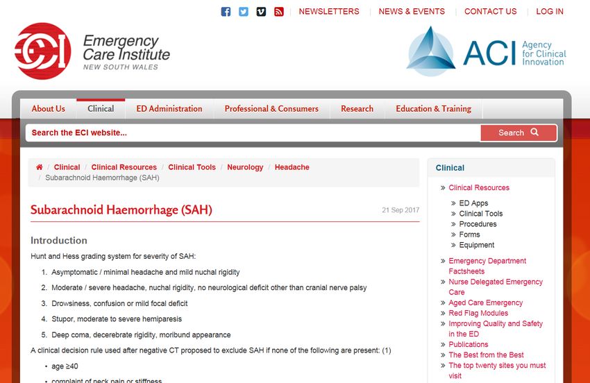
Thanks for joining
    House rules
Agenda • 3 Case discussions - Headache • Review clinical context • Learning points and take home messages • Next meeting • We encourage participation, comments or questions throughout – we want to reflect YOUR needs and interests
Case 1
• 46 male, presented to hospital A, on Sunday @1722hrs
• PC: Frontal headache, nausea, neck stiffness and
  vomiting at triage
• Nil significant PMHx given at triage
• Afebrile, strong regular pulse, assigned ATS Category 3
Case 1 - assessment
• Seen by JMO 2055hrs
• Mild, gradual onset headache that morning whilst lying
  down
• 1400hours, worsening of headache – ‘agony’
• Vomited at home, associated with photophobia, neck
  stiffness, post occular pain and subjective fever
• Pain 8-9 earlier that day, currently 4-5/10
Case 1 – pmhx/ exam
• No history migraines, but recurrent sinusitis reported
• No foreign travel/ coryzal symptoms
• A headache 1/52 prior, which lasted
Thoughts
   ??
Case 1 – progress
• DW FACEM – need CT +/- LP
• “CT showed no obvious abnormalities”. Advised that
  absence of photophobia means not SAH
• Given ibuprofen/ oxycodone, t/f to ESSU for analgesia
• 0530, pt wanted to leave – seen by RMO, d/c with
  panadeine forte and ibuprofen and told to contact GP
Case 1 – re-present
• 15 days later, referred by GP to hospital B for ‘acute
  work up of headaches’
• No abnormal clinical signs, 3/52 history of headache,
  worse during sexual activity and felt like ‘being hit with
  a bat’. Radiation to occiput/ lower back
• Seen by JMO and DW medical registrar – provisional
  diagnosis chronic headache on background of overuse
  of Panadeine forte.
• No need for LP. Previous CT ‘normal’ – will not repeat
• Discharged
Thoughts
   ??
Case 1 – catastrophe
• 25 days later – collapse/ headache whilst walking
• Decorticate positioning and obstructive breathing
• Large left frontal ICH
• TF to ICU – unsurvivable haemorrhage, declared brain
  dead. Proceeded to organ donation
• Review of first CT scan showed ‘subtle signs of
  subarachnoid blood and an anterior communicating
  artery aneurysm’
What contributed to this outcome?
System
• Diagnostic anchoring
• Premature closure

Skills
• CT reporting
• Understanding the literature on SAH

Staffing
• Senior supervision and review – ‘represent’ = high
  risk
Would you have
 managed this
  differently
       ??
The problem • Up to 5% of ED presentations are with headache • Vast majority of pathologies are benign – how do we screen for a identify the sinister ones?
Subarachnoid haemorrhage –
       the evidence
• Still controversial – who needs an LP in SAH?
• CT within 6 hours of onset and interpreted by
  neuroradiology = up to 100% sensitive
• After 6 hours, sensitivity drops – negative CT under 6
  hours, discuss pros/cons with patient
• LP may be helpful in providing alternative diagnosis
• Lack of symptoms/ signs at time of assessment is
  NOT predictive for excluding SAH
ECI clinical tool
Case 2
•   15 year old Aboriginal male to rural ED
•   Complex psychosocial history
•   Daily marijuana use, irregular school attendance
•   Headache/ fever/ nausea/ vomiting/ meningism
Case 2 – initial management
• Treated empirically for meningitis – iv ceftriaxone and
  acyclovir
• LP from ED – opening pressure 28.5mmH2O, clear,
  colourless fluid, negative fungus India Ink stain, Entero-
  Virus PCR positive
• Treated for viral encephalitis – discharged when
  clinically improved
Thoughts
   ??
Case 2 - progress
• Re-presented 5 more times with ongoing intractable
  headache, neck stiffness, vomiting
• Had one presentation by ambulance with headache and
  haemoptysis
• Haemoptysis not documented
• Handed over to adult medical team as deemed
  unsuitable for paediatric admission given social
  situation
• Repeat LP – no significant findings. ICP pressure within
  range
Case 2 - progress
• Re-presented a further 4 times, eventually admitted
  under adult cardiologist – t/f care to tertiary paediatric
  neurologist due to intractable pain
• Following t/f – had repeat LP and grew Cryptococcal
  Gatti
• MRI showed cerebellar abscess and evidence of raised
  ICP
• Left lower lung lobe mass was identified with
  cryptococcal origins
Case 2 – learning points
• Cognitive bias
- Psychosocial situation – rationale for re-presentations
  not fully addressed
- Found one positive test (enterovirus), diagnostic
  anchoring
• Failure to review all evidence – eg abnormal ICP
• Communication failure
- Haemoptysis was important clue, confirmation bias
• High risk population – high index of suspicion
Case 2 - outcome
• TF back to rural hospital and then d/c home 3 days later
  with outpatient follow up
• Represented to rural ED 2 days later with seizures and
  raised ICP
• TF back to tertiary hospital and VP shunt inserted
• Uncomplicated recovery
• Re-presented to ED once for r/v after trip and fall with
  minor head injury
• No further presentations to any ED facility within LHD
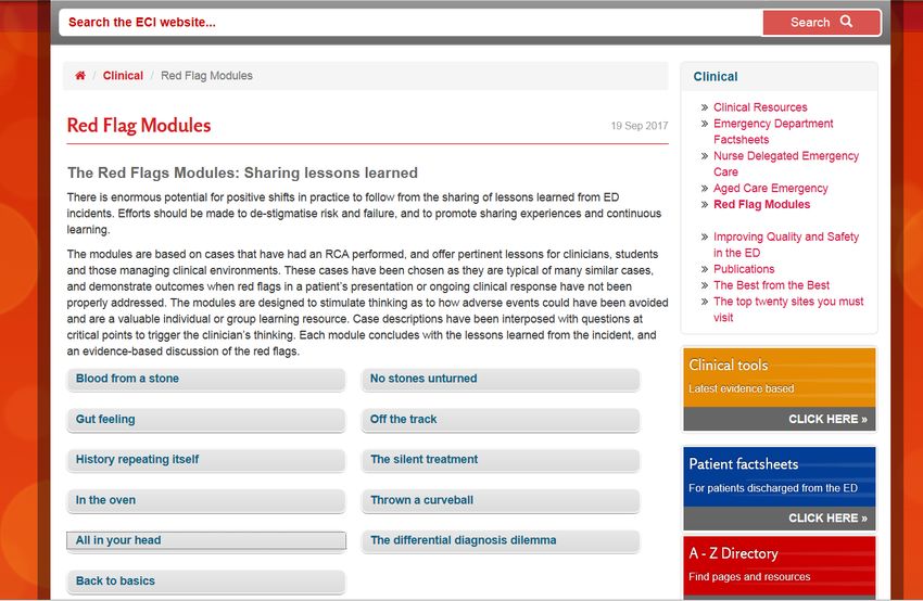
Red flag modules
• There are often recurring themes when reviewing
  adverse events
• Not all adverse events or poor outcomes may be
  predicted or prevented, but there may often be ‘red
  flags’
• Re-presentation with the same symptom is a ‘red
  flag’ and the patient should be reviewed carefully,
  ideally by a senior medical officer and all
  investigations and results revisited and reviewed
ECI Red flag modules
Case 3
• 32 year old female, 15 weeks pregnant
• 0157hrs: BIBA in with sudden onset of severe headache
• With ambulance crew, patient was able to walk, equal
  strength all limbs
• No history of headaches
• Ambulance officers administered 5mg morphine iv for
  pain
• ATS category 3 and placed in side room
• GCS at triage = 14/15
Case 3 - progress
• 0215 – HR 75, BP 138/74, RR 10, sats 97% RA
• Nursing staff noted patient was pale, rousable to voice
  but not opening her eyes. Breathing heavily and unable
  to squeeze hand to command
• Transferred to resuscitation room
• 0228hrs, RR 7, GCS 7/15
• 0256 – collateral history from husband: patient vomited
  and complained of blurred vision at home. Examined by
  medical officer
Differentials at this
       point
        ??
Case 3 - progress • 0330: GCS 6/15 • 0356: 400micrograms naloxone administered • 0416: GCS 6/15 and RR 11 • 0514: right pupil more dilated than left. MO aware. 400micrograms naloxone administered • 0530: GCS 5/15 • 0600: CT brain showed large cerebellar haemorrhage
What contributed to this Cognitive error outcome? • IV Morphine attributed as cause for respiratory depression/ drowsiness – diagnostic anchoring • Failure to review all evidence eg husband’s information – information synthesis Patient factors • Pregnancy + radiation – what is best for mother is best for foetus Skills/ staffing • “Ostrich effect” – 0157 arrival CT scan 0600
Summary • Headache is common presentation in ED • High index of suspicion is required • Severity of pathology does not necessarily correlate well with degree of pain or absence of neurological deficit • What processes or pathways do you have in your ED that might help navigate this difficult diagnosis? • ECI clinical tool (neurology headache): https://www.aci.health.nsw.gov.au/networks/eci/clinical/clinical- resources/clinical-tools/neurology/headache
E-QuESTs so far
•   Atypical Chest Pain - ACS
•   Sepsis in the elderly
•   Abdominal Pain in the elderly - AAA & Ischaemic gut
•   Acute scrotum
Suggested future topics
•   Minor head injury
•   Paediatrics
•   Eye emergencies
•   Transfer/ retrieval issues, including clinical handover

• Any feedback on these topics or other suggestions?
You can also read