ENAMEL THICKNESS MEASUREMENTS ON 3D RECONSTRUCTIONS OF TEETH FOR PALEONTOLOGICAL APPLICATIONS - The International Archives of the ...
←
→
Page content transcription
If your browser does not render page correctly, please read the page content below
The International Archives of the Photogrammetry, Remote Sensing and Spatial Information Sciences, Volume XLIV-2/W1-2021
4th Int. Worksh. on “Photogrammetric & computer vision techniques for video surveillance, biometrics and biomedicine”, 26–28 April 2021, Moscow, Russia

   ENAMEL THICKNESS MEASUREMENTS ON 3D RECONSTRUCTIONS OF TEETH
                FOR PALEONTOLOGICAL APPLICATIONS

                              A.V. Gaboutchian 1∗, V. A. Knyaz2,3 , E.N. Maschenko4 , D.V. Korost5 , A.A. Kudaev5
                     1
                     Peoples’ Friendship University of Russia, 117198, Moscow, Russia – armengaboutchian@mail.ru
                2
                 State Research Institute of Aviation System (GosNIIAS), 125319 Moscow, Russia – knyaz@gosniias.ru
                             3
                               Moscow Institute of Physics and Technology (MIPT), Dolgoprudny, Russia
          4
            Borissiak Paleontological Institute of Russian Academy of Sciences, 117647, Moscow, Russia – evmash@mail.ru
        5
           Faculty of Geology, Moscow State University, 119234, Moscow, Russia – dkorost@mail.ru ; a.a.kudaev@gmail.com

                                                            Commission II, WG II/10

KEY WORDS: enamel thickness, odontometry, micro-computed tomography, palaeontology, LangTrank cave, Sunghir

ABSTRACT:

Findings of teeth play a significant role in palaeoanthropology. And excavations in Vietnamese LangTrank cave serve as a vivid example
and evidence of this statement. Teeth constitute the majority of the paleontological material dated to Middle and Late Pleistocene
periods. This is to some extent the result of dietary preferences of porcupines as these rodents include in their diets bones of animals
however avoiding extremely hard coronal parts of teeth. Under such circumstances teeth serve a key to taxonomic differentiation of
findings as genetic analysis is often hindered by a lack of preserved DNA at such dating of material. However morphological analysis
is difficult in some cases either, as teeth can be worn out or broken. In that case enamel thickness measurements become an effective
study instrument as this feature varies between species. In the current study two teeth with clear signs of expressed dental wear,
presumably upper fourth premolars of wild boar required more detailed analysis. Thus they were reconstructed after micro-computed
tomography scanning similarly to other upper teeth picked for comparison: orang-utan tooth from the same location and two teeth
from the Upper Palaeolithic Sunghir (they have been scanned earlier). This study required new approaches to image processing and
measurement methodology due to marked attrition of the samples. The workflow and results of enamel thickness assessments which
facilitated taxonomical differentiation of the findings are presented in the article.

                         INTRODUCTION                                         new impetus to enamel thickness measurement has been given
                                                                              with the introduction of 3d techniques, especially those based
Enamel thickness measurements have a well-known historical and                on micro-CT. They enable greater accuracy in reconstruction of
methodological background. They serve to shed light on taxo-                  external and internal surfaces of enamel. In addition the stud-
nomic differentiation, adaptation and other issues referred to a              ied samples’ integrity can be completely preserved regardless of
wide variety of studies in natural science (Alvesalo et al., 2009;            numbers and directions of obtained sections.
Olejniczak et al., 2007; Smith et al., 2009; Zanolli et al., 2017).
And notably in paleontological research, when teeth can account               It should be noted that teeth are difficult object to measure. Of
for the majority of excavated fossil material (largely due to their           course, there are morphological regularities which are put in the
durability), the mentioned assessments are of high demand. The                bases of dental measuring techniques. At the same time a lot
obtained measurements results, in line with dental or other mor-              irregularities, curvatures with complex topography and a large
phological studies, are able to facilitate differentiation of the find-       number of varieties exist as well. Under such circumstances re-
ings. However in some cases, like in the present study, enamel                sults become dependent to a large extent on orientation of the
thickness measurements start playing the leading role when teeth,             teeth being measured. Hence nearly all studies of sizes of teeth,
due to their condition, loose their important morphological fea-              including enamel thickness measurements, require correct orien-
tures.                                                                        tation of the measured objects.

There are principally two alternative approaches to enamel thick-             Usually expert based traditional approaches are used in odon-
ness measurements. One is sectioning teeth and the other requires             tometric studies. However, orientation of teeth and measure-
x-ray application. The first of traditionally implemented tech-               ments on contours are a matter of concern and discussions in
niques is destructive as direct sectioning of studied samples de-             odontological community (Suwa and Kono, 2005; Zanolli et al.,
stroys them. Taking into consideration that paleontological find-             2010). In anthropological and paleontological research based on
ings are unique it would be preferable to preserve them. Regard-              3D reconstructions from micro-CT scans several approaches are
ing conventional planar x-ray imaging, their implementation is                used. Thus enamel thickness measurement can be carried out
limited due to significant distortions in depiction of tooth enamel           after orientating tooth by a plane approximated with respect to
as its thickness varies across the tooth volume. It should be                 enamel cervical margin (Olejniczak et al., 2008). As an alterna-
noted that, in contrast to the mentioned above techniques, micro-             tive, dentinal horns or lowest depressions on tooth occlusal sur-
computed tomography does not have such disadvantages. Thus a                  face can serve for orientation purposes as well (Benazzi et al,
   ∗ Corresponding   author                                                   2014; Zanolli et al., 2017). According to the mentioned planes

                                                         This contribution has been peer-reviewed.
                         https://doi.org/10.5194/isprs-archives-XLIV-2-W1-2021-61-2021 | © Author(s) 2021. CC BY 4.0 License.                       61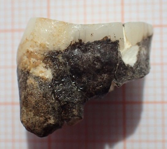
The International Archives of the Photogrammetry, Remote Sensing and Spatial Information Sciences, Volume XLIV-2/W1-2021
4th Int. Worksh. on “Photogrammetric & computer vision techniques for video surveillance, biometrics and biomedicine”, 26–28 April 2021, Moscow, Russia

teeth are sectioned for obtaining contours and conducting mea-
surements on them. Tooth contour analysis usually requires addi-
tional constructions based on available and morphologically rel-
evant landmarks (Martin, 1983).

The specific feature of the current study is that the teeth under ex-
amination were found to be characterised by very different degree
of preservation. The two samples from the LangTrank cave (lt 8
and lt 9) had a high degree of wear, with their occlusal surface
relief having been completely lost its morphologically essential
features. Their enamel surface has cracks and chippings involv-
ing the area of cervical margins as well. In contrast to these two
samples, all the other studied teeth, including the orang-utan up-
per molar from the LangTrank (o u) and both upper molars from
Sunghir (s 17 and s 27), possess a rather complete morphology.
The above-mentioned circumstances, pertaining to the poor con-
dition of some teeth on the one hand and divergent conditions of
all samples - on the other, are a hindering factor in the application              Figure 1. Excavations near the entrance to lower eastern
of the aforementioned techniques that are commonly used.                                          galleries of LangTrank.

Thus if we refer to structures necessary for orientation, neither             One of the most common species of mammals in this location in
enamel margin nor dentin horns have been preserved on the lt 8                Pleistocene has been the wild boar (Sus scrofa Linnaeus, 1758).
and lt 9 samples. For this reason development of non-standard                 Thus coronal parts of these animals’ teeth account for around
approaches to measurements was required. Consequently it has                  25% of all findings during expeditions. There are teeth which
become necessary to pick areas on the teeth studied where the                 can be referred to animals of different ages: from new-borns with
layer of enamel had been preserved in relatively similar condi-               deciduous dentitions to very old specimen with severely worn
tion on all of the studied teeth. Hence an unusual for this kind of           out distal molars (M3). The majority of difficulties in differen-
studies area of buccal enamel was picked for comparative studies.             tiation and classification of wild boars’ dental remains are tradi-
It should be noted that buccal cervical enamel is not characterised           tionally related to their upper fourth premolars (P4), especially if
by high stability. These areas relatively often can be affected by            their morphology had been changes by the process of functional
carious or non-carious (caused by stress) lesions (Margherita et              wear, as their sizes largely correspond to possible morphologi-
al., 2017; Nascimento et al., 2016). However, the studied sam-                cal variability of human upper molars. Two teeth of that kind,
ples possessed intact enamel on the cervical portion of buccal                preliminary identified as human, were subjected to currently pre-
groove. Thus under such conditions of limited choice the re-                  sented studies (Figure 2a,b). More detailed morphological data
search direction was chosen with respect to this uniting all the              has showed that they could exactly refer to boars.
above-mentioned teeth feature.

A number of our previously conducted studies were held by means
of a technique specially developed for measuring teeth – auto-
mated digital odontometry (Knyaz et al., 2016, Gaboutchian et
al., 2020). Taking into consideration the unusual sample set and
study requirements we did not use completely automated mea-
surement and orientation algorithms for the current study.

         1. PALEONTOLOGICAL BACKGROUND
                                                                                              (a)                                  (b)
The studied paleontological dental samples are findings from Lang-
Trank cave situated in the northern Vietnamese Province of Thanh
Hoa. It is a multilevel solutional cave in the limestone and dolomite          Figure 2. Studied samples of teeth from LangTrank: lt 8 view
rock body related to the Devonian period. The lowermost eastern                from distal surface (a) and lt 9 view from occlusal surface (b).
galleries of the cave, which were filled at the turn of the Middle
                                                                              Nevertheless this kind of comparative studies of enamel thickness
and Late Pleistocene periods by loamy red beds with mixture car-
                                                                              and micromorphology of coronal enamel and dentin surfaces of
bonates, contain skeletal and dental remains of mammals (Figure
                                                                              extinct mammalian teeth is one of the first experiences in terms
1). Findings from this location have been studied by palaeon-
                                                                              of the applied methodology as well. Enamel thickness of the
tologists and classified as consistent with more than 30 species
                                                                              studied teeth was assessed in comparison to orang-utan upper
of large mammals (Lopatin et al., 2019). It should be noted
                                                                              tooth (Pongo sp.) from the same location (sample o u) as well
that teeth of an extinct during the Pleistocene period continen-
                                                                              as two upper teeth of an adolescent individual from Upper Palae-
tal species orang-utan (Pongo. sp) in particular have been found
                                                                              olithic Sunghir which is well-known archaeological site situated
in the LangTrank cave. The reason explaining that the majority of
                                                                              in Vladimir Oblast, Russian Federation (samples s 17 and s 27).
findings were represented by teeth of mammal is in habitation of
porcupines in that area in Pleistocene (their remains were found
as well during the excavations – South-Asian species of Hystrix                                           2. METHOD
kiangsenensis Wang, 1931). These rodents include in their diets
bones of animals, however avoiding consumption of covered by                  Software which was applied for the current study has been spe-
enamel and for this reason extremely hard coronal parts of teeth.             cially developed at GosNIIAS (Russian Federation). Initially it

                                                        This contribution has been peer-reviewed.
                        https://doi.org/10.5194/isprs-archives-XLIV-2-W1-2021-61-2021 | © Author(s) 2021. CC BY 4.0 License.                        62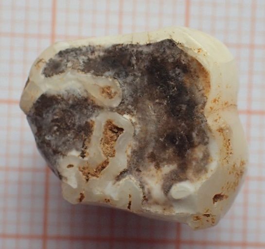
The International Archives of the Photogrammetry, Remote Sensing and Spatial Information Sciences, Volume XLIV-2/W1-2021
4th Int. Worksh. on “Photogrammetric & computer vision techniques for video surveillance, biometrics and biomedicine”, 26–28 April 2021, Moscow, Russia

has been used for conducting measurement on digital reconstruc-
tions of teeth in a research related to prosthetic dentistry (Knyaz
et al., 2007). The software allows uploading 3D images, setting
landmark position, different modes of sectioning for contour ob-
taining and distance measurements on contours. Measurement
results can be subsequently saved for further analysis. These pro-
cedures run partially in automated mode as well as in “manual”
mode.

The process of 3d reconstruction obtainment was based on X-Ray
imaging through micro-computed tomographic scanning. Sam-
ples s 17 and s 27, being antimeres within a single dental arch,
have been previously scanned as a whole with the complete skull
on General Electric Phoenix v|tome|x Metrical Edition. Their
reconstructions were performed after extraction of their images
from the entire stack. Voxel edge for these two human upper sec-
ond molars accounts for 43 µm. Samples lt 8, lt 9 and o u as sep-
arate teeth (i.e. objects of significantly smaller size than skull)
could be scanned in higher resolutions. Thus they were recon-
structed after micro-computed tomographic scanning in SkyScan
                                                                                 Figure 3. Reconstruction of enamel and pulpal chamber of
1172 at 10,3 µm inter-slice distance. In both cases 3d reconstruc-
                                                                                           sample lt 8 with initial landmarks set.
tions were obtained from images stacks consisting of 8-bit depth
.bmp format files.

Further image and reconstruction processing procedures were per-
formed by use of Avizo 9.01 software. Segmentation procedures
of the studied objects were based on variety in X-ray absorption
by different dental tissues: external highly-mineralised enamel,
massive bulk of dentin and internal cavity of pulpal chamber. Ar-
eas with same absorption level were sorted and morphologically
irrelevant ones were excluded and cleaned. At the final stage
models’ sizes were reduced in order to allow easy software run-
ning and subsequently they were converted to .stl and .x formats.

Enamel thickness measurements were performed on reconstruc-                      Figure 4. Reconstruction of enamel and pulpal chamber of
tions of enamel or combined enamel/pulpal chamber. The fol-                                 sample lt 8 with “sliced” contours.
lowing procedures provided necessary orientation of slices, sec-
tioning for contour obtaining, setting landmarks and orientation              buccal cusp outer surface contours. The second is analogous sin-
on contours and measurements. On the surfaces of digitally re-                gularity of another line parallel to the above-mentioned line and
constructed enamel models corresponding to their buccal grooves               simultaneously tangent to the deepest point of the groove con-
two points were set: the first was localised in the area of inter-            tour. The next stage of constructions on 2d contours is combined
cusp (or inter-root) protrusion of cervical enamel portion and the            with measurements of enamel thickness. It sets the distance be-
other – on the projection of buccal groove. As a matter of con-               tween the two points: groove maximal deepening (localised at
venience these points were marked on inner surface of enamel                  the previous stage of constructions) and the most protruded point
(Fig. 4). The above-mentioned landmark setting is an expert-                  on the internal, faced to dentin contour of enamel. This point, in
based “manually” conducted procedure.                                         its turn, is also localised in compliance with parallelism of lines
                                                                              constructed on the measured contours (Figure 5b). The obtained
Further stages run automatically by means of the used software.               result corresponds to the required enamel thickness on the current
The constructions include 10 parallel slicing planes which are                contour and the process is repeated on the next contour.
perpendicular to line connecting initially set landmarks on the
3d reconstruction of the dental enamel. The planes are equally
spaced and serve for contour obtaining (Figure 5).                                           3. RESULTS AND DISCUSSION

The contours which were obtained and visualised in 2d mode,                   The suggested partially automated algorithms including orienta-
served for enamel thickness measurements in the buccal groove                 tional and measuring stages were used for a rather specific re-
area. Planar geometric constructions combined with measure-                   search task. At the same time application of ready for use fully
ments were conducted in two stages by means of a properly de-                 automated algorithms has been hampered by the lack on two of
veloped for such studies software tool – a distance measurer oper-            the studied teeth of morphologically significant structures. Pos-
ating between two parallel lines. The first construction providing            sibly in the future new algorithms can be suggested which would
orientation was performed on the external contour of enamel in                automatically measure teeth with damaged surfaces as well. How-
order to localise the point corresponding the maximal deepening               ever in the current study we had to rely on partially automated
of the groove which would allow precise setting of the subsequent             methods which possess a certain degree of subjectivity at the ini-
constructions’ direction (Figure 5a). This is a manually operated             tial stage of marking the studied 3d reconstructions for orien-
procedure based the following two conditions. The first is singu-             tation. However accuracy and objectivity of measurements has
larity of line tangent simultaneously to both (mesial and distal)             been achieved through combination of the used high-resolution

                                                        This contribution has been peer-reviewed.
                        https://doi.org/10.5194/isprs-archives-XLIV-2-W1-2021-61-2021 | © Author(s) 2021. CC BY 4.0 License.                        63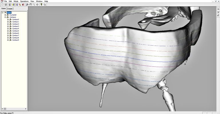
The International Archives of the Photogrammetry, Remote Sensing and Spatial Information Sciences, Volume XLIV-2/W1-2021
4th Int. Worksh. on “Photogrammetric & computer vision techniques for video surveillance, biometrics and biomedicine”, 26–28 April 2021, Moscow, Russia

                                                                                    Contour           Enamel thickness on contours, mm
                                                                                                 lt 8       lt 9     ou      s 17     s 27
                                                                                       1         0,676     0,379 0,112 0,282 0,233
                                                                                       2         0,905     0,574 0,123 0,497 0,421
                                                                                       3         0,981     0,888 0,394 0,562 0,518
                                                                                       4         1,018      0,89    0,506 0,759 0,661
                                                                                       5         1,085     0,969 0,559 0,939 0,895
                                                                                       6         1,207     1,052 0,582 1,052 1,187
                                                                                       7         1,346     1,142 0,645 1,303 1,387
                                                                                       8         1,499     1,119 0,591 1,474 1,508
                                                                                       9         1,541      1,12    0,786    1,64    1,708
   (a) Orientational geometric construction on a sample lt 8 contour
                                                                                       10        1,598      1,08    1,011 1,728 1,863
                                                                                                       Average enamel thickness , mm
                                                                                                 1,186      0,92    0,53    1,024 1,038

                                                                               Table 1. Parameters of enamel thickness; individually maximal
                                                                                             values are in grey-shaded cells.

                                                                              dental wear degree on the two (lt 8 and lt 9) teeth. The other fac-
                                                                              tor that could determine such patters can depend on orientational
                                                                              landmark setting as their positions are determined by inclination
                                                                              of buccal surface which is an individual feature on every separate
                                                                              tooth.
     (b) Enamel thickness measurement on a sample lt 8 contour                If we summarise the results of dental enamel thickness measure-
                                                                              ments we can define that the thinnest enamel layer has the o u
    Figure 5. Orientation algorithms before and after manual                  sample which the upper molar of orang-utan from Plectocene lay-
                          adjustments.                                        ers of the LangTrank cave. High degree of similarity of enamel
                                                                              thickness patterns is characteristic for antimeres from Sungirian
imaging technique and the suggested uniform approach to mea-                  upper dental arch (s 17 and s 27 samples). These human teeth
surements on all of the studied samples. It should mention that               have more bulky enamel in average if compared with the pongine
enamel thickness varies depending to the localisation of the stud-            molar as well as highest maximal values on separate sections
ied contour. Thus measurements were taken on each of the stud-                among all of the studied teeth. The most significant differences
ied teeth on 10 contours obtained by parallel equally spaced sec-             can be marked between samples lt 8 and lt 9. The first of these
tioning planes. The results of enamel thickness measurements on               two teeth has the highest among the studied samples average
the five upper teeth are presented in Table 1.                                enamel thickness. In addition its layer of enamel in the cervi-
                                                                              cal area significantly thicker than on lt 8, however thick cervical
The samples of teeth which were involved in the current study                 enamel is uniting feature for these two teeth.
belong to different species. However even within one individual
                                                                              The presented measurement and enamel thickness analysis re-
difference can be found on two antimere teeth. In addition de-
                                                                              sults were used for further studies of paleontological findings and
spite the developed measurement technique directed to achieving
                                                                              taxonomical interpretations.
similarity of measurements on the studied samples, it cannot be
expected that the mentioned similarity can become identical. For
instance, orientation definitely differs on objects having variety                                    4. CONCLUSION
of shapes; and the third contour on any tooth would never cor-
respond by its location to the same contour number on all teeth.              The applied in the current study imaging and image processing
This means that there are too many factors to consider a single               techniques significantly facilitate for increasing research objec-
measurement consistent. This is the reason why average enamel                 tivity, and provide important data in terms of accuracy and de-
thickness parameters (arithmetic mean of results obtained on 10               tailed visualisation. 3D reconstructions of the teeth allowed to
contours) are more reliable for assessments and comparisons in                develop and use new non-invasive methods and to receive reliable
such studies. These values referred to each of the studied teeth              and as close as possible to objective data on enamel thickness.
are presented in the table.                                                   Such information serves as an important additional support, in
                                                                              line with other traditional morphological assessment methods, in
However every measurement is essential as it allows to trace the              taxonomic differentiation of paleontological findings, especially
tendency of changes across the tooth crown. Thus maximal val-                 in cases of controversy. This study is an encouraging example
ues for each tooth are in highlighted cells as well. Areas with               of combining 3d imaging and image processing techniques with
the thinnest enamel layer are located on cervical (closest to roots)          dental metrics and their implementation in paleontological re-
portions of dental crowns on all the teeth. Thickness of enamel               search, which has a potential for further development.
grows gradually with increasing distance from cervical area to-
wards cusp tips. Maximal enamel thickness values are registered                                   ACKNOWLEDGEMENTS
on the most distally located from tooth cervices contours. The
only exception is the lt 9 sample, having the peak of enamel                  We would like to express our gratitude to the Shared Research
thickness on the 7th contour. Nevertheless this fact does not                 Facilities “Palaeoantropological collections Institute of Ethnol-
change the general pattern, as it can be referred to difference of            ogy and Anthropology of Russian Academy of Sciences” and

                                                        This contribution has been peer-reviewed.
                        https://doi.org/10.5194/isprs-archives-XLIV-2-W1-2021-61-2021 | © Author(s) 2021. CC BY 4.0 License.                        64
The International Archives of the Photogrammetry, Remote Sensing and Spatial Information Sciences, Volume XLIV-2/W1-2021
4th Int. Worksh. on “Photogrammetric & computer vision techniques for video surveillance, biometrics and biomedicine”, 26–28 April 2021, Moscow, Russia
Russian-Vietnamese Scientific Centre for providing the study ma- thickness in Neandertal and modern human molars. Journal of
terials. human evolution. 55. 12-23. 10.1016/j.jhevol.2007.11.004.
Smith et al., 2009. Smith, T., Olejniczak, A., Kupczik, K., Laz-
REFERENCES zari, V., Vos, J., Kullmer, O., Schrenk, F., Hublin, J-J., TEUKU,
J., Tafforeau, P. Taxonomic assessment of the Trinil molars us-
Alvesalo et al., 2009. Alvesalo, L., Tammisalo, E., Hakola, ing non-destructive 3D structural and development analysis. Pa-
P. Enamel thickness in 47, XYY males’ permanent teeth. An- leoAnthropology. 2009. 117-129.
nals of Human Biology - ANN HUM BIOL. 12. 421-427. Suwa and Kono, 2005. Suwa, G., Kono R.T. A Micro-CT Based
10.1080/03014468500007981. Study of Linear Enamel Thickness in the Mesial Cusp Section of
Benazzi et al., 2014. Benazzi, S., D. Panetta, C. Fornai, M. Human Molars: Reevaluation of Methodology and Assessment of
Toussaint, G. Gruppioni, and J. Hublin (2014). Technical Note: Within-Tooth, Serial, and Individual Variation. Anthropological
Guidelines for the Digital Computation of 2D and 3D Enamel Science, 113: 273–289
Thickness in Hominoid Teeth. American Journal of Physical An- Zanolli et al., 2010. Zanolli, C., Bayle, P., Macchiarelli, R. Tissue
thropology, 153: 305–313 proportions and enamel thickness distribution in the early Middle
Gaboutchian et al., 2020. Gaboutchian, A. V., Knyaz, V. Pleistocene human deciduous molars from Tighenif, Algeria. C.
A., Novikov, M. M., Vasilyev, S. V., Leybova, N. A., Ko- R. Palevol 9 (2010) 341–348. doi:10.1016/j.crpv.2010.07.019
rost, D. V., Cherebylo, S. A., and Kudaev, A. A.: Auto- Zanolli et al., 2017. Zanolli C., Bayle, P., Bondioli, L., Dean,
mated Digital Odontometry: Measurement Data Analyses In M., Le Luyer, M., Mazurier, A., Morita, W., Macchiarelli, R. Is
Cases Of Complicated Dental Morphology, Int. Arch. Pho- the deciduous/permanent molar enamel thickness ratio a taxon-
togramm. Remote Sens. Spatial Inf. Sci., XLIII-B2-2020, specific indicator in extant and extinct hominids? Comptes Ren-
851–856, https://doi.org/10.5194/isprs-archives-XLIII-B2-2020- dus Palevol. 16. 10.1016/j.crpv.2017.05.002.
851-2020, 2020
Knyaz et al., 2007. Knyaz V., Zheltov S., Gabuchyan A., Bol-
shakov G. Photogrammetric system for automated teeth arches
3D models generation and teeth occlusion analysis. Optical 3D
Measurement Techniques VIII, Zurich, 2007, Vol. I, pp. 299-304
Knyaz et al., 2016. Knyaz V. A. and Gaboutchian, A. V.:
PHOTOGRAMMETRY-BASED AUTOMATED MEASURE-
MENTS FOR TOOTH SHAPE AND OCCLUSION ANALY-
SIS, Int. Arch. Photogramm. Remote Sens. Spatial Inf. Sci.,
XLI-B5, 849–855, https://doi.org/10.5194/isprs-archives-XLI-
B5-849-2016, 2016.
Lopatin et al., 2017. Lopatin A.V., Maschenko E.N., Vis-
lobokova I.A., Serdyuk N.V., Dac L.X. Pleitocene Mam-
mals from Lang Trang Cave (Vietnam): New Data. Doklady
Akademii Nauk. Nauki o Zhizni. 2021. Vol. 496, pp 5-9, doi:
10.31857/s2686738921010170 (in Russian)
Margherita et al., 2017. Margherita, C., Oxilia, G., Barbi, V.,
Panetta, D., Hublin, J-J., Lordkipanidze, D., Meshveliani, T.,
Jakeli, N., Matskevich, Z., Bar-Yosef, O., Belfer-Cohen, A., Pin-
hasi, R., Benazzi, S. News and Views Morphological description
and morphometric analyses of the Upper Palaeolithic human re-
mains from Dzudzuana and Satsurblia caves, western Georgia.
Journal of Human Evolution. 113. 10.1016/j.jhevol.2017.07.011.
Martin, 1983. Martin L.B. The relationships of the later Miocene
Hominoidea. PhD Thesis, University College London, London.
1983
Nascimento et al., 2016. Nascimento, M., Dilbone, D., Pereira, P.,
Geraldeli, S., Delgado, A., Duarte, W. (2016). Abfraction lesions:
Etiology, diagnosis, and treatment options. Clinical, Cosmetic
and Investigational Dentistry. 8. 79. 10.2147/CCIDE.S63465
Olejniczak et al., 2007. Olejniczak, A., Grine, F., Martin, L.
Micro-computed tomography of primate molars: Methodologi-
cal aspects of three-dimensional data collection. Vertebrate Pale-
obiology and Paleoanthropology. 01/2007, doi: 10.1007/978-1-
4020-5845-5 7
Olejniczak et al., 2008. Olejniczak, A., Smith, T., Feeney, R.,
Macchiarelli, R., Mazurier, A., Bondioli, L., Rosas, A., Fortea,
J., Rasilla, M., Garcia-Tabernero, A., Radovcić, J., Skinner, M.,
Toussaint, M., Hublin, J-J. Dental tissue proportions and enamel
This contribution has been peer-reviewed.
https://doi.org/10.5194/isprs-archives-XLIV-2-W1-2021-61-2021 | © Author(s) 2021. CC BY 4.0 License. 65You can also read