Endometriosis and Deep endometriosis - 16/01/2018 Dott Matteo Generali AUSL Modena Carpi U.O. Ostetricia e Ginecologia
←
→
Page content transcription
If your browser does not render page correctly, please read the page content below
Endometriosis and Deep
    endometriosis

            16/01/2018

       Dott Matteo Generali
        AUSL Modena Carpi
    U.O. Ostetricia e Ginecologia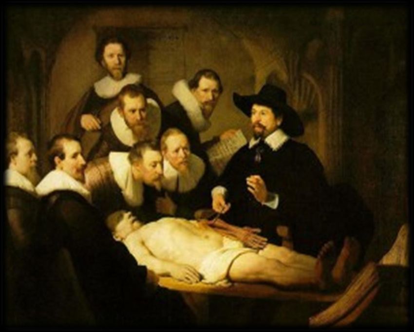
Definizione
 L’endometriosi è una patologia
 ginecologica, benigna, cronica,
 ormonodipendente caratterizzata
 dalla presenza di tessuto
 endometriale componente
 stromale e ghiandolare,
 al di fuori della cavità uterina

Von Rokitansky C. Ueber uterusdrusen-neubildung in uterus and ovarilsarcomen. Z Ges Aerzte Wein 1860; 37: 577–93

Sampson J. Peritoneal endometriosis due to menstrual dissemination of endometrial tissue into the
peritoneal cavity. 1927;
Incidenza

        Giudice LC, Endometriosis 2004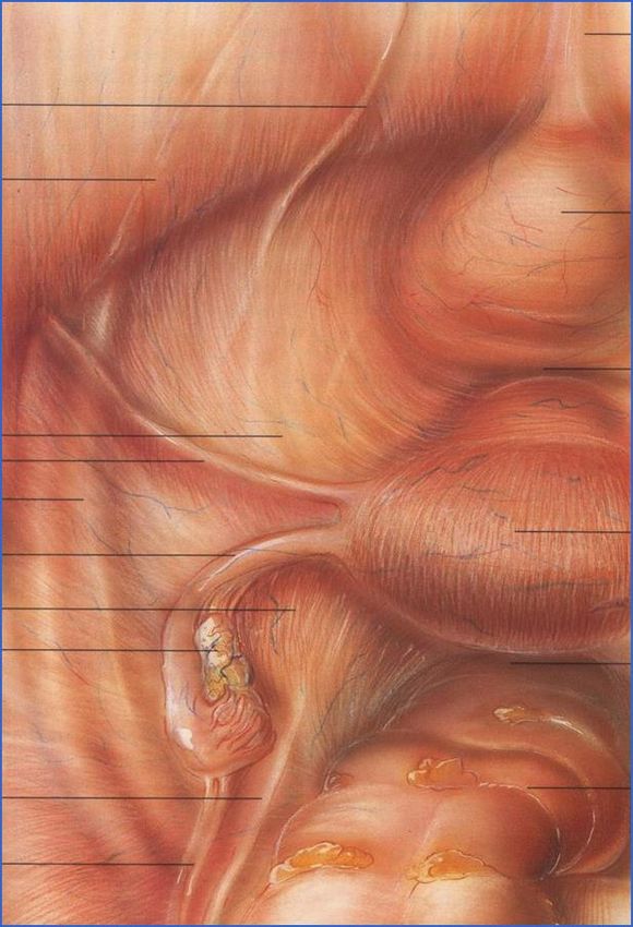
Localizzazioni pelviche

ESHRE guideline for the diagnosis and treatment of endometriosis 2005
Sintomatologia

90%
      60%
             50%
                      45%

                                10%       20%

      Chapron C Endometriosis and infertility: pathophysiology and management 2010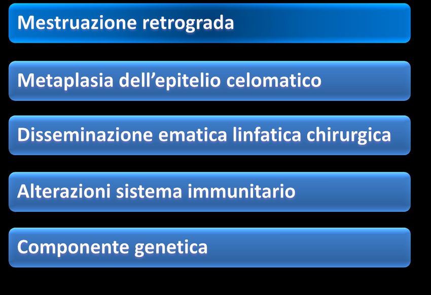
Time to diagnosis

Hadfield R Delay in the diagnosis of endometriosis: a survey of women from the USA and the UK. 1996;
Etiopatogenesi

ESHRE guideline for the diagnosis and treatment of endometriosis 2005
Le cellule endometriosiche
                                     Migrano

                                       Aderiscono

                                    Invadono la matrice
                                       extracellulare

                                             Neovascolarizzano

Starzinsky.The putative role of cell adhesion molecules in endometriosis: can we learn from tumor metastasis?
1999;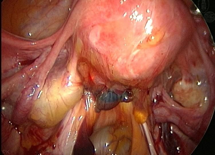
3 forme di endometriosi
ovarica Peritoneale-aderenze Deep endometriosis
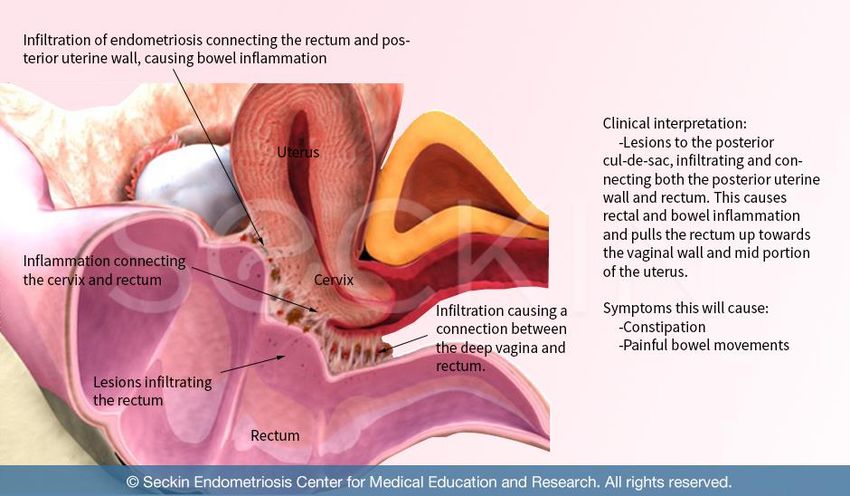
Lesione infiltrante il peritoneo o
retro peritoneale di almeno 5 mm;
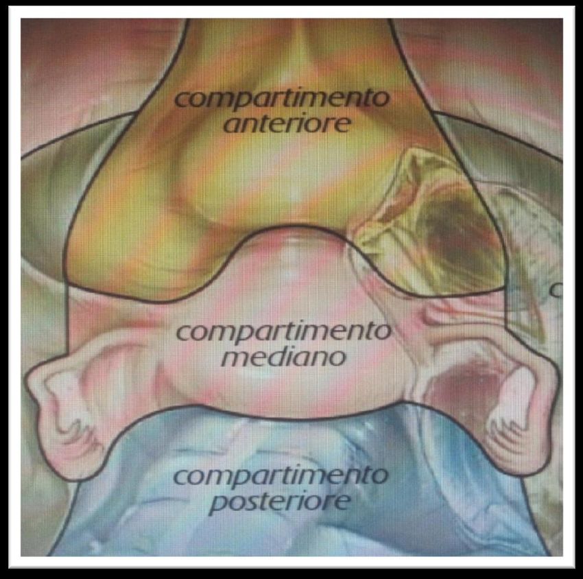
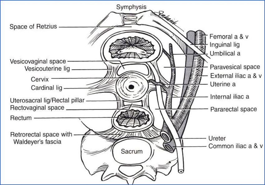
4-37% donneCompartimenti pelvici Anteriore Centrale Posteriore Vescica ureteri Utero ed annessi Douglas Rettosigma
Compartimenti pelvici
Anteriore                 Centrale                     Posteriore
Vescica ureteri          Utero ed annessi                 Retto

                                            Centrale

                                            Utero ed
                                            annessi
La mia conoscenza della pelvi
Lezione di anatomia del dottor Tulp 1632 RembrandtLa mia conoscenza della patologia
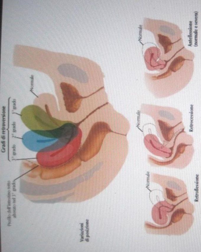
Compartimento centrale
        utero ed annessi

Descrivere dimensioni, morfologia, ecostruttura

   Valutazione della mobilità e dei rapporti
       anatomici con visceri adiacenti

    Cercare la collaborazione della paziente
             nell’evocare i sintomi
Utero
Ovaio
Hard Marker endometrioma

Typical ultrasound appearance of an ovarian endometrioma:

a unilocular cyst with (less than 5 locules)

ground glass echogenicity

and little to moderate peripheral vascularisation.

Note the normal ovarian tissue around the cyst.
Endometrioma atipico

       Atypical ultrasound appearance of an ovarian endometrioma:
       Unilocular cyst
       Ground glass echogenicity,
       Internal papillation
       No vascularisation in the papillary projection.

       This is not a true papillations but hyperechoic tissue consisting of
       blood clots or fibrin lying adjacent to the cyst wall

Almost 50% of the endometriomas had other ultrasound characteristics than the
typical ‘unilocular cyst with ground glass echogenicity of the cyst fluid’.
Guerriero S, Ajossa S, Mais V, et al. The diagnosis of endometriomas using colour Doppler energy imaging. Hum Reprod 1998;6:1691–5.
Endometriod borderline tumor
Ultrasound characteristics of endometriomas differ in pre- and postmenopausal
women.

Masses in postmenopausal women, whose cystic contents have a ground glass
appearance, have a high risk of malignancy.

Endometriomas could serve as precursors of endometrioid borderline ovarian tumours.

Endometrioid borderline ovarian tumours have the potential to progress to low-grade
invasive carcinoma.

Borderline tumours and carcinomas arising from endometrioid cysts show a
vascularised solid component at ultrasound examination
Endometriosi e tube

Endometriosis that affects the ovary and Fallopian tubes can create a tubo–
ovarian complex, in which the ovaries and tubes are identified and recognised,
but the ovaries cannot be separated by pushing the tube with the vaginal probe
Endometriosi e aderenze

Ovarian endometriomas are frequently associated with other endometriotic lesions ,
such as adhesions and DIE, which are not easy to diagnose. Underestimation of
extensive adhesions in women with endometriomas before surgery is one of the main
reasons why surgery is often incomplete leading to repeat operations
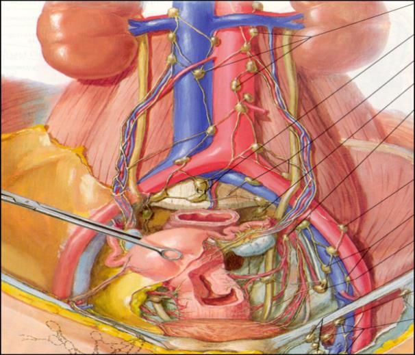
Compartimento posteriore
        D.I.E.
Morphological features - Hypoechoic nodules - Irregular margins « indian hairdresser sign » - No vascularization or minal peripheral spots
Compartimento posteriore

     Introduzione della sonda
Compartimento posteriore
63%
                                   interessamento
Specificita 97%    Donne con POD      intestinale
VPN        92.6%    obliteration           Vs
                                      1.4% senza
                                     obliterazione
Legamenti uterosacrali
Legamenti uterosacrali
Intestino

            10-15cm

                      30 cm
M Muscolare ipoecogeno (rivestito da linea
iperecogena che delinea la parte esterna
ed interna del muscolo) M
SM Strato sottomucoso iperecogeno SM
Mu
Mu Mucosa ipoecogenaSchematic and ultrasound image of a nodule of deep infiltrating endometriosis in the upper rectum (arrow). Nodules located above the level of a virtual line (red line) passing through the insertion of the uterosacral ligaments on the cervix are considered to be located in the upper rectum or recto-sigmoid junction.
Water-contrast in the rectum during transvaginal sonography is performed by
injecting saline solution into the rectal lumen during transvaginal ultrasound
examination. Note the presence of the deep infiltrating endometriosis nodule bulging
into the bowel lumen. The lesion clearly reduces the rectal lumen. It infiltrates only
the muscle layer of the bowel. The lesion is covered by the hyperechogenic
submucosa and hypoechogenic mucosa.
Compartimento anteriore
E gli ureteri???
Vescica… ureteri…
…e Reni
You can also read