Examination of the morphometric features and three-dimensional modelling of the skull in Van cats by using computed tomographic images
←
→
Page content transcription
If your browser does not render page correctly, please read the page content below
Ankara Univ Vet Fak Derg, 68, 213-222, 2021
DOI: 10.33988/auvfd.775971
Examination of the morphometric features and three-dimensional
modelling of the skull in Van cats by using computed tomographic
images
Osman YILMAZ1,a,, İsmail DEMİRCİOĞLU2,b
1Van Yüzüncü Yıl University, Faculty of Veterinary Medicine, Department of Anatomy, Van; 2Harran University, Faculty of
Veterinary Medicine, Department of Anatomy, Şanlıurfa, Turkey.
aORCID: 0000-0003-2013-9213; bORCID: 0000-0002-0724-3019
Corresponding author: osman_40_5@hotmail.com
Received date: 30.07.2020 - Accepted date: 08.09.2020
Abstract: This study was conducted to make the three-dimensional modelling of the skull in Van cats by using computed
tomographic images and to determine the morphometric features between sexes. The skulls of 16 adult Van cats were used in the study.
The skulls of the anesthetized animals were scanned by using a Computed Tomography (CT) device and their images were obtained.
These images were converted to a three-dimensional structure using MIMICS 20.1 software and their morphometric measurements
were calculated. It was determined in the study that total length of the skull (TLS), facial length (FCL), upper neurocranium length
(UNCL), greatest length of the nasal (GLN), maximum zygomatic width (MZW), condylobasal length (CBL), basal length (BL),
median palatal length (MPL), palatal length (PL), least palatal breadth (LPB), length of the cheek tooth row (LCR), greatest inner
height of the orbit (GIHO), skull height (SH), and volumetric measurement values were statistically significantly higher in the male
cats; whereas, breadth dorsal to the external auditory meatus (BEAM) and neurocranium length (NL) measurement values were
statistically significantly higher in the female cats (P
214                                            Osman Yılmaz - İsmail Demircioğlu

heterochromia), moderately long straight nose, round face,      from Van Yüzüncü Yıl University Van Cat Research and
pointed ear, triangular-shaped head, superior learning          Application Center. Drinking water and standard cat food
ability, and intelligence (3, 16, 47).                          were given ad libitum to the Van cats until one day before
      Skull is a strong structure composed of many bones        the study. This study was approved by Van Yüzüncü Yıl
which are paired, but some of them which are median-            University Animal Experiments Local Ethics Committee
located and single. This structure includes the sense           (Decision no: 2020/02 and Date: 27. 02. 2020).
organs such as sight, olfactory, balance, and taste as well           Anesthesia: The Van cats included in the study were
as upper respiratory and digestive tracts together with the     numbered and they were not given any food one day
brain. Cranial bones are joined with each other through         before the study. On the day of examination, the
sutures and with mandibulae and apparatus hyoideus              combination of Ketamine (15 mg/kg, IM, Ketasol® 10%
through joints (12). The morphometric and morphological         injectable, İnterhas Veterinary Drugs, Ankara) and
studies conducted on the skull not only reflect the effects     Xylazine (1-2 mg/kg, IM, Alfazyne® 2% injectable, Ege-
of the genetic and environmental factors (such as genetic       Vet Veterinary Drugs, İzmir) was used for the anesthesia
and ecophenotypic variations in general) on individual          of the cats.
development but also underlie the clinical and surgical               Computed tomography imaging: The 16-slice
applications related to the region (43). Also, the              computed tomography (CT) device (Somatom Sensation
morphometric measurement values of skull may show               16; Siemens Medical Solutions, Erlangen, Germany)
many individual differences among species and including         available in Van Yüzüncü Yıl University Faculty of
breed, age, and the structure specific to the animal (5).       Medicine Radiology Department was used for the
      Significant changes have taken place in anatomy           Computed Tomography (CT) examinations of the cats.
education by means of the technological developments in         The cats under anesthesia were positioned symmetrically
medical imaging and computer-aided learning fields,
                                                                in a prone head-first position on a "disposable" cover
various software developed, and three-dimensional
                                                                spread in the gantry of the machine. During scan, the CT
modelling (2). Especially in small pets such as cats, the
                                                                device parameters were determined as KV / Effective mAs
computed tomography and three-dimensional modelling
                                                                / Rotation time (sec) values 120 / 120 / 0.75; gantry
programs are used as a standard imaging method in the
                                                                rotation period 420 ms; physical detector collimation, 16
imaging of skull and the anatomic structures around it and
                                                                × 0.6 mm; section thickness, 0.5 mm; final section
monitoring the changes in these structures (14, 17). Also,
                                                                collimation 32 × 0.63 mm; feed/rotation, 6 mm; Kernel,
skull and various extracranial and intracranial
                                                                U90u; increment 0.5 mm; and resolution 512 × 512 pixels.
pathological structures around it may be determined and
                                                                Dosage parameters and scanning were performed in
the effectiveness of treatment may be evaluated using
                                                                accordance with standard protocols found in published
these imaging methods and three-dimensional modelling
                                                                literature (11, 31). The images obtained were recorded in
software (17, 44). In addition, these imaging methods are
                                                                DICOM format.
commonly used in anthropological studies such as
determining the osteometric features of the crania                    Three-dimensional modelling of the images and
obtained in the excavation works conducted in various           performing their measurements: Then, the modelling of
regions and obtaining their measurement values (32, 33).        these images was performed by transferring them to
      The measurements obtained from the skull using            MIMICS 20.1 (The Materialise Group, Leuven, Belgium),
computed tomography (CT) and three-dimensional                  a three-dimensional modelling software. Osteometric
modelling software may be mostly used in revealing the          measurements were taken for 34 different parameters on
biometric differences between sexes (34). For this              the skulls whose three-dimensional modelling was
purpose, many studies have been conducted in veterinary         performed. The morphometric measurements were taken
medicine (7, 23, 37). No study was found on the skull of        based on the measurement points specified in the literature
Van cats in the literature reviews.                             (4, 20, 21, 25, 42). After completing the morphometric
      This study was conducted to perform the three-            measurements, the surface area and volumetric values of
dimensional modelling of the skull in Van cats by using         the remaining part of the skull other than mandibulae were
computed tomography, display their anatomic structures,         calculated. Nine different indices were calculated using
obtain their morphometric measurement values, and               osteometric measurements. While Table 1 shows the
reveal the biometric differences of these measurement           linear measurement points obtained from the dorsal,
values between sexes.                                           lateral, and ventral surfaces of the skull, Table 2 shows the
                                                                formula of the indices calculated by using these
                Material and Methods                            measurement points. Nomina Anatomica Veterinaria was
      Animal materials: In the study, a total of 16 adult Van   used as terminology in the study (15). Also, a digital scale
cats (8 males and 8 females) which had a weight of 4810 -       (TESS®, RP - LCD, Çomak Terazi, İstanbul) was used for
7050 gram, were aged between 3 and 8 years, were procured       the weight measurements of the cats used in the study.
Ankara Univ Vet Fak Derg, 68, 2021                                                     215

Table 1. Studied cranial parameters (mm).
 Parameter         Abbreviation     Definition
 1                 TLS              Total length of the skull: the distance between akrokranion-prosthion
 2                 FCL              Facial length: frontal midpoint-prosthion
 3                 UNCL             Upper neurocranium length: akrokranion-frontal midpoint
 4                 CL               Cranial length: akrokranion-nasion
 5                 VL               Viscerocranial length: nasion-prosthion
 6                 GLN              Greatest length of the nasals: nasion-rhinion
 7                 LBO              Least breadth between the orbits: entorbitale-entorbitale
 8                 GFB              Greatest frontal breadth: ectorbitale-ectorbitale
 9                 LBS              Least breadth of skull: breadth at the postorbital constriction
 10                MWN              Maximum width of neurocranium: euryon-euryon
 11                MZW              Maximum zygomatic width: zygion-zygion
 12                CBL              Condylobasal length: caudal border of occipital condyles-prosthion
 13                BL               Basal length: basion-prosthion
 14                MPL              Median palatal length: staphylion-prosthion
 15                LHP              Length of the horizontal part of the palatine: staphylion-palatinoorale
 16                LHP-1            Length of the horizontal part of the palatine-1: the median point of intersection of the line joining
                                    the deepest indentations of the choana-palatinoorale
 17                PL               Palatal length: the median point of intersection of the line joining the deepest indentations of the
                                    choana-prosthion
 18                GBP              Greatest breadth of the palate: maximum width of the distal ends of the alveolus of upper P3
 19                LPB              Least palatal breadth
 20                BCA              Breadth at the canine alveoli
 21                LPR              Length of the premolar row
 22                LMR              Length of the molar row
 23                LCR              Length of the cheektooth row
 24                GDAB             Greatest diameter of the auditory bulla: from the most aboral point of the bulla on the suture with
                                    the jugular processes up to the external carotid foramen
 25                BEAM             Breadth dorsal to the external auditory meatus: breadth between external auditory porus
 26                GIHO             Greatest inner height of the orbit
 27                NL               Neurocranium length: basion-nasion
 28                SH               Skull height: basion-external occipital crest
 29                HOT              Height of the occipital triangle: akrokranion-basion
 30                HFM              Height of the foramen magnum: basion-opisthion
 31                GWFM             Greates breadth of the foramen magnum
 32                GBOC             Greatest breadth of the occipital condyles
 33                GBJP             Greatest breadth of the bases of the jugular processes
 34                GMB              Greatest mastoid breadth: otion-otion

Table 2. Indices and formulas of the skulls.
 Studied indexes           Formulas
 Skull index               Greatest frontal breadth (var.8) / total length of the skull (var. 1) x 100.
 Cranial index             Maximum width of neurocranium (var. 10) / Cranial length (var. 4) x 100.
 For. magnum index         Height of the for. magnum (var. 30) / greatest breadth of the for. magnum (var. 31) x 100.
 Facial index-1            Maximum zygomatic width (var. 11) / Viscerocranial length (var. 5) x 100.
 Facial index-2            Greatest breadth of the palate (var. 18) / greatest length of the nasals (var. 6) x 100.
 Basal indeks-1            Maximum width of neurocranium (var. 10) / basal length (var. 13) x 100.
 Basal indeks-2            Maximum zygomatic width (var. 11) / basal length (var. 13) x 100.
 Palatal index-1           Greatest breadth of the palate (var. 18) / median palatal length (var. 14 ) x 100.
 Palatal index-2           Greatest breadth of the palate (var. 18) / palatal length (var. 17) x 100.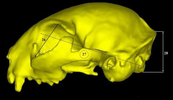
216                                           Osman Yılmaz - İsmail Demircioğlu

      Statistical analysis: Shapiro-Wilk test (n
Ankara Univ Vet Fak Derg, 68, 2021 217
Figure 4. Measurement points of the skull in Van cats (caudal view).
29- HOT: Height of the occipital triangle; 30- HFM: Height of the foramen
magnum; 31- GWFM: Greatest breadth of the foramen magnum; 32- GBOC:
Greatest breadth of the occipital condyles; 33- GBJP: Greatest breadth of the
bases of the jugular processes; 34- GMB: Greatest mastoid breadth.
Table 3. Descriptive statistics of the measurements of the skull according to gender.
MALE FEMALE
Parameter Mean±S.E. Min. Max. Mean±S.E. Min. Max. *P
Age(A) 5.00±2.00 3.00 8.00 5.00±2.00 3.00 8.00 1.00
Weight(W) kg 6.08±0.70 5.30 7.05 5.28±0.28 4.81 5.63 0.027
TLS 96.16±6.38 89.38 108.44 87.38±2.71 83.93 93.08 0.002
FCL 52.14±2.99 49.08 58.85 48.91±2.77 43.27 52.23 0.036
UNCL 56.41±2.93 53.95 61.53 51.32±1.93 48.49 53.37 0.001
CL 73.32±3.05 68.79 78.17 72.12±2.76 67.80 74.83 0.600
VL 26.38±1.67 23.78 28.72 24.79±2.39 22.10 29.62 0.074
GLN 20.37±2.11 18.43 24.95 17.77±1.14 15.54 19.33 0.003
LBO 21.82±1.08 20.44 23.40 20.16±2.02 16.90 23.07 0.074
GFB 49.48±2.41 46.08 52.62 48.28±2.34 45.85 51.85 0.248
LBS 32.04±1.62 30.00 34.97 32.37±1.04 30.95 34.07 0.599
MWN 41.87±1.44 40.19 45.01 46.37±10.39 38.99 64.56 0.834
MZW 66.64±3.66 62.50 72.24 56.11±8.81 41.38 62.33 0.001
CBL 86.42±4.55 78.52 93.39 80.83±4.64 71.16 86.74 0.027
BL 79.86±4.34 73.43 87.36 73.96±2.99 69.53 79.08 0.012
MPL 37.39±1.54 34.63 39.70 34.37±2.13 32.20 38.44 0.015
LHP 15.78±2.76 12.95 21.08 14.50±0.96 13.33 16.34 0.462
LHP-1 13.65±2.33 11.11 17.06 13.35±1.27 12.14 16.11 0.916
PL 34.63±2.32 30.21 37.95 32.83±2.06 30.17 37.09 0.046
GBP 37.78±2.28 34.89 41.56 37.33±2.44 34.42 41.88 0.636
LPB 22.96±1.00 21.93 24.68 21.64±.90 20.19 23.37 0.009
BCA 22.17±1.67 20.08 25.09 21.49±1.56 18.94 23.87 0.753
LPR 16.46±1.13 14.34 17.78 15.46±1.12 13.86 17.21 0.083
LMR 7.83±0.89 6.77 9.31 8.62±0.97 7.34 9.64 0.115
LCR 23.52±1.10 22.12 25.26 21.49±1.72 18.77 23.77 0.016
GDAB 18.85±1.48 17.61 21.05 18.60±0.87 17.06 19.85 0.916
BEAM 33.36±2.45 29.85 36.37 37.35±1.26 34.97 38.71 0.002
GIHO 25.67±0.50 24.79 26.22 24.47±1.12 23.17 26.38 0.027
NL 61.36±2.52 58.44 64.43 64.34±3.72 59.22 71.23 0.046
SH 29.72±2.68 25.65 32.99 24.25±1.76 21.74 27.02 0.002
HOT 26.04±0.73 25.01 27.10 25.27±1.08 22.88 26.28 0.115
HFM 11.47±1.09 10.25 13.09 12.38±0.59 11.54 13.57 0.126
GWM 13.69±0.75 12.85 14.88 13.49±0.79 12.43 14.67 0.640
GBOC 20.87±1.19 19.40 22.42 21.08±1.18 19.26 22.67 0.605
GBJP 28.00±1.82 25.69 30.78 28.13±2.00 25.39 31.57 0.916
GMB 41.39±1.66 38.73 43.21 40.21±1.06 38.25 41.72 0.093
Volume(cm3) 28.44±4.36 25.28 35.56 22.96±2.03 19.96 26.20 0.003
Area (cm2) 329.41±26.07 290.04 375.02 303.80±37.49 226.30 354.75 0.172
*P218 Osman Yılmaz - İsmail Demircioğlu
Table 4. Correlation between skull measurements according to gender.
MALE FEMALE
Parameter A W A W
W r 0.957** 0.390
TLS r 0.781* 0.814* -0.195 0.667
FCL r 0.537 0.611 0.000 0.738*
UNCL r 0.732* 0.707 -0.123 0.240
CL r 0.927** 0.946** 0.000 0.262
VL r 0.146 0.096 0.245 0.024
GLN r 0.878** 0.898** 0.049 0.310
LBO r 0.732* 0.635 0.439 0.262
*
GFB r 0.732 0.683 0.293 0.119
LBS r -0.146 -0.407 0.515 -0.144
MWN r 0.732* 0.707 -0.098 -0.286
MZW r 0.586 0.623 0.390 0.619
CBL r 0.781* 0.886** -0.146 0.262
BL r 0.634 0.755* 0.195 0.357
MPL r 0.293 0.263 0.195 0.381
LHP r -0.195 -0.252 -0.244 -0.214
LHP-1 r -0.293 -0.431 0.439 0.071
PL r 0.781* 0.766* 0.146 0.500
*
GBP r 0.781 0.826* 0.634 0.286
LPB r 0.634 0.515 -0.172 -0.707
BCA r 0.683 0.503 0.098 0.095
LPR r 0.293 0.419 -0.146 -0.262
LMR r -0.390 -0.431 0.098 -0.500
LCR r 0.146 0.036 -0.244 -0.167
GDAB r 0.732* 0.659 0.390 0.500
BEAM r -0.098 -0.084 -0.244 0.024
GIHO r 0.390 0.383 0.293 0.238
NL r 0.830* 0.946** -0.491 0.311
SH r 0.488 0.575 -0.098 -0.333
HOT r 0.927** 0.898** 0.390 0.167
HFM r -0.293 -0.419 -0.293 -0.429
GWM r -0.049 -0.084 -0.195 -0.143
GBOC r 0.683 0.671 -0.439 0.190
GBJP r 0.736* 0.795* 0.146 0.429
GMB r 0.933** 0.934** 0.342 0.095
**PAnkara Univ Vet Fak Derg, 68, 2021 219
Table 3 shows the morphometric measurement and volumetric values of the skull using CT and three-
values of the skull based on sex. Accordingly, it was dimensional modelling in adult Van cats and reveal the
observed that W, TLS, FCL, UNCL, GLN, MZW, CBL, biometric differences of these values between the males
BL, MPL, PL, LPB, LCR, GIHO, SH, and volumetric and the females.
measurement values were higher in the male cats In general, it has been determined that the
compared to the female cats. Furthermore, it was craniometric measurement values are higher mostly in
determined that BEAM and NL measurement values were males compared to females in both humans and animals
higher in the female cats compared to the male cats. These (8, 24, 26, 30, 34, 46). It was also determined that 28
differences in the male and female Van cats were measurement parameters among 37 measurement
statistically significant (P220 Osman Yılmaz - İsmail Demircioğlu measurement parameters and a positive significant the volume of the skull was determined to be 22.96 ± 2.03 correlation was observed only between body weight and cm3 in females and 28.44 ± 4.36 cm3 in males. Its surface FCL measurement value (P
Ankara Univ Vet Fak Derg, 68, 2021                                              221

                          References                                19. Olude MA, Olopade JO, Fatola IO, et al. (2009): Some
1.    Breiman RS, Beck JW, Korobkin M, et al (1982): Volume             aspects of the neurocraniometry of the African giant rat
      determinations using computed tomography. Am J                    (Cricetomys gambianus Waterhouse). Folia Morphol, 68,
      Roentgenol, 138, 329-333.                                         224-227.
2.    Brenton H, Hernandez J, Bello F, et al (2007): Using          20. Onar V (1999): A morphometric study on the skull of the
      multimedia and web 3D to enhance anatomy teaching.                German shepherd dog (Alsatian). Anat Histol Embryol, 28,
      Comput Educ, 49, 32-53.                                           253-256.
3.    Cak B (2017): Turkish Van cat and Turkish Angora cat: a       21. Onar V, Belli O, Owen PR (2005): Morphometric
      review. J Agric Sci Technol A, 7, 151-159.                        examination of red fox (Vulpes vulpes) from the Van-
4.    Dayan MO, Gürbüz İ, Demiraslan Y, et al (2017):                   Yoncatepe Necropolis in Eastern Anatolia. Int J Morphol,
      Craniometric measurements of the male Eurasian Lynx               23, 253-260.
      from Turkey. Anim Vet Sci, 5, 15-20.                          22. Onar V, Güneş H (2003): On the variability of skull shape
5.    Dyce KM, Sack WO, Wensing CJG (2010): Textbook of                 in German Shepherd (Alsatian) puppies. Anat Rec A Discov
      Veterinary Anatomy. 32-99. 4th Edition, Saunders Elsevier         Mol Cell Evol Biol, 272, 460-466.
      Inc, Missouri, United States.                                 23. Onar V, Kahvecioğlu KO, Cebi V (2002): Computed
6.    Farhadinia MS, Kaboli M, Karami M, et al (2014):                  tomographic analysis of the cranial cavity and
                                                                        neurocranium in the German shepherd dog (Alsatian)
      Patterns of sexual dimorphism in the Persian Leopard
                                                                        puppies. Vet Arh, 72, 57-66.
      (Panthera pardus saxicolor) and implications for sex
                                                                    24. Onar V, Mutuş R, Kahvecioğlu KO (1997):
      differentiation. Zool Middle East, 60, 195-207.
                                                                        Morphometric analysis of the foramen magnum in German
7.    Gordon CR, Marchant TW, Lodzinska J, et al (2018):
                                                                        shepherd dogs (Alsatians). Ann Anat, 179, 563-568.
      Morphological variation of the caudal fossa of domestic cat
                                                                    25. Onar V, Ozcan S, Pazvant G (2001): Skull typology of
      skulls assessed with CT and geometric morphometrics
                                                                        adult male Kangal dogs. Anat Histol Embryol, 30, 41-48.
      analysis. J Feline Med Surg, 20, 752-758.
                                                                    26. Özkadif S, Eken E (2016): Craniometric measurements of
8.    Gündemir O, Duro S, Jashari T, et al (2020): A study on
                                                                        New Zealand rabbits skull from three-dimensional
      morphology and morphometric parameters on skull of the
                                                                        reconstruction images. ARC J Anim Vet Sci, 2, 9-14.
      Bardhoka autochthonous sheep breed in Kosovo. Anat
                                                                    27. Özkan ZE (2007): Macro-anatomical investigations on the
      Histol Embryol, 49, 365-371.
                                                                        skeletons of mole-rat (Spalax leucodon Nordmann) III.
9.    İnce NG, Demircioğlu İ, Yılmaz B, et al (2018):
                                                                        Skeleton axiale. Vet Arh, 77, 281-289.
      Martılarda (Laridae spp.) cranium’un üç boyutlu
                                                                    28. Papadopoulos MA, Jannowitz C, Boettcher P, et al
      modellemesi. Harran Univ Vet Fak Derg, 7, 98-101.
                                                                        (2005): Three-dimensional fetal cephalometry: an
10.   Kahvecioğlu KO, Onar V, Alpak H, et al (2000): The
                                                                        evaluation of the reliability of cephalometric measurements
      morphometry of the foramen magnum in rabbits and its
                                                                        based on three-dimensional CT reconstructions and on dry
      correlation with craniometric measurements. Folia Vet, 44,
                                                                        skulls of sheep fetuses. J Cranio Maxill Surg, 33, 229-237.
      62-69.
                                                                    29. Piechocki R (1990): Die Wildkatze Felis Silvestris - Die
11.   Kalra MK, Maher MM, Toth TL, et al (2004): Strategies
                                                                        Neue Brehm Bücherei A. 232. Ziemsen Verlag, Wittenberg.
      for CT radiation dose optimization. Radiology, 230, 619-
                                                                    30. Pitakarnnop T, Buddhacha K, Euppayo T, et al (2017):
      628.
                                                                        Feline (Felis catus) skull and pelvic morphology and
12.   Liebich HG, König HE (2007): Axial skeleton. 49-85. In:
                                                                        morphometry: gender-related difference? Anat Histol
      Veterinary Anatomy of Domestic Mammals: Text Book and
                                                                        Embryol, 46, 294-303.
      Colour Atlas. 3rd Edition, Schattauer, Germany.
                                                                    31. Prokop M (2003): General principles of MDCT. Eur J
13.   Mo G, Zotti A, Agnesi S, et al (2009): Age classes and sex
                                                                        Radiol, 45, S4-S10.
      differences in the skull of the Mediterranean monk seal,
                                                                    32. Raghina DT, Perlea P, Marinescu M (2017): Forensic
      Monachus monachus (Hermann, 1779). A study based on
                                                                        anthropology from skeletal remains to CT scans: a review
      bone shape and density. Anat Rec, 292, 544-556.
                                                                        on sexual dimorphism of human skull. Rom J Leg Med, 25,
14.   Moselhy AA, Mahdy EA (2019): Comparative three-
                                                                        287-292.
      dimensional computed tomography (CT) scans and                33. Ramamoorthy B, Pai MM, Ullal S, et al (2020):
      anatomical investigation of rabbit (Oryctolagus cuniculus)        Discriminant function analysis of craniometric traits for
      and cat (Felis domestica) skull. Slov Vet Res, 56, 365-379.       sexual dimorphism and its implication in forensic
15.   Nomina Anatomica Veterinaria (2017): International                anthropology. J Anat Soc India, 68, 260-268.
      Committee on Veterinary Gross Anatomical Nomenclature         34. Rooppakhun S, Surasith P, Vatanapatimakul N, et al
      (ICVGAN), Published by the Editorial Committee,                   (2010): Craniometric study of Thai skull based on three-
      Hannover.                                                         dimensional computed tomography (CT) data. J Med Assoc
16.   Odabaşıoğlu F, Ateş CT (2000): Van Cats. 1st Edition,             Thai, 93, 90-98.
      Selcuk University Printing Office, Konya, Turkey.             35. Saber A, Cacec T, Gummow B, et al (2016):
17.   Ohlerth S, Scharf G (2007): Computed tomography in                Morphometric studies on the skull of the Australian
      small animals-basic principles and state of the art               domestic cat (F. catus) and its clinical implications for
      applications. Open Vet J, 173, 254-271.                           regional anesthesia. J Vet Med, 9, 1-24.
18.   Olopade JO, Onwuka SK (2005): Morphometric study of           36. Saber AS, Gummow B (2015): Skull morphometry of the
      the skull of the West African dwarf goat from South West          lion (Panthera leo), dog (Canis lupus familiaris) and cat
      Nigeria. Niger Vet J, 26, 18-21.                                  (Felis catus). J Vet Med, 8, 13-30.
222 Osman Yılmaz - İsmail Demircioğlu
37. Segura V (2015): A three-dimensional skull ontogeny in the 43. Wehausen JD, Ramey RR (2000): Cranial morphometric
bobcat (Lynx rufus) (Carnivora: Felidae): a comparison and evolutionary relationships in the nothern range of Ovis
with other carnivores. Can J Zool, 93, 225-237. canadensis. J Mammal, 81, 145-161.
38. Shukla BB, Kumar R, Upadhyay AK, et al (2003): 44. Wisner ER, Zwingenberger AL (2015): Atlas of small
Relationship of age with body weight in orphaned leopard animal CT and MRI. 55-68. Willey-Blackwell Publishing,
cubs. Zoos Print J, 18, 1058. USA.
39. Simoens P, Poels P, Lauwers H (1994): Morphometric 45. Yahaya A, Olopade JO, Kwari HD (2013):
analysis of the foramen magnum in Pekingese dogs. Am J Morphological analysis and osteometry of the foramen
Vet Res, 55, 34-39. magnum of the one-humped camel (Camelus dromedarius).
40. Travaini A, Juste J, Novaro AJ, et al (2000): Sexual Anat Histol Embryol, 42, 155-159.
dimorphism and sex identification in the South American 46. Yılmaz B, Demircioğlu İ, Bozkaya F, et al (2020): Three-
culpeo fox, Pseudalopex culpaeus (Carnivora: Canidae). dimensional tomographic reconstruction and morphometric
Wildlife Res, 27, 669-674. analysis of skull in gazelles (Gazella subgutturosa). Ankara
41. Uddin M, Sarker MHR, Hossain ME, et al (2013): Univ Vet Fak Derg, 67, 161-168.
Morphometric investigation of neurocranium in domestic 47. Yilmaz O, Soyguder Z, Yavuz A, et al (2020): Three-
cat (Felis catus). Bangl J Vet Med, 11, 69-73. dimensional computed tomographic examination of pelvic
42. Von Den Driesch A (1976): A guide to the measurement of cavity in Van Cats and its morphometric investigation. Anat
animal bones from archaeological sites. Peabody Museum Histol Embryol, 49, 60-66.
Bulletin 1. Cambridge, MA, Harvard University.You can also read