Blood profile in captive adult male leopard geckos (Eublepharis macularius)
←
→
Page content transcription
If your browser does not render page correctly, please read the page content below
Original Paper                                                      Veterinarni Medicina, 64, 2019 (04): 172–177

                                                                       https://doi.org/10.17221/164/2018-VETMED

Blood profile in captive adult male leopard geckos
(Eublepharis macularius)
Zora Knotkova1*, Manuel Morici2, Matteo Oliveri3, Zdenek Knotek1
1
  Avian and Exotic Animal Clinic, Faculty of Veterinary Medicine, University of Veterinary
  and Pharmaceutical Sciences Brno, Czech Republic
2
  Veterinary Service, Safari Park Pombia, Novara, Italy
3
  Clinic Polivet, Centro Veterinario Animali Esotici, Roma, Italy
*Corresponding author: knotkovaz@vfu.cz

Citation: Knotkova Z, Morici M, Oliveri M, Knotek Z (2019): Blood profile in captive adult male leopard geckos (Eublepharis
macularius). Veterinarni Medicina 64, 172–177.

Abstract: The aim of this study was to determine blood profile data in captive adult male leopard geckos. Animals
were manually restrained with the head and neck extended. The right external jugular vein was punctured with
a pre-heparinised needle and insulin syringe. The means and standard deviations for haemoglobin concentration,
packed cell volume, total red blood cell count, total white blood cell count and counts for heterophils, basophils,
eosinophils, monocytes, azurophils and lymphocytes for 20 healthy male leopard geckos were 72.58 ± 11.03 g/l,
25.40 ± 3.68%, 0.85 ± 0.14 1012/l, 10.47 ± 2.58 109/l, 1.83 ± 0.92 109/l, 1.67 ± 1.04 109/l, 0.29 ± 0.33 109/l, 0.48 ± 0.40
109/l, 2.03 ± 1.07 109/l and 4.17 ± 2.12 109/l, respectively. The means and standard deviations for total protein,
albumin, globulins, glucose, uric acid, aspartate aminotransferase, creatine kinase, calcium, phosphorus and potas-
sium for 20 healthy adult captive male leopard geckos were 55.60 ± 7.52 g/l, 16.45 ± 2.37 g/l, 39.15 ± 5.74 g/l,
6.18 ± 1.35 mmol/l, 67.95 ± 42.63 µmol/l, 0.83 ± 0.42 µkat/l, 25.40 ± 29.46 µkat/l, 3.05 ± 0.18 mmol/l, 1.4 ± 0.23 mmol/l,
and 5.78 ± 0.58 mmol/l, respectively. This is the first study to report blood haematology and biochemistry values
for a group of captive adult male leopard geckos.

Keywords: reptiles; lizards; venepuncture; haematology; blood chemistry

  The leopard gecko (Eublepharis macularius) is                et al. 2011). Blood collection from the ventral tail
one of the most common species of captive liz-                 vein is difficult and can potentially lead to caudal
ards and the most traded species of the family                 autotomy. Ventral abdominal vein venepuncture
Eublepharidae. Blood analysis in geckos has been               has been described by some authors (Redrobe
a subject of interest for some authors (Sacchi et al.          and MacDonald 1999; Hernandez-Divers 2006).
2007; Mayer at al. 2011; Olayemi 2011; Salamat et              However, for this technique, the geckos have to be
al. 2013). Results of previous studies showed vari-            anaesthetised and placed in dorsal recumbency;
ations, which were due to animal selection and the             the needle is then inserted through the skin in
methods used for venepuncture and the process-                 the ventral midline with a risk of lacerating the
ing of blood samples (Redrobe and MacDonald                    vessel and the formation of haematoma (Redrobe
1999, Pejrilova et al. 2004; Hernandez-Divers 2006;            and MacDonald 1999; Hernandez-Divers 2006).
Knotkova et al. 2005; Knotkova et al. 2008; Mayer              Recently, a newly developed method for safe blood

Supported partially by the Faculty of Veterinary Medicine, University of Veterinary and Pharmaceutical Sciences Brno
(IGA/130/2016 FVL).

172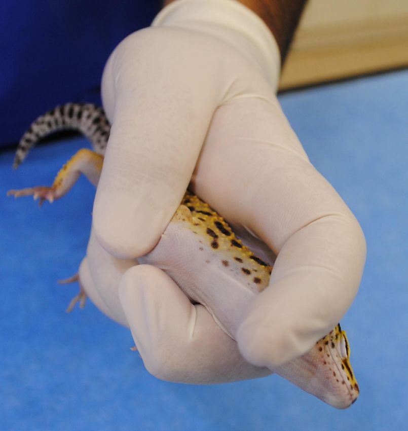
Veterinarni Medicina, 64, 2019 (04): 172–177 Original Paper
https://doi.org/10.17221/164/2018-VETMED
collection from leopard geckos has been published the operator was placed on the head just behind the
(Morici et al. 2016). The aim of this study was to right eye while the left middle finger was positioned
establish reference data for blood parameters in on the left side of the neck. The head of the gecko
a group of healthy adult male leopard geckos in was slightly rotated to the left, exposing the area
captivity. of the right jugular vein (Figure 1). The skin on the
right side of the neck was disinfected with a diluted
alcohol solution. The needle of the pre-heparinised
MATERIAL AND METHODS (heparinum natricum, Heparin inj., 5000 IU/ml,
Léčiva Praha, Czech Republic) insulin syringe
Animals. Forty-four (44) clinically healthy adult (0.5 ml – 29G insulin syringe, BD medical, France)
male leopard geckos (Eublepharis macularius) kept was gently inserted rostro-caudally into the right
in captivity were included in this study. They origi- jugular vein (Figure 2). After the total volume of
nated from one large captive bred population in the
Czech Republic and ranged in age from 15 months
to 16 months. All geckos in this study were kept
and used in accordance with directive 2010/63/EU
and ethical approval was obtained. Geckos were
housed in glass terraria (90 × 45 × 45 cm) with air
temperature maintained at 30–31 °C, and air hu-
midity at 40–50%. The substrate consisted of folded
paper towel. Recycled paper box and paper towel
rolls were offered to geckos in order to provide
more surface area for climbing and as hiding spots.
A heat pad (RH-6, ReptiTherm®, Zoo Med, USA)
was placed under each terrarium as a heat source,
while a UVA/UVB light bulb (Reptisun 5.0 UVB
Mini Compact Fluorescent Bulb, Zoo Med, USA)
was placed inside each terrarium as a light and UV
light source. The geckos were maintained under
a 12/12 light/dark regime. Feeding was performed
three times a week with crickets or cockroaches.
The geckos were acclimatised in terraria for Figure 1. Leopard gecko manually restrained for jugular
14 months before the procedures were undertak- vein venepuncture
en. Twenty-two geckos (group A) were subjected
to blood collection for haematology analyses, and
twenty-two geckos (group B) were used for blood
collection for blood biochemistry analyses. The
body condition of each gecko was evaluated in
a standard clinical examination. The body weight
of each of the geckos was measured on digital scales
(Kern and Sohn GmbH, Balingen, Germany). The
mean body weight of the 44 leopard geckos was
37.6 ± 5.9 g (range 28.6–54.7 g). Geckos were fasted
for 24 hours prior to blood collection. Access to
water was not limited.
Blood collection from the jugular vein. The
room air temperature was maintained at 26 °C.
Geckos were manually restrained with the head
and neck extended. Blood was collected using the
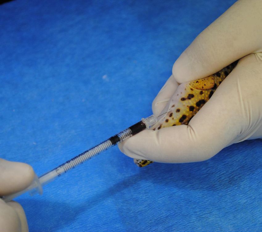
method published recently by Morici et al. (2016) Figure 2. Jugular vein venepuncture in a leopard gecko
and Morici et al. (2018). Briefly, the left index of (Eublepharis macularius)
173Original Paper Veterinarni Medicina, 64, 2019 (04): 172–177
https://doi.org/10.17221/164/2018-VETMED
0.15–0.20 ml of blood was collected the needle was Windows 7 (Microsoft ®, USA). For all data, the
gently withdrawn and a cotton swab was pushed mean, median, standard deviation and minimum-
against the neck to prevent bleeding and haema- maximum (min-max) were calculated.
toma formation. The phlebotomy was conducted
by the same person (M.M.) for all animals. Blood
samples were immediately transferred to a labora- RESULTS
tory in the same building for processing.
Haematological analyses. All analyses were In two leopard geckos from group A the blood
conducted by the same person (Z.K.). Haematocrit collection from both the right and the left jugular
(packed cell volume) measurements were per- veins was unsuccessful. These two animals were
formed using the micro-haematocrit method excluded from the study. Blood was collected easily
(Pejrilova et al. 2004). Haemoglobin concentra- from the right jugular vein in 12 of the 20 leopard
tion was determined spectrophotometrically us- geckos. In seven animals, collection of blood from
ing a standard cyanmethaemoglobin method with the right jugular vein was difficult, and the blood
one modification: the samples were centrifuged samples had to be collected from both the right
following red blood cell lysis to remove the nu- and left jugular vein. In one animal, our attempts
clear and cytoplasmic debris. The total red blood to collect blood from the right jugular vein were
cell count and white blood cell count were deter- not successful and the blood sample had to be col-
mined manually using a haemocytometer with lected from the left jugular vein.
Natt and Herrick’s stain. Air-dried blood smears In group B, blood was collected from the right
were stained using the Pappenheim method (May- jugular vein easily in 17 leopard geckos. In two
Grünwald + Giemsa-Romanowski stains, Pejrilova animals, we failed to collect the whole volume of
et al. 2004). The leukocyte differential counts were blood necessary for analysis from the right jugular
analysed using an Olympus BX 51TF light micro- vein, and blood samples had to be collected from
scope and documented with an Olympus C 3030 di- the right and left jugular veins. In one animal, our
gital camera. Two hundred cells were evaluated for attempts to collect blood from the right jugular vein
the differential count. White blood cells were clas- were not successful and the blood sample had to be
sified as heterophils, basophils, eosinophils, mono- collected from the left jugular vein only.
cytes, azurophils or lymphocytes. In five cases, only small haematomas were pre-
Biochemical analyses. The biochemical analyses sent immediately after the venepuncture, which
were performed with the use of the Avian/Reptilian persisted for between one and two days. The effect
Profile Plus rotor on the VetScan VS 2 ® analyser of the phlebotomy was monitored for a time span of
(Abaxis, Inc., Union City, CA). All samples were six to twelve weeks and all animals were considered
run with exactly 100 μl of plasma. The following healthy without any complications.
parameters were measured: total protein, albu- Blood samples collected from two animals in
min, globulins, glucose, bile acids, aspartate ami- group B had to be excluded from the study due
notransferase, creatine kinase, calcium, sodium, to haemolysis. The total number of blood sam-
potassium, phosphorus and uric acid. ples used for laboratory analyses was therefore
Statistics. The distribution of data was evaluated 20 in group A and 20 in group B. The mean body
with the use of a standard Excel program test for weight of the 20 leopard geckos in group A and
Table 1. Haematology profiles of 20 male leopard geckos (Eublepharis macularius)
Haemoglobin PCV RBCs WBCs Heterophils Basophils Eosinophils Monocytes Azurophils Lymphocytes
Parameter
(g/l) (%) (10 /l) (109/l)
12
(%) (109/l) (%) (109/l) (%) (109/l) (%( (109/l) (%) (109/l) (%) (109/l)
Mean 72.58 25.40 0.85 10.47 17.6 1.83 16.6 1.67 2.7 0.29 4.7 0.48 19.5 2.03 39 4.17
SD 11.03 3.68 0.14 2.58 8.1 0.92 10.8 1.04 2.7 0.33 4.1 0.40 8.7 1.07 14.7 2.12
Max 96.05 31.00 1.08 12.50 34 3.36 41 3.62 8 1.2 15 0.40 36 4.50 66 9.24
Min 54.94 18.00 0.48 6.00 4 0.40 2 0.14 0 0 0 0 6 0.54 15 1.12
Median 70.07 25.00 0.83 10.00 17.5 1.91 14 1.47 2 0.14 3 0.35 22.5 1.94 36.5 3.86
PCV = packed cell volume; RBCs = red blood cells; SD = standard deviation; WBCs = white blood cells
174Veterinarni Medicina, 64, 2019 (04): 172–177 Original Paper
https://doi.org/10.17221/164/2018-VETMED
Table 2. Blood chemistry profiles of 20 male leopard geckos (Eublepharis macularius)
TP Albumin Globulin Glucose UA AST CK Ca P K+
Parameter
(g/l) (g/l) (g/l) (mmol/l) (µmol/l) (µkat/l) (µkat/l) (mmol/l) (mmol/l) (mmol/l)
Mean 55.60 16.45 39.15 6.18 67.95 0.83 25.40 3.05 1.40 5.78
SD 7.52 2.37 5.74 1.35 42.63 0.42 29.46 0.18 0.23 0.58
Max 67.00 23.00 47.00 10.10 160.00 1.80 117.30 3.44 1.87 7.00
Min 42.00 13.00 29.00 4.30 18.00 0.30 1.20 2.76 1.14 4.50
Median 56.50 15.50 40.00 6.20 54.00 0.70 15.75 3.00 1.30 5.90
AST = aspartate aminotransferase; Ca = calcium; CK = creatine kinase; K+ = potassium; Na+ = sodium; P = phosphorus;
SD = standard deviation; TP = total protein; UA = uric acid
20 leopard geckos in group B was 36.7 ± 5.2 g (range other gecko species (Sacchi et al. 2007; Mayer et
28.6–49.7 g) and 38.5 ± 6.7 g (range 29.2–54.7 g), al. 2011; Olayemi 2011; Salamat et al. 2013). The
respectively. effects of age, sex, reproductive activity, altitude,
Haematology values and blood biochemistry season and feeding activity on lizard blood pro-
profiles of male leopard geckos are presented in files have been well documented (Knotek et al.
Tables 1 and 2. The most common leukocytes in 2003; Pejrilova et al. 2004; Knotkova et al. 2005;
the peripheral blood were lymphocytes; azurophils, Gonzalez-Morales et al. 2015, 2017; Guadarrama
heterophils and basophils were relatively common, et al. 2019) and confirmed for different gecko spe-
monocytes were uncommon and eosinophils were cies (Sacchi et al. 2007; Mayer et al. 2011; Olayemi
the least common. The calcium to phosphorus ratio 2011). Based on these findings, only captive adult
(Ca : P) was about 2 in all leopard geckos. The re- males in good health condition were included in
sults of bile acids in all 20 male leopard geckos were the present study. Similar to Mayer et al. (2011), the
the same with bile acids < 3 5 µmol/l. Therefore, presented data have been generated from a single
bile acids was excluded from the blood biochem- colony and we acknowledge that care has to be tak-
istry analysis. en in interpreting the results as a reference range.
In comparison with the other methods for ve-
nepuncture in geckos that have been suggested
DISCUSSION by other authors (e.g. cardiocentesis, phleboto-
my of the ventral abdominal vein (Redrobe and
Leopard geckos are very commonly kept as a pet MacDonald 1999; Hernandez-Divers 2006)), the
species and have become a lizard model for re- technique of blood collection from the jugular vein
search purposes. Despite the fact that the leopard was not stressful or dangerous for any of the leop-
gecko is one of the three most common species of ard geckos used in the present study. In only two
captive lizards in Europe (with the veiled chame- of 44 animals was the method not successful. In
leon Chamaeleo clyptratus and the inland bearded 11 animals, the blood sample had to be collected
dragon Pogona vitticeps), data dealing with hae- from the right and/or left jugular vein. Blood col-
matological and biochemical blood profiles of lection using this method proved to be easy, reliable
captive leopard gecko have not been reported. To and repeatable, and the geckos did not show any
the best of our knowledge, this is the first study to adverse effects.
report haematology and blood biochemistry values Our mean values for white blood cell counts
for a group of captive adult male leopard geckos. were similar to the results of Mayer et al. (2011),
The study incorporated a large population of male but in the present study the range for the white
leopard geckos kept in a standardised environment, blood cell counts was narrower. The most com-
with monitoring of the animals’ health and activity mon leukocytes in the peripheral blood of 20 male
performed daily. leopard geckos were lymphocytes, azurophils and
The present study differs from similar studies that heterophils. In accordance with the study of Mayer
have been focused on haematology and biochem- at al. (2011), lymphocytes were the most common
istry in wild or laboratory-housed populations of leukocyte seen on the blood smears. The classifica-
175Original Paper Veterinarni Medicina, 64, 2019 (04): 172–177
https://doi.org/10.17221/164/2018-VETMED
tion of leukocytes in reptiles is difficult since these productive activity (Knotek et al. 2003; Pejrilova et
cells show morphological variation among species al. 2004; Knotkova et al. 2005; Mayer et al. 2011). In
(Sacchi et al. 2007). Olayemi (2011) identified only accordance with this knowledge and the expected
four types of leukocytes in the peripheral blood differences in blood biochemistry profiles between
of house geckos (Hemidactylus frenatus): hetero- males and females, the evaluation of blood profiles
phils, lymphocytes, eosinophils and monophils, for female leopard geckos within different repro-
while Sacchi et al. (2007) and Mayer et al. (2011) ductive activity periods is needed. This research is
identified five different types of leukocytes: hetero- currently ongoing.
phils, basophils, eosinophils, monocytes and lym-
phocytes. In the present study, six different types
of leukocytes were identified in leopard geckos: Acknowledgement
heterophils, basophils, eosinophils, monocytes,
lymphocytes and azurophils. In reptiles, azuro- The author thanks to Dr. William Lewis for his
phils differ from monocytes and lymphocytes by valuable comments.
the presence of typical azurophilic granules in the
cytoplasm (Knotkova et al. 2002; Pejrilova et al.
2004). The percentage of the most common leu- REFERENCES
kocytes, lymphocytes, was similar to results for
Moorish geckos (Sacchi et al. 2007), but the per- Gonzalez-Morales JC, Quintana E, Diaz-Albiter H, Gue-
centages of heterophils and eosinophils were lower vara-Fiore P, Fajardo V (2015): Is erythrocyte size a strat-
and the percentages of basophils and monocytes egy to avoid hypoxia in Wiegmann´s Torquate Lizards
were higher for the leopard gecko. (Sceloporus torquatus)? Field evidence. Canadian Journal
The use of the VetScan VS 2® automatic chemistry of Zoology 93, 377–382.
analyser with the Avian/Reptilian Profile Plus ro- Gonzalez-Morales JC, Beamonte-Barrientos R, Bastiaans
tor for clinical practice and research with reptiles E, Guevara-Fiore P, Quintana E, Fajardo V (2017):
has been well documented (Knotkova et al. 2010; A mountain or a plateau? Hematological traits vary non-
Mayer et al. 2011). Similar to results from previous linearly with altitude in a Highland Lizard. Physiological
studies (Knotkova et al. 2010; Mayer et al. 2011), and Biochemical Zoology 90, 638–645.
bile acid concentrations in all blood samples from Guadarrama SS, Dominguez-Vega H, Diaz-Albiter HM,
leopard geckos in the present study were reported Quijano A, Bastiaans E, Carrillo-Castilla P, Manjarrez J,
as < 35 µmol/l. The lower end of the dynamic range Gomez-Ortiz Y, Fajardo V (2019): Hypoxia by altitude
of the bile acids on the rotor is < 35 µmol/l. These and welfare of captive Beaded Lizards (Heloderma hor-
results were therefore excluded from the blood bio- ridum) in Mexico: Hematological approaches. Journal of
chemistry analysis. In cases where the true bile acid Applied Animal Welfare Science 9, 1–9.
concentration is required another analyser must be Hernandez-Divers SJ (2006): Diagnostic techniques. In:
used. The ability to measure the concentration of bile Mader DR (ed.) Reptile Medicine and Surgery. 2nd edn.
acids in the peripheral blood proved interesting and Saunders Elsevier, St. Louis, 490–532.
feasible for veterinary practice with chelonians and Knotek Z, Jekl V, Knotkova Z, Dorrestein GM (2007): Serum
medium-sized lizards (Knotek et al. 2007; Knotkova bile acid concentrations in red-eared slider females with
et al. 2008; Knotek et al. 2009). The concentration of active folliculogenesis. Proceedings 43 rd… International
bile acids can be determined with the use of different Symposium on Diseases of Zoo and Wild Animals, Ed-
automatic analyser systems. However, the volume inburgh, 107–109.
of blood sample that could be easily collected from Knotek Z, Knotkova Z, Doubek J, Pejrilova S, Hauptman K
small reptile species like the leopard gecko (e. g. (2003): Plasma biochemistry in female Green Iguanas
0.100–0.200 µl) is not large enough for the use of (Iguana iguana) with calcium metabolism disorders. Acta
standard laboratory analysers. Further studies fo- Veterinaria Brno 72, 83–189.
cused on feasible methods for determining bile acid Knotek Z, Knotkova Z, Hrda A, Dorrestein GM (2009):
concentrations in small reptile species are necessary. Plasma bile acids in reptiles. 30 th Annual Conference
Blood concentrations of total protein, albumin, AAV, AEMV, ARAV, Milwaukee, 124–127.
globulins, cholesterol, uric acid and total calcium Knotkova Z, Dorrestein GM, Jekl V, Janouskova J, Knotek
in female lizards are strongly influenced by their re- Z (2008): Fasting and 24-hour postprandial bile acids of
176Veterinarni Medicina, 64, 2019 (04): 172–177 Original Paper
https://doi.org/10.17221/164/2018-VETMED
healthy female red-eared terrapins (Trachemys scripta Morici M, Di Giuseppe M, Spadola F, Oliveri M, Knotkova
elegans). Veterinary Record 163, 510–514. Z, Knotek Z (2018): Intravenous alfaxalone anaesthesia
Knotkova Z, Doubek J, Knotek Z, Hajkova P (2002). Blood in leopard geckos (Eublepharis macularius). Journal of
cell morphology and plasma biochemistry in Russian Exotic Pet Medicine 27, 11–14.
tortoises (Agrionemys horsfieldi). Acta Veterinaria Brno Olayemi OA (2011): Hematological parameters of House
71, 191–198. Gecko (Hemidactylus frenatus) in Ibadan Metropolis,
Knotkova Z, Knotkova E, Hrda A, Knotek Z (2010): Com- Nigeria. Veterinary Research 4, 77–80.
parison of blood plasma of Green Iguana (Iguana iguana) Pejrilova S, Knotkova Z, Knotek Z, Vrbas S (2004): Age-
by Cobas Mira Plus, AA Series Spectrometer, and VetScan related changes of the haematological profile in Green
VS2 analyser (in Czech). Veterinarni klinika 7, 55–58. Iguana (Iguana iguana rhinolopha). Acta Veterinaria Brno
Knotkova Z, Pejrilova S, Trnkova S, Matouskova O, Knotek 73, 305–312.
Z (2005): Influence of reproductive season upon plasma Redrobe S, MacDonald J (1999): Sample collection and
biochemistry values in Green Iguanas. Acta Veterinaria clinical pathology of reptiles. Veterinary Clinics of North
Brno 74, 515–520. America: Exotic Animal Practice 2, 709–730.
Mayer J, Knoll J, Mitchell MA (2011): Characterizing the Sacchi R, Pupin F, Zuffi MAL, Scali S, Boncompagni E,
hematologic and plasma chemistry profiles of captive Binda A, Galeotti P, Fasola M (2007): Blood cell morphol-
Crested Geckos (Rhacodactylus ciliatus). Journal of Her- ogy of the Moorish Gecko, Tarentola mauritanica. Am-
petological Medicine and Surgery 21, 68–75. phibia-Reptilia 28, 503–508.
Morici M, Di Giuseppe M, Spadola F (2016): Preliminary study Salamat MA, Vaissi S, Fathipour F, Sharifi M, Parto P (2013):
on intravenous alfaxalone anaesthesia in Leopard Geckos Morphological observations on the erythrocyte and
(Eublepharis macularius). Proceedings 10th Congress In- erythrocyte size of some gecko species, Iran. Global Vet-
ternational on Exotic-Yaboumba World, Paris, 47–48. erinaria 11, 248–251.
Received: December 7, 2018
Accepted after corrections: March 26, 2019
177You can also read