Feline Sino-orbital Fungal Infection Caused by Aspergillus and Scopulariopsis - seer ufrgs
←
→
Page content transcription
If your browser does not render page correctly, please read the page content below
Acta Scientiae Veterinariae, 2019. 47(Suppl 1): 383.

                                                                     CASE REPORT                                                      ISSN 1679-9216
                                                                      Pub. 383

       Feline Sino-orbital Fungal Infection Caused by Aspergillus and Scopulariopsis

    Fernanda Vieira Amorim da Costa1, Andreia Spanamberg2, Ricardo Araujo3,4, Juliana Werner5 & Laerte Ferreiro2

                                                                   ABSTRACT

Background: Deep fungal infections of the orbit and nasal passages causing rhinitis and ulcerative keratomycosis are
uncommonly reported in cats. Hyalohyphomycetes and phaeohyphomycetes have rarely been associated with this disorder.
Sino-orbital fungal diseases are emerging and more invasive than sino-nasal fungal diseases with poor response to therapy
and a worse prognosis. Brachycephalic feline breeds seem to be at increased risk for development of upper respiratory
fungal diseases. Diagnosis is based on the demonstration of fungal hyphae by cytology or histology and definitive confir-
mation by fungal culture and molecular methods. This is the first case report of a cat with clinical mixed fungal ball with
Aspergillus and Scopulariopsis in Brazil.
Case: A 3-year-old male Persian cat, in São José city, Santa Catarina, Brazil, was presented with exophthalmos and cor-
neal ulcer of the left eye and protrusion, hyperemia, quemosis and fibroses of the left third eyelid. The retropulsion of the
globe was negative in this eyeball and a presumptive diagnosis of a retrobulbar mass was made. The patient underwent
a surgical procedure for inspection and collection of samples for bacterial and mycological culture. Culture revealed no
bacterial growth, however, unique and abundant growth of Aspergillus spp. was present. A subconjunctival enucleation of
the left eye was made and the mass was sent for histopathology examination. Histology showed inflammatory proliferative
necrotizing pyogranulomatous reaction; with the presence of severe fungal infection evidenced by large number of hyaline
septated regular and irregular mold hyphae. Molecular identification was performed using panfungal primers (ITS3-F /
ITS4-R). Patient was treated with systemic itraconazole associated with amphotericin B and topical clotrimazole. A mass
started to grow rapidly in the left pterygopalatine fossa and was surgically removed, but recurrence occurred seven days
after. After 22 days of treatment, the animal died suddenly with a history of acute inspiratory dyspnea and cyanosis at
the time prior to death. The diagnosis of sino-orbital fungal disease in the feline was based on clinical signs, mycological
culture, histopathology and molecular methods.
Discussion: Sino-orbital fungal diseases rare in cats and can result in significant injuries to the upper respiratory tract
and eyes, sometimes resulting in enucleation and death. It seems feasible that a brachycephalic facial conformation may
be an important risk factor for the development of sino-nasal fungal diseases in cats. Despite using selected drugs and
eye enucleation to treat the disease, the cat developed a rapid growing oral mass that probably caused acute inspiratory
dyspnea and death. Since no controlled studies exist on the treatment of feline fungal diseases, these cases are a challenge
to the feline practitioner and this type of clinical manifestation should be included in the differential diagnosis of upper
respiratory and ocular diseases.
Keywords: Aspergilllus, cat, fungal disease, retrobulbar mass, upper respiratory disease.

                                                           DOI: 10.22456/1679-9216.91581
Received: 8 December 2018                                      Accepted: 25 March 2019                                        Published: 10 April 2019
1
 Setor de Medicina Felina do Hospital de Clínicas Veterinárias (HCV) & Setor de Micologia Veterinária, Faculdade de Veterinária (FaVet), Universidade
                                                                      2

Federal do Rio Grande do Sul (UFRGS), Porto Alegre, RS, Brazil. 3i3S - Instituto de Investigação e Inovação em Saúde, Porto, Portugal. 4Department of
Medical Biotechnology, College of Medicine and Public Health, Flinders University, Adelaide, South Australia, Australia. 5Werner & Werner - Labo-
ratório de Patologia Veterinária, Curitiba, PR, Brazil. CORRESPONDENCE: F.V.A. Costa [fernanda.amorim@ufrgs.br - Tel.: +55 (51) 3308-9643].
Setor de Medicina Felina - HCV, UFRGS. Av. Bento Gonçalves n. 9090. Bairro Agronomia. CEP 91540-000 Porto Alegre, RS, Brazil.

                                                                          1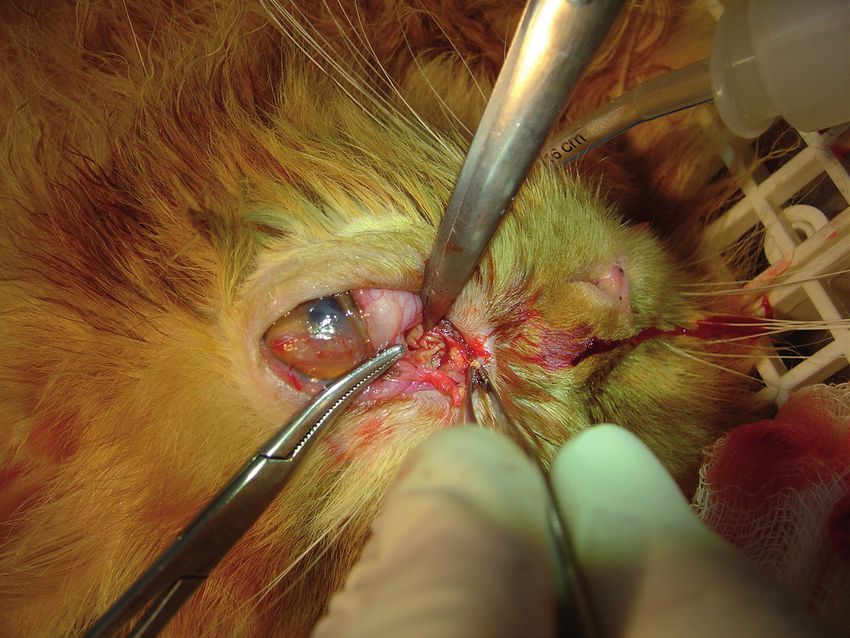
F.V.A. Costa, A. Spanamberg, R. Araujo, J. Werner & L. Ferreiro. 2019. Feline Sino-orbital Fungal Infection Caused by
Aspergillus and Scopulariopsis. Acta Scientiae Veterinariae. 47(Suppl 1): 383.
INTRODUCTION However, the retropulsion of the globe was negative in
Sino-nasal and sino-orbital fungal disease can be this eyeball. The left superior cornea had an irregular
caused by hyalohyphomycetes (hyaline septate hyphae) form, corneal edema, neovascularization and retained
and phaeohyphomycetes (pigmented septate hyphae). fluorescein stain (Fluoresceína Strips ®)4 [Figure 2].
Investigations suggested that the majority of infections A presumptive diagnosis of a retrobulbar mass was
were caused by Aspergillus species. In cats, the disea- made. Furthermore, the cat had mucopurulent nasal
se occurs in two main forms: sino-nasal aspergillosis and ocular discharge from the same side.
(SNA) and sino-orbital aspergillosis (SOA) [5,16,29]. The patient had no fever or weight loss, but had
SNA is characterized by signs of chronic nasal infection, a history of poor appetite and prostration. Immunoas-
such as sneezing, uni or bilateral serous to mucopurulent says for the detection of feline immunodeficiency virus
nasal discharge, and sometimes epistaxis. SOA is the and feline leukemia virus (FIV/FeLV) were negative.
more invasive form because the fungus invades the sub- Complete blood count and serum biochemical assay
mucosal tissue of the nasal cavity and sinus and extends were also performed, which showed only leukopenia
through the orbital bone and into the retrobulbar space. and lymphopenia. Initial supportive therapy consisted
SNO clinical signs include unilateral exophthalmos, of fluid and electrolyte therapy, eye drops of flurbipro-
third eyelid prolapse, conjunctival hyperaemia and fen, moxifloxacin and chondroitin sulfate, in addition
keratitis. Response to therapy is less successful in SNO to oral amoxicillin and cyproheptadine for 15 days.
aspergillosis and the prognosis is generally worse [16]. After clinical improvement, the patient un-
Definitive diagnosis requires a combination of derwent a surgical procedure for inspection and col-
imaging, visualization of fungal plaques by rhinoscopy, lection of samples for cytology, culture, antimicrobial
and identification of fungal structures on culture and sensitivity profile and histopathological analysis of
cytology, or histopathology [4,29]. Treatment of this the mass. During the procedure, there was a purulent
form of the disease should be multifactorial, including and caseous tissue in the third eyelid conjunctiva, oc-
orbitotomy or eye enucleation and a combination of cupying the medial and retrobulbar region (Figure 3).
systemic antifungal treatment with local therapy using Cytology showed the presence of red blood
intranasal infusions under general anesthesia [16]. cells, predominance of degenerate neutrophils and few
This is the first case report of a cat with cli- lymphocytes, and free and intracellular bacteria. Tissue
nical mixed fungal infection with Aspergillus and fragments were subjected to aerobic culture at 37°C
Scopulariopsis in Brazil showing details of the clinical on blood agar and MacConkey agar, and incubated at
presentation, diagnosis, medical and surgical manage- 30°C on Sabouraud dextrose agar. Culture revealed
ment of the disease. no bacterial growth, however, unique and abundant
growth of Aspergillus spp. was present.
CASE
The patient was then submitted to radiographic
A 3-year-old male Persian cat, weighing 3.8 kg investigation, which showed increased radiopacity in
was seen at Green Cross Veterinary Clinic, São José, the left sinus and loss of definition of the turbinate
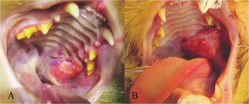
Santa Catarina, Brazil, presenting with exophthalmos, bones. The cat underwent a surgical procedure for
lagophthalmos and asymmetry of the frontal and dor- subconjunctival enucleation of the left eye (Figure
sal face. Severe protrusion, hyperemia, chemosis and 4), which was sent for histopathology examination.
fibrosis of the left third eyelid were noted (Figure 1). Histology showed inflammatory proliferative necro-
The cat had normal vision in both eyes; vision was tizing pyogranulomatous reaction, with presence of
diminished in the left eye showing limited response septate branching hyphae both regular and irregular,
to the menace reflexe in this eyeball. The Schirmer morphologically compatible with Aspergillus sp. and
I Tear Test (STT strips)1 was 16 mm/min in the right other hyaline septated mold (Figure 5). Special stai-
eye and 3mm/min in the left eye. Intraocular pressure ning with Ziehl Nielsen techinique was performed for
(IOP) was measured using applanation tonometry (To- mycobacteria investigation, but the result was negative.
nopen® XL)2, after application of a topical ophthalmic The DNA was extracted from the formalin
anesthetic solution (Anestalcon®)3. The IOP was 17 fixed paraffin embedded (FFPE) sample using the
mmHg in the right eye and 48 mmHg in the left eye. QIAamp® FFPE tissue kit (Qiagen)5. DNA extracted
2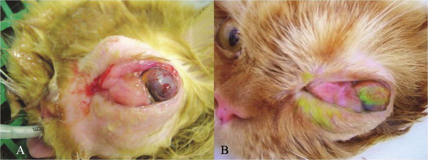
F.V.A. Costa, A. Spanamberg, R. Araujo, J. Werner & L. Ferreiro. 2019. Feline Sino-orbital Fungal Infection Caused by
Aspergillus and Scopulariopsis.                                             Acta Scientiae Veterinariae. 47(Suppl 1): 383.

was detected with panfungal PCR using ITS3-F                    Ringer’s solution at the maintenance dose of 50 mL/
(5’-GCATCGATGAAGAACGCAGC-3’) and ITS4-                          kg/day. After 2 days of treatment, the animal died
R (5’- TCCTCCGCTTATTGATATGC-3’ [34] for                         suddenly with a history of acute inspiratory dyspnea
amplification of internal transcribed spacer 2 (ITS2)           and cyanosis at the time prior to death. The animal’s
region. PCR amplification was performed in a 25 μL              owner declined to perform a necropsy.
containing 1 μL of DNA extract, 12.5 μL Qiagen Taq
                                                                                      DISCUSSION
PCR master mix (Qiagen)5 and 0.5 μL of each primer
(for a 0.2 μM final concentration of each primer). After                 Sino-orbital fungal disease is rare in cats and
a preincubation at 94°C for 15 min, the amplification           can result in significant injuries to the upper respira-
was performed for a total of 35 cycles as follows: de-          tory tract and eyes, sometimes resulting in enucleation
naturation at 94°C for 30 s, annealing at 57°C for 90           and death. In this report, the patient was a Persian cat,
s, extension at 72°C for 1 min, and a final extension           agreeing with previous reports that 8 out of 12 cats
step of 10 min at 72°C. The primers ITS3 and ITS4               treated for nasal or ocular aspergillosis were Persian
amplify a fragment of between 300 and 400 bp. PCR               or Himalayan [2,12,15,18,23,31,35]. It seems feasible
product was separated on a 2% agarose. PCR product              that a brachycephalic facial conformation may be an
was purified using PuriLink® PCR Purification Kit               important risk factor for the development of sino-nasal
(Invitrogen)6, and sequencing to confirmed the pres-            fungal disease in cats [29], because fungal organisms
ence of fungal elements in the tissue sample.                   are a part of the normal feline ocular and nasal flora
         The sequencing analysis of the fragments re-           [6,19] and it is possible that the brachycephalic airway
vealed a clear mix of two fungal sequences (two peaks           of Persian and Himalayan cats may at times become
for each position). Aspergillus was initially cultured          overwhelmed in its ability to clear Aspergillus spp.
from the tissue, therefore the consensus profile for ITS2       from the cat’s upper airway [2]. Tomsa et al. [31]
of 100 Aspergillus sequences was subtracted from the            presupposed that susceptibility to localized fungal
sequencing graph to obtain the sequence for the second          infection can occur as a consequence of the possible
fungus (e.g. for a sequencing position with A and C, if         altered airflow observed in pronounced brachycephaly.
the nucleotide of consensus Aspergillus sequence was            This could cause changes in the vasomotor action of
A, then the second position was considered to be C).            the vasculature and decrease mucociliary clearance.
At the end of this detailed analysis of each nucleotide         Wilkinson et al. [36] reported two cats with sino-nasal
position, two sequences were obtained. Basic local              disease who also had involvement of the orbit. In one
alignment search tool (BLAST®)7 was performed in                case this was associated with fungal sinusitis. Our
both sequences giving the results 100% match for As-            patient had both presentations of this disease, with
pergillus and Scopulariopsis respectively. No fungal            the same clinical signs as reported for the sino-orbital
species could be identified using these ITS2 sequences,         (SOA) and SNA forms. The disease probably extended
as these region was common (>99% match) to multiple             beyond the sinonasal cavity and involved the adjacent
species within each genus.                                      orbital structure and palate. Therefore, the first presen-
         The patient was treated with intranasal clotrim-       ting signs in cats are referable to orbital invasion [3].
azole once a week and systemic itraconazole at a dose           In this case, the patient presented first orbital involve-
of 50 mg orally per day. After 20 days of treatment, the        ment, and the signs were exophthalmos, high intraocu-
patient showed a mass of 2 cm in diameter on the left           lar pressure, severe protrusion and inflammation of the
side of the soft palate (Figure 6A), which was removed          third eyelid. All of these signs were likely secondary
surgically. However, recurrence occurred 7 days after           to an invasive retrobulbar fungal granuloma that was
surgery (Figure 6B), and the patient began to show              pushing and compressing the eyeball, characterizing
labored breathing due to obstruction of the oropharynx.         the SOA form [2,3,15,35]. Furthermore, the patient
         Due to the rapid evolution of the disease and          showed mucopurulent nasal and ocular discharge from
poor response to treatment, the cat was treated with            the same side, corroborating with Hamilton et al. [15]
subcutaneous amphotericin B at 0.5 mg/kg diluted in             and Barachetti et al. [2], which said that these signs
350 mL of warm 0.45% sodium chloride and 2.5%                   are commonly found in nasal aspergillosis. The corneal
glucose, and intravenous fluid therapy with lactated            and lacrimal film alterations, including the corneal

                                                            3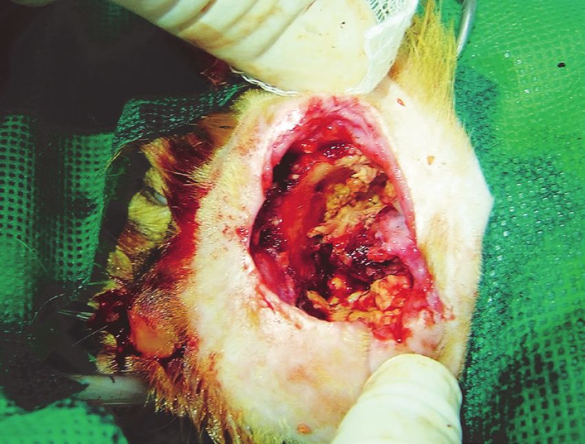
F.V.A. Costa, A. Spanamberg, R. Araujo, J. Werner & L. Ferreiro. 2019. Feline Sino-orbital Fungal Infection Caused by
Aspergillus and Scopulariopsis.                                             Acta Scientiae Veterinariae. 47(Suppl 1): 383.

                                                                               ulcer and low STT in the left eye, were possibly due to
                                                                               corneal exposure, a consequence of the exophthalmos
                                                                               and lagophthalmos [2,5,15,35].
                                                                                       Disseminated aspergillosis is apparently an
                                                                               opportunistic infection in immunosuppressed cats [26].
                                                                               In contrast, incompetence of the immune system is not
                                                                               believed to be required for nasal infection. Only 2 of
                                                                               16 previously reported cats with sinonasal or orbital
                                                                               aspergillosis had a confirmed immunosuppressive
                                                                               condition [12,15,18,23,31,35]. In this study, the animal
                                                                               had no history of trauma or previous infections and was
                                                                               negative for FIV and FeLV infection, both important
                                                                               causes of immunosuppression in cats.
Figure 1. Patient showing protrusion, hyperemia, chemosis of the third
eyelid and exophthalmos in the left eye. It’s possible to note nasal and
                                                                                       Diagnostic imaging for orbital disease typically
ocular mucopurulent discharge in the same side.                                includes skull radiography and orbital ultrasonography,
                                                                               however, CT and magnetic resonance imaging (MRI)
                                                                               have been successfully used to aid in the diagnosis of

Figure 2. A- Lagophthalmos, corneal edema, neovascularization and mydriasis in the left eye. B- Visible corneal ulcer after the lesion retained fluo-
rescein stain.

                                                                               Aspergillus spp. infections [15,16,25,29]. Radiography
                                                                               of the nasal cavity and frontal sinuses may show abnor-
                                                                               malities such as increased soft-tissue densities, loss of
                                                                               turbinates [9,16,31,35] and possible bone erosion/lysis
                                                                               [2,9], as the patient showed in this report. CT imaging
                                                                               is important for assessing the extent of the disease
                                                                               [35] because of its ability to generate cross-sectional
                                                                               images [8]. This technique is also useful to detect cal-
                                                                               cium accumulation within the fungus colony or bone
                                                                               destruction [25]. However, MRI is the best technique
                                                                               to diagnose soft tissue lesions and vascular invasion
                                                                               [16,25]. It can be used to evaluate more critically the
                                                                               optic chiasm, differentiate neoplasia and determine
                                                                               the full extent of the disease [2,25]. Occasionally,
Figure 3. Inspection and biopsy of medial region of the left eye showing
                                                                               cytology of nasal discharge or nasal flush will show
purulent and caseous tissue.                                                   septate hyphae. The presence of hyphae in the tissues

                                                                           4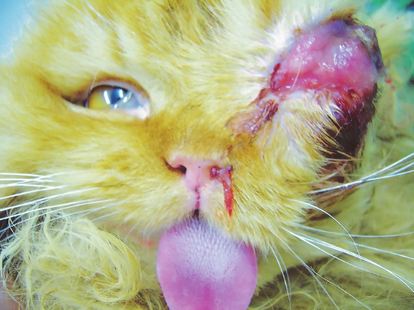
F.V.A. Costa, A. Spanamberg, R. Araujo, J. Werner & L. Ferreiro. 2019. Feline Sino-orbital Fungal Infection Caused by
Aspergillus and Scopulariopsis.                                             Acta Scientiae Veterinariae. 47(Suppl 1): 383.

Figure 4. Left orbital cavity after the enucleation with purulent and case-       Figure 5. Microscopy of sino-orbital lesion showing necrotic tissue with
ous tissue.                                                                       multiple irregular, septate branching hyphae, morphologically compatible
                                                                                  with Aspergillus sp. and other hyaline septated mold [PAS; 200x].

Figure 6. After treatment with antifungal therapy. A- Mass of 2 cm in diameter in the soft palate on the left side. B- Recurrence of the lesion in the soft
palate 7 days after surgery.

will aid in differentiating diagnosis of Aspergillus spp.                         by septate hyaline hyphae (suggesting both regular
from Mucor spp., and Candida spp. Infections [9].                                 and irregular structures), some with dichotomous
However, sometimes it can be inconclusive [15,23],                                branching suggestive of Aspergillus sp. corroborating
as in the case reported here.                                                     with previous reports [2,17,31,35]. Histopathology
         Occasionally, the results of fungal culture do                           has been used as the primary ante mortem diagnostic
not distinguish between Aspergillus spp. colonization                             method of feline aspergillosis [10]. Biopsy samples
and active infection [26]. False negatives can also occur                         are more likely to reveal the organisms, but false ne-
[36]. Nevertheless, when a sample can be obtained                                 gative biopsies occur if only the superficial aspect of
directly from the site of visible fungal infection, a                             the lesion is biopsied [35].
positive fungal culture would provide useful adjunct                                       Molecular methods are needed for correct fungal
diagnostic information [23,31]. In this report, culture                           identification when the conventional methodologies
from biopsied material revealed unique and abundant                               (histopathology and culture) does not allow the complete
growth of Aspergillus spp.                                                        clarification. In our study, although the Aspergillus sp.
         In this case, histology showed inflammatory                              has grown in culture, PCR panfungal was required to
proliferative necrotizing pyogranulomatous reaction,                              confirm fungal identification and a possible case of co-
with the presence of severe fungal infection evidenced                            -presence of molds, due to histopathology suggesting

                                                                              5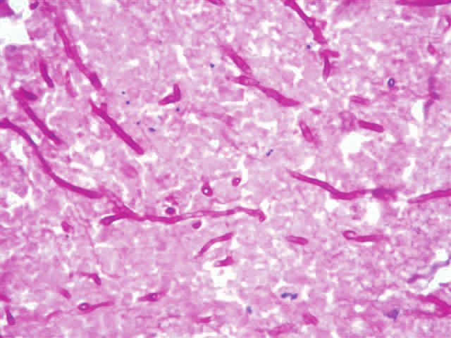
F.V.A. Costa, A. Spanamberg, R. Araujo, J. Werner & L. Ferreiro. 2019. Feline Sino-orbital Fungal Infection Caused by
Aspergillus and Scopulariopsis.                                             Acta Scientiae Veterinariae. 47(Suppl 1): 383.

multiple fungi. The presence of another fungus, Sco-            of clotrimazole is considered the treatment of choice
pulariopsis, was confirmed by sequencing analysis of            for canine nasal aspergillosis. The cure rate has been
ITS2. In tissue the features of hyaline septated molds          as high as 94%.28 This technique has been reported
were similar to those seen with aspergillosis and Sco-          effective in three cats with fungal rhinitis [12,31].
pulariopsis mycosis [13].                                       Two other cats were treated with a topical infusion of
         The most common causes of chronic nasal                clotrimazole in previous reports [15,23]. One had no
discharge in cats include nasal neoplasia, fungal               response to treatment and the other responded initially
sinorhinitis -Aspergillosis [6] and cryptococcosis              but was euthanatized later because of the development
[21,32] - upper respiratory complex [1,5,30,33] and             of new clinical signs of dysphagia and anorexia. Both
bacterial, allergic and idiopatic rhinitis [9]. The drugs       of these cats had also SOA, similar to the case repor-
available to treat fungal infections of the eyeball are         ted here, while the cats that responded successfully to
restricted because eyedrops have limited penetration            clotrimazole had SNA.
into the eyeball [11]. One option is topical itracona-                   In this case, the animal died suddenly. Cer-
zole 1% which may be useful to treat corneal ulcers             tainly, the brachycephalic conformation decreases
caused by Aspergillus fumigatus [22]. In this case, only        sinus aeration and drainage of respiratory secretions
supportive therapy was prescribed until a definitive            secondary to infection, polyps, and allergic rhinosi-
diagnosis was made, that’s why a systemic treatment             nusitis, which have been identified as risk factors for
was preferably chosen.                                          invasive SNA aspergillosis in human beings [3]. As
         Itraconazole generally has good in vitro activi-       was observed, the patient had a mass in the soft palate,
ty against Aspergillus spp., and itraconazole-resistant         which was causing the difficulty to breath and feeding.
strains are uncommonly seen [24].Azoles are by far              This was probably the reason for death, because he
the most ineffective agents, and amphotericin B has             showed acute inspiratory dyspnea and cyanosis at his
limited activity against Scopulariopsis spp. [27,28].           final hours.
In this patient, oral itraconazole was initially selected                Orbital aspergillosis/hialohyphomycosis is rare
because of its documented efficacy against a variety of         in cats and can result in significant injuries to the eyes,
deep mycotic infections in cats and its relative safety         sometimes resulting in enucleation. Nasal aspergillosis
in this species [7]. But its effectiveness for treatment        is also uncommon and can cause dyspnea and death of
of aspergillosis has controversial results. Itraconazole        cats through nasopharyngeal asphyxia. The association
was effective in four cats with SNA aspergillosis, but          of both forms can occur and, unfortunately, the treat-
three of them had recurrence of clinical signs follo-           ment is ineffective so far. Further studies are required
wing discontinuation of treatment [31,35]. However,             in order to better understand and efficiently treat fungal
itraconazole was ineffective for a cat with orbital as-         sino-orbital infections in this species.
pergillosis [24]. In two studies of SOA, there was no
clinical improvement with use of itraconazole and then          MANUFACTURERS
amphotericin B systemically [18,23]. Amphotericin B             1
                                                                 Schering Plough Anima Health. Union, NJ, USA.
has been used for more than 40 years in aspergillosis           2
                                                                 Mentor O & O Inc. Norwell, MA, USA.
                                                                3
                                                                 Allergan Pharmaceuticals. São Paulo, SP, Brazil.
treatment and also targets the fungal cell membrane.            4
                                                                 Ophthalmos Pharmaceuticals. São Paulo, SP, Brazil.
Its less toxic lipid formulations have been used in             5
                                                                 Qiagen. Hilden, Germany.
cases of aspergillosis refractory to azole antifungals          6
                                                                 Invitrogem. Carlsbad, CA, USA.
[20]. Resistance to AMB is a rare phenomenon in A.              7
                                                                 U.S. National Library of Medicine. Rockville, MD, USA.
fumigatus, however A. terreus is intrinsically resistant        Declaration of interest. The authors report no conflicts of
and A. flavus has reduced sensitivity to this antifungal        interest. The authors alone are responsible for the content and
[14]. Topical therapy with a non-invasive infusion              writing of paper.

                                                            6
F.V.A. Costa, A. Spanamberg, R. Araujo, J. Werner & L. Ferreiro. 2019. Feline Sino-orbital Fungal Infection Caused by
Aspergillus and Scopulariopsis. Acta Scientiae Veterinariae. 47(Suppl 1): 383.
REFERENCES
1 Andrew S.E. 2001. Ocular manifestations of feline herpesvirus. Journal Feline Medicine Surgery. 3: 9-16.
2 Barachetti L., Mortellaro C.M., Di Giancamillo M., Giudice C., Martino P., Travetti O. & Miller P.E. 2009.
Bilateral orbital and nasal aspergillosis in a cat. Veterinary Ophthalmology. 12: 176-182.
3 Barrs V.R., Beatty J.A., Lingard A.E., Malik R., Krockenberger M.B., Martin P., O’Brien C., Angles J.M.,
Dowden M. & Halliday C. 2007. Feline sino-orbital aspergillosis: an emerging clinical syndrome. Australian Veteri-
nary Journal. 85(3):23.
4 Barrs V.R. & Beatty J.A. 2010. Upper respiratory tract aspergillosis. In: August J.R. (Ed). Consultations in Feline
Medicine. 6th edn. St. Louis: Saunders Elsevier, pp.36-52.
5 Barrs V.R. & Talbot J.J. 2014. Feline aspergillosis. The Veterinary Clinics of North America Small Animal Practice.
44(1): 51-73
6 Benitah N. 2006. Canine Nasal Aspergillosis. Clinical Techniques Small Animal Practice. 21: 82-88.
7 Boothe D.M., Herring I., Calvin J., Way N. & Dvorak J. 1997. Itraconazole disposition after single oral and intra-
venous and multiple oral dosing in healthy cats. America Journal Veterinary Research.58: 872-877.
8 Choi M.Y., Bae I.H., Lee J.H. & Lee S.J. 2002. Aspergillosis presenting as an optic neuritis. Korean Journal of
Ophthalmology.16: 119-123.
9 Codner E.C., Lurus A.G., Miller J.B., Gavin P.R., Gallina A. & Barbee D.D. 1993. Comparison of computed
tomography with radiography as a noninvasive diagnostic technique for chronic nasal disease in dogs. Journal of the
American Veterinary Medical Association. 202: 1106-1110.
10 Davies C. & Troy G. 1996. Deep mycotic infections in cats. Journal of the American Veterinary Medical Association.
32: 380-391.
11 Freda R. 2006. Use of oral voriconazole as adjunctive treatment of severe cornea fungal infection: case report. Arquivos
Brasileiros de Oftalmologia. 69: 431-434.
12 Furrow E. & Groman R.P. 2009. Intranasal infusion of clotrimazole for the treatment of nasal aspergillosis in two
cats. Journal of the American Veterinary Medical Association. 235: 1188-1193.
13 Guarner J. & Brandt M.E. 2011. Histopathologic diagnosis of fungal infections in the 21st century - Clinical micro-
biology reviews. Journal of the American Veterinary Medical Association. 213: 501-506.
14 Gonçalves S.S., Souza A.C.R., Chowdhary A., Meis J.F. & Colombo A.L. 2016. Epidemiology and molecular
mechanisms of antifungal resistance in Candida and Aspergillus. Mycoses. 59: 198-219.
15 Hamilton H.L., Whitley R.D. & McLaughlin S.A. 2000. Exophthalmos secondary to aspergillosis in a cat. Journal
of the American Animal Hospital Association. 36: 343-347.
16 Hartmann K., Lloret A., Pennisi M.G., Ferrer L., Addie D., Belák S., Boucraut-Baralon C., Egberink H., Frymus
T., Gruffydd-Jones T., Hosie M.J., Lutz H., Marsilio F., Möstl K., Radford A.D., Thiry E., Truyen U. & Horzinek
M.C. 2013. Aspergillosis in cats: ABCD guidelines on prevention and management. Journal of Feline Medicine and
Surgery. 15(7): 605-610.
17 Hazell K.L.A., Swift I.M. & Sullivan N. 2011. Successful treatment of pulmonary aspergillosis in a cat. Australian
Veterinary Journal. 89(3): 101-104.
18 Kano R., Itamoto K., Okuda M., Inokuma H., Hasegawa A. & Balajee S.A. 2008. Isolation of Aspergillus udagawae
from a fatal case of feline orbital aspergillosis. Mycoses. 51: 360-361.
19 Labelle A.L., Hamor R.E., Barger Anne M., Maddox C.W. & Breaux C.B. 2009. Aspergillus flavus keratomycosis
in a cat treated with topical 1% voriconazole solution. Veterinary Ophthalmology. 12: 48-52.
20 Linden P.K. 2003. Amphotericin B lipid complex for treatment of invasive fungal infections. Expert Opinion on
Pharmacotherapy. 4: 2099-2110.
21 Malik R., Wigney D.I., Muir D.B. & Love D.N. 1997. Asymptomatic carriage of Cryptococcus neoformans in the
nasal cavity of dogs and cats. Journal of Medical and Veterinary Mycology. 35: 27-31.
22 Martínez-Ramos M., Claros-B J.A., Vale-Oviedo M.A., Siso-Villarroel E., Padilla R., Santiago A. & Simón
J.A. 2008. Effect of the vehicle on the topical itraconazole efficacy for treating corneal ulcers caused by Aspergillus
fumigatus. Clinical and Experimental Ophthalmology. 36: 335-338.
23 McLellan G.J., Aquino S.M., Mason D.R., Kinyon J.M. & Myers R.K. 2006. Use of posaconazole in the manage-
ment of invasive orbital aspergillosis in a cat. Journal of the American Animal Hospital Association. 42: 302-307.
7F.V.A. Costa, A. Spanamberg, R. Araujo, J. Werner & L. Ferreiro. 2019. Feline Sino-orbital Fungal Infection Caused by
Aspergillus and Scopulariopsis. Acta Scientiae Veterinariae. 47(Suppl 1): 383.
24 Moore C.B., Sayers N., Mosquera J., Slaven J. & Denning D.W. 2000. Antifungal drug resistance in Aspergillus.
Journal of Infection. 41: 203-220.
25 Morgan R.V. 2011. Aspergillosis, Nasal (Zoonotic). Available at: www.vin.com/Members/Associate/Associate.
plx?DiseaseId=2088. [Accessed online in February 2011].
26 Sharp N.J.H. 1998. Canine nasal aspergillosis-penicilliosis. In: Greene C.E. (Ed). Infectious diseases of the dog and
cat. 2nd edn. Philadelphia: WB Saunders Co, pp.404-409.
27 Skóra M., Bulanda M. & Jagielski T. 2015. In vitro activities of a wide panel of antifungal drugs against various
Scopulariopsis and Microascus species. Antimicrobial Agents and Chemotherapy. 59(9): 5827-5829.
28 Skóra M., Macura A.B. & Bulanda M. 2014. In vitro antifungal susceptibility of Scopulariopsis brevicaulis isolates.
Medical Mycology. 52(7): 723-727.
29 Sykes J.E. 2014. Canine and Feline Infectious Diseases. St. Louis: Elsevier Saunders, 915p.
30 Thiry E., Addie D., Belák S., Boucraut-Baralon C., Egberink H., Frymus T., Gruffydd-Jones T., Hartmann
K., Hosie M.J., Lloret A., Lutz H., Marsilio F., Pennisi M.G., Radford A.D., Truyen U. & Horzinek M.C. 2009.
Feline herpesvirus infection. ABCD guidelines on prevention and management. Journal Feline Medicine Surgery. 11:
547-555.
31 Tomsa K., Glaus T.M., Zimmer C. & Greene C.E. 2003. Fungal rhinitis and sinusitis in three cats. Journal of the
American Veterinary Medical Association. 222: 1380-1384.
32 Trivedi S.R., Sykes J.E., Cannon M.S., Wisner E.R., Meyer W., Sturges B.K., Dickinson P.J. & Johnson L.R. 2011.
Clinical features and epidemiology of cryptococcosis in cats and dogs in California: 93 cases (1988-2010). Journal of
the American Veterinary Medical Association. 239: 357-369.
33 Volopich S., Benetka V., Schwendenwein I., Möstl K., Sommerfeld-Stur I. & Nell B. 2005. Cytologic findings, and
feline herpesvirus DNA and Chlamydophila felis antigen detection rates in normal cats and cats with conjunctival and
corneal lesions. Veterinary Ophthalmology. 8: 25-32.
34 White T.J., Bruns T., Lee S. & Taylor J. 1990. Amplication and direct sequencing of fungal ribosomal RNA genes
for phylogenetics. In: Innis M.A., Gelfand D.H., Sninsky J.J. & White T.J. (Eds). PCR protocols: a guide to methods
and applications. San Diego: Academic Press Inc., pp.315-322.
35 Whitney B.L., Broussard J. & Stefanacci J.D. 2005. Four cats with fungal rhinitis. Journal Feline Medicine Surgery.
7: 53-58.
36 Wilkinson G.T., Sutton R.H. & Grono L.R. 1982. Aspergillus spp. infection associated with orbital cellulitis and
sinusitis in a cat. Journal of Small Animal Practice. 23: 127-131.
CR383
http://seer.ufrgs.br/ActaScientiaeVeterinariae
8You can also read