Frontal Fibrosing Alopecia - Case Report
←
→
Page content transcription
If your browser does not render page correctly, please read the page content below
Case Report

                                   Frontal Fibrosing Alopecia
                                                  -A Case Report
                                           Jin-Bon Hong1 Sung-Jan Lin1,2

       Clinically, frontal fibrosing alopecia (FFA) is characterized by band-like scarring alopecia anterior to
the recessed frontotemporal hairline in a progressive course and often accompanied by eyebrow loss. The
unique feature of follicular keratosis is often observed at the active margin. Histologically, it resembles li-
chen planopilaris (LPP). To date, more than 80 cases of FFA have been described in the literature. The ma-
jority of FFA was observed in postmenopausal women although this disease has also been found in males
and premenopausal females. Hererin, we describe a case of FFA in a premenopausal woman and discuss
the differential diagnosis, histopathogenesis, and treatment. FFA is probably an under-recognized alopecia.
With increasing reports to delineate this disease, the confusion with other types of alopecia can be avoided.
(Dermatol Sinica 26: 28-33, 2008)

Key words: Frontal fibrosing alopecia, Premenopausal hair loss

INTRODUCTION                                                      with swollen painful joints of fingers 10 years
      Frontal fibrosing alopecia (FFA) was first                  ago. She was initially treated with prednisolone
proposed by Kossard in 1994 with the term                         for 2 years and then the rheumatoid arthritis re-
“postmenopausal frontal fibrosing alopecia”. 1                    mitted. She denied any other disorders of scalp
FFA is an acquired scarring alopecia presenting                   and hair prior to the onset of hair loss and still
as symmetrical recession of frontal and tempo-                    had regular menstruation.
ral hairlines as well as eyebrow loss. The exact                        Upon examination, symmetrical band-
pathophysiology of FFA has not been elucidated                    like hair loss was revealed on the frontal and bi-
and the optimal treatment is still unknown. The                   temporal areas (Fig. 1A, 1B, 1C). The frontal
majority of the cases are postmenopausal women                    hair line was about 4.0 cm above the original
although this disease has been found in males and                 level. She also had prominent eyebrow loss bi-
premenopausal females.2-5 Herein, we describe a                   laterally and was tattooed in these areas for a
case of FFA in a premenopausal woman and re-                      better cosmetic appearance. The hairless skin in
view the relevant literature.                                     the involved scalp was pale, smooth and slightly
                                                                  shiny without visible follicular orifices and the
CASE REPORT                                                       marginal hair density was decreased. Folliculo-
                                                                  centric keratosis was revealed in the remaining
       A 46-year-old woman had suffered from
                                                                  hair follicles along the regressed anterior hairline
progressive frontal hair loss and eyebrow loss for
                                                                  (Fig. 1D). In addition to the scalp and eyebrow
about one year without other associated symp-
                                                                  hair loss, the patient also had slightly decreased
toms before she visited our department. She had
                                                                  hair density in the axillae, forearms, lower legs
a history of rheumatic arthritis, initially presented

From the Department of Dermatology, National Taiwan University Hospital and College of Medicine1 and Institute of Biomedical
Engineering, National Taiwan University 2
Accepted for publication: October 30, 2007
Reprint requests: Sung-Jan Lin, Department of Dermatology, National Taiwan University Hospital, No.7, Chung-Shan South Road,
Taipei, Taiwan.
TEL:886- 2-23562141 FAX:886- 2-23934177 E-mail: drsjlin@ntu.edu.tw

28
Frontal fibrosing alopecia
and the pubic area. sheath was discernible. There were also infundib-
In her laboratory studies, complete blood ular dilatation and follicular hyperkeratosis. The
count, serum ferritin level and thyroid func- eccrine glands were generally spared. There was
tion test were within normal ranges. The test of neither interfollicular epidermal change nor in-
VDRL gave a negative result. The serum hor- tense interfollicular dermal inflammation. The
mone levels of estrodiol and testosterone were horizontal section revealed perifollicular inflam-
within normal ranges. Rheumatoid factor was mation with mild lamellar fibrosis. Based on the
examined two years prior to the onset of alopecia clinical and histological findings, frontal fibros-
and revealed the following results: RF-IgM 47.3 ing alopecia was diagnosed. Topical treatment
U/ml (reference range: 1.3~12.0), RF-IgG 37.4 with 2% minoxidil solution twice daily was given
U/ml (reference range 1.3~50), and RF-IgA 12.5 for 4 months but failed to stop the progression of
(reference range 3.0~15.0). frontal hair loss. Then, oral finasteride (2.5mg/
Four-mm punch biopsies were obtained day) was administered to prevent further hair
from her frontal hair line for direct immunofluo- loss.
rescence and histology examinations. The direct
immunofluorescence study gave a negative result. DISCUSSION
The histology showed a lichenoid perifollicular Frontal fibrosing alopecia (FFA), a distinc-
lymphocytic infiltration around infundibulum and tive pattern of frontal hair loss, was first reported
isthmus of hair follicles with focal basal vacuolar by Kossard in 1994 using the term “postmeno-
change of the outer root sheath (Fig. 2). Scattered pausal frontal fibrosing alopecia”.1 To date, more
eosinophilic necrosis of cells in the outer root
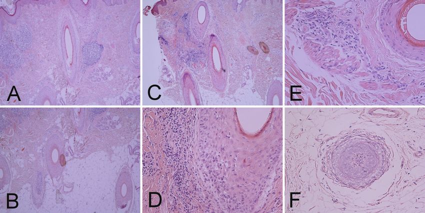
Fig. 1
(A, B, C) Band-like symmetrical hair loss was noted on the frontal and bi-temporal areas with prominent eyebrow involve-
ment (cosmetic tattoos on the eyebrow areas were present). The hairless scalp skin was pale, smooth, and slightly shiny.
The follicular orifices anterior to the frontal hairline were obviously diminished. (D) Folliculocentric keratosis can be more
clearly appreciated along the shaved regressing hairline before the biopsy.
Dermatol Sinica, Mar 2008 29Jin-Bon Hong and Sung-Jan Lin
Fig. 2
(A, B, C) There was lichenoid interface infiltration around the isthmus and infundibulum, sparing the lower part of the
follicle and hair bulb. (H&E, original magnification x40) (D) Lymphocytic lichenoid infiltration around the isthmus and
infundibulum was decernible with focal basal vacuolar degeneration and scattered dyskeratosis. (H&E, original magnifica-
tion x200) (E) The horizontal section in the dermis revealed perifollicular inflammation with mild lamellar fibrosis. (H&E,
original magnification x200) (F) The inflammatory process does not extend to the lower part of the follicle in the subcutis.
(H&E, original magnification x200)
than 80 cases of FFA have been described in hyperkeratosis on histological examination can
the literature.5-10 The majority of the cases were be prominent, corresponding to clinical follicular
postmenopausal women. 2-5 FFA is a band-like keratosis. Perifollicular lamellar fibrosis can be
cicatricial alopecia characterized by symmetri- seen in late-stage lesions. The interfollicular epi-
cal frontotemporal hairline recession. Although dermis in FFA is always devoid of the lichenoid
the presence of scarring is typical of FFA, this inflammation, which is commonly present in
scarring may be less remarkable than the other lichen planopilaris (LPP).10 The interfollicular
cicatricial alopecia. Follicular keratosis with vari- dermis and eccrine glands remain uninvolved.1,10
able perifollicular erythema is usually found at The histological findings in our case showed
the active margin of hair loss. Prominent loss of these typical features mentioned above with mild
eyebrows is often associated with FFA.2,5 There perifollicular fibrosis (Fig. 2). Normally, remark-
is variable hair loss on other body parts, includ- able perifollicular lamellar fibrosis will develop
ing axillae, extremities, and pubic area. To our at a late stage following the lichenoid inflamma-
knowledge, only one Asian patient of Korean tion.
ethnicity has been described in the English litera- Due to the histological similarities, FFA
ture.11 The rarity of reported cases from Asia may is commonly considered a variant of LPP in a
be due to either under-recognition of this disease number of reports. However, Poblet et al. indi-
or an ethnic difference. cated that, compared with LPP, FFA tended to
Histologically, FFA is featured by lichenoid show less inflammation around the infundibulum
lymphocytic infiltration around the upper portion and isthmus, less superficial perivascular lym-
of the follicle with focal basal vacuolar degen- phohistiocytic infiltrate and less interfollicular
eration and individual cell necrosis in the outer lichenoid changes.10 Still, it is unlikely to dif-
root sheath. Infundibular dilatation and follicular ferentiate between FFA and LPP based on the
30 Dermatol Sinica, Mar 2008Frontal fibrosing alopecia
histological findings alone. Furthermore, the site lead to perifollicular lamellar fibrosis.
and stage of the sampled lesion will affect the In other cicatricial alopecic diseases, the
histological results. Thus, the major difference is neutrophilic types are usually quite inflamed clin-
determined on the clinical ground. Both LPP and ically. Among the lymphocytic types of cicatri-
FFA are characterized by perifollicular erythema cial alopecia, discoid lupus erythematosus (DLE)
and follicular keratosis clinically but the unique usually presents as patchy erythema followed by
distribution of hair loss is only present in FFA.6,10 scarring or depression. The pathological features
Additionally, patients with FFA do not have other of interfollicular interface dermatitis, deep peri-
mucocutaneous lichenoid lesions as frequently as follicular infiltration and perieccrine inflamma-
with LPP.4,10,12 tion can help to distinguish DLE from FFA. The
Though distinctive clinically, FFA may direct immunofluorescence study of DLE lesions
sometimes be confused with non-scarring alope- at times shows granular deposition of IgG, IgM
cia, such as telogen effluvium, androgenetic and and C3 along the epidermal and follicular basal
alopecia areata. 2,5 Unlike FFA, non-cicatricial membrane zone, which is not present in FFA as
alopecia preserves follicalar orifices. Further, an- in our case.2
drogenetic alopecia does not cause eyebrow loss The treatment of FFA is difficult. It is re-
and hair miniaturization is not a feature of FFA.5 calcitrant to topical minoxidil solution treatment.
The frontal hairline is usually preserved in andro- The responses to plaquenil and either topical,
genetic alopecia in women. Although FFA most intralesional, or systemic corticosteroids are vari-
frequently occurs in women, male patients have able or limited. Due to the age of onset and pat-
been described in the literature.8 The Hamilton- terned hair loss on the frontal area, androgen hor-
type hair loss of male androgenetic alopecia can mone is a suspected etiology of FFA although no
easily be distinguished from FFA by the presence proof of hormone abnormality has been shown.2
of hair miniaturization, relative preservation of Finasteride 2.5 mg per day was reported to be
central frontal hair line, slow disease progression able to stabilize frontal recession in some cases.6
and lack of follicular erythema and keratosis. Whether finasteride is indeed superior to the oth-
Alopecia areata can also present with sud- er treatment is not known. Our patient was put on
den-onset and progressive hair loss with or with- oral finasteride 2.5 mg per day after the treatment
out eyebrow involvement. However, the fibrosing failure of topical 2% minoxidil solution. The pa-
nature, follicular signs of erythema and keratosis tient is now followed up in our department.
are not supposed to appear in alopecia areata. Of
note is the ophiasis variant of alopecia areata, in REFERENCES
which hair loss can extend from posterior and 1. Kossard S: Postmenopausal frontal fibrosing
bitemporal scalp to the frontal area symmetri- alopecia. Scarring alopecia in a pattern distribu-
cally. Although FFA rarely involves the occipital tion. Arch Dermatol 130: 770-774, 1994.
scalp, some patients who do not have prominent 2. Kossard S, Lee MS, Wilkinson B: Postmeno-
pausal frontal fibrosing alopecia: a frontal vari-
follicular signs may be misdiagnosed as ophiasis ant of lichen planopilaris. J Am Acad Dermatol
variant alopecia areata. 13 A skin biopsy is of great 36: 59-66, 1997.
help for a correct diagnosis in this condition. 3. Zinkernagel MS, Trueb RM: Fibrosing alopecia
Alopecia areata is characterized by peri-bulbar in a pattern distribution: patterned lichen plano-
lymphocytic inflammation with follicular minia- pilaris or androgenetic alopecia with a lichenoid
tissue reaction pattern? Arch Dermatol 136:
turization and telogen arrest. By comparison, the 205-211, 2000.
hallmark of FFA is lichenoid interface infiltration 4. Faulkner CF, Wilson NJ, Jones SK: Frontal
around the isthmus and infundibulum, sparing fibrosing alopecia associated with cutaneous
the lower third of the follicle and hair bulb. In the lichen planus in a premenopausal woman. Aus-
late stage, alopecia areata can make follicules re- tralas J Dermatol 43: 65-67, 2002.
5. Moreno-Ramirez D, Camacho Martinez F: Fron-
duced and replaced by fibrous tracts but FFA will
Dermatol Sinica, Mar 2008 31Jin-Bon Hong and Sung-Jan Lin
tal fibrosing alopecia: a survey in 16 patients. J 10. Poblet E, Jimenez F, Pascual A, et al.: Frontal
Eur Acad Dermatol Venereol 19: 700-705, 2005. fibrosing alopecia versus lichen planopilaris:
6. Tosti A, Piraccini BM, Iorizzo M et al.: Frontal a clinicopathological study. Int J Dermatol 45:
fibrosing alopecia in postmenopausal women. J 375-380, 2006.
Am Acad Dermatol 52: 55-60, 2005. 11. Lee WS, Hwang SM, Ahn SK: Frontal fibrosing
7. Mirmirani P, Willey A, Headington JT, et al.: alopecia in a postmenopausal woman. Cutis 60:
Primary cicatricial alopecia: histopathologic 299-300, 1997.
findings do not distinguish clinical variants. J 12. Mehregan DA, Van Hale HM, Muller SA: Li-
Am Acad Dermatol 52: 637-643, 2005. chen planopilaris: clinical and pathologic study
8. Kossard S, Shiell RC: Frontal fibrosing alopecia of forty-five patients. J Am Acad Dermatol 27:
developing after hair transplantation for andro- 935-942, 1992.
genetic alopecia. Int J Dermatol 44: 321-323, 13. Kwong RA, Kossard S: Alopecia areata mas-
2005. querading as frontal fibrosing alopecia. Australas
9. Clark-Loeser L, Latkowski JA: Frontal fibrosing J Dermatol 47: 63-66, 2006.
alopecia. Dermatol Online J 11: 6, 2005.
32 Dermatol Sinica, Mar 2008Frontal fibrosing alopecia
前額纖維化禿髮
-病例報告
1 1, 2
洪楨邦 林頌然
1
國立台灣大學醫學院皮膚科與附設醫院皮膚部
2
國立台灣大學醫學工程研究所
「前額纖維化禿髮」的臨床特徵是發生於前額至兩側顳部之進行性帶狀疤痕性禿髮,常
伴隨眉毛掉髮。在掉髮活性高的邊緣地帶常可以見到具特色之毛孔角化。此病症之組織學變
化則與毛孔扁平苔癬類似。直至今日,全世界已有超過80個案例被報告,其中大部分的前額
纖維化禿髮發生於停經之女性,但亦可發生於停經前之女性,甚至是男性都曾被報告過。在
此,我們報告一例發生於停經前女性之前額纖維化禿髮並討論此疾病的鑑別診斷、組織病理
學變化及其治療。前額纖維化禿髮是一種以往較少被辨認出的禿髮疾病,隨著越來越多相關
的報告來描繪此疾病,應該可以避免將前額纖維化禿髮與其它禿髮的診斷混淆。(中華皮誌
26: 28-33, 2008)
Dermatol Sinica, Mar 2008 33You can also read