Case Report Obstructive Fecalomas in an Infant Treated with Successful Endoscopic Disimpaction
←
→
Page content transcription
If your browser does not render page correctly, please read the page content below
Hindawi
Case Reports in Pediatrics
Volume 2021, Article ID 8815907, 4 pages
https://doi.org/10.1155/2021/8815907

Case Report
Obstructive Fecalomas in an Infant Treated with Successful
Endoscopic Disimpaction

          Risa Kanai ,1 Kengo Nakaya ,1 Koji Fukumoto ,1 Masaya Yamoto ,1
          Hiromu Miyake ,1 Akiyoshi Nomura ,1 Susumu Yamada ,1 Akihiro Makino ,1
          Hideto Iwafuchi ,2 and Naoto Urushihara 1
          1
            Department of Pediatric Surgery, Shizuoka Children’s Hospital, Address: 860, Urushiyama Aoi-ku Shizuoka-shi,
            Shizuoka 420-8660, Japan
          2
            Department of Pathology, Shizuoka Children’s Hospital, Address: 860, Urushiyama Aoi-ku Shizuoka-shi, Shizuoka 420-8660,
            Japan

          Correspondence should be addressed to Risa Kanai; leokanai15@gmail.com

          Received 5 August 2020; Revised 21 January 2021; Accepted 27 January 2021; Published 3 February 2021

          Academic Editor: Georg Singer

          Copyright © 2021 Risa Kanai et al. This is an open access article distributed under the Creative Commons Attribution License,
          which permits unrestricted use, distribution, and reproduction in any medium, provided the original work is properly cited.
          A fecaloma is a mass of accumulated feces with a consistency much harder than that of a fecal impaction. It is most frequently
          observed in the rectum and sigmoid area, and associated complications include colonic obstruction, ulceration, bleeding, and
          perforation. A one-year-old, previously healthy boy with no history of chronic constipation was admitted because of vomiting and
          abdominal distension. An abdominal computed tomography scan showed small and large bowel distension due to multiple
          obstructive fecalomas in the transverse colon. As the fecalomas could not be resolved by laxatives, enemas, or colonic lavage,
          endoscopic disimpaction under general anesthesia was attempted. Repeatedly shaving the fecalomas with biopsy forceps finally
          resulted in gradual fragmentation with subsequent passage. Gastrointestinal food allergy was later suggested as the cause because
          eosinophilic infiltration was found in a biopsy specimen of the colon wall. Endoscopic disimpaction is an effective treatment
          approach for addressing fecalomas to avoid more invasive surgical intervention.

1. Introduction                                                        2. Case Presentation
A fecaloma is a laminated mass of accumulated feces and has            A one-year-old, previously healthy boy with no history of
often been reported in patients suffering from chronic con-             chronic constipation presented with a five-day history of
stipation [1–11]. The typical consistency of a fecaloma is more        frequent vomiting, constipation, and abdominal distension.
than that of a fecal impaction due to coprostasis [6]. Most cases      He was 74 cm in height and weighed 9 kg at the time of
in the literature have been reported in adults, whereas pediatric      presentation. Upon examination, he was tachycardic but
cases with no medical history are rare, especially for infants         otherwise relatively stable. His abdomen was considerably
[7, 8, 11]. Fecalomas can trigger serious complications such as        distended but nontender and filled with palpable stool, yet a
colonic obstruction, ulceration, bleeding, and perforation,            rectal exam revealed no stool. An abdominal X-ray scan
sciatica, urinary retention, and deep vein thrombosis                  demonstrated multifocal air-fluid levels (Figure 1). Sus-
[2–4, 7–9]. Often, they can be treated by conservative methods;        pecting mechanical obstruction, we performed an abdom-
however, intractable cases sometimes require endoscopic                inal computed tomography scan. It revealed multiple
disimpaction or surgical intervention. Herein, we report a case        fecalomas in the transverse colon, wall thickening of the
of multiple fecalomas with small and large bowel obstructions          descending colon, bowel distension from the jejunum to the
in an infant, suspected of having a gastrointestinal food allergy,     transverse colon, moderate ascites, and no evidence of free
which was successfully resolved by endoscopic disimpaction.            air (Figure 2). A gastrografin enema examination revealed
2 Case Reports in Pediatrics
Figure 3: A gastrografin enema examination revealed expansion
failure of the descending colon and the presence of impacted
fecalomas in the transverse colon (arrows), which could not be
resolved by colonic lavage.
Figure 1: An abdominal X-ray scan demonstrated multifocal air-
fluid levels.
was not imminent, and we continued conservative obser-
vation. However, the vomiting and inability to pass the stool
continued even on the following day, and his abdominal
distension had worsened upon nasogastric tube drainage. He
was taken to the operating room for an emergency endo-
scopic fecal disimpaction under general anesthesia.
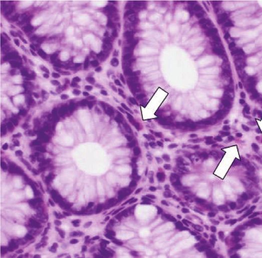
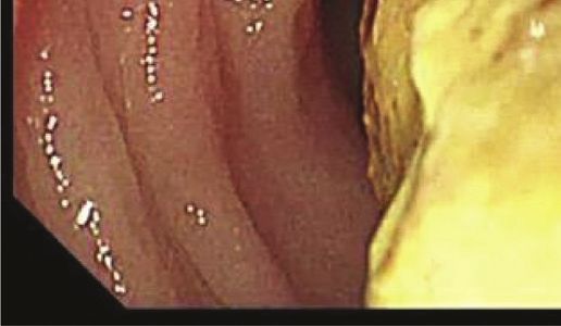
A colonoscopy was performed using a scope of the upper
gastrointestinal tract (GIF TYPE Q260, 9.2 mm in diameter;
Olympus, Tokyo, Japan) with insufflation of carbon dioxide.
Edematous mucosa of the descending colon and multiple
giant brown fecalomas were observed to be occupying the
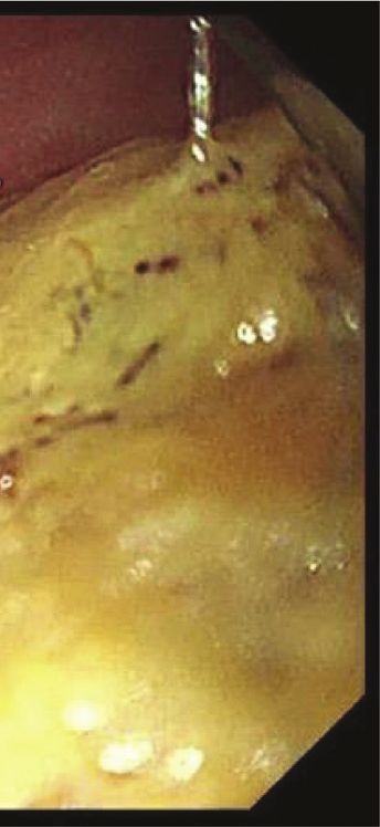
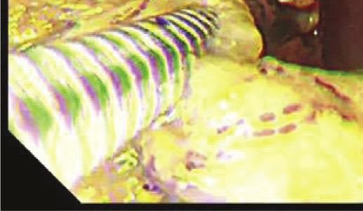
lumen of the transverse colon (Figure 4). Although the
surface of the fecaloma was hard, large, and slippery, re-
peated shaving of the fecalomas with biopsy forceps resulted
in gradual fragmentation. Finally, after 88 minutes, all
fecalomas had been broken into fragments of a size that we
thought would be able to pass through the anus. Samples
were collected from the colon wall to explore the cause of the
situation using biopsy. After endoscopic disimpaction, the
patient experienced an intermittent bowel movement im-
mediately, and his abdomen was less distended obviously
thereafter. He was able to consume meals after recovery and
was successfully dismissed from hospital care 18 days after
the endoscopic treatment. Later, the pathology of a biopsy
sample taken from the descending colon wall showed 22
eosinophils per high-power field (HPF), while all investi-
gations conducted for intestinal motility disorders such as
Hirschsprung’s disease were negative (Figure 5). He was
considered to have a food protein-induced enterocolitis
Figure 2: An abdominal computed tomography revealed fecalo-
syndrome, which is a kind of gastrointestinal food allergy. It
mas in the transverse colon (arrow), bowel distension from the
jejunum to the transverse colon, and moderate ascites.
was determined that, before the described event, he had
eaten barley for the first time in his life. Because an allergy
load test is thought to be risky in the context of severe
expansion failure of the descending colon and the presence gastrointestinal allergy, we are considering performing the
of impacted fecalomas in the transverse colon, which could test to confirm his possible barley allergy once he reaches
not be resolved by colonic lavage (Figure 3). His condition about three years old.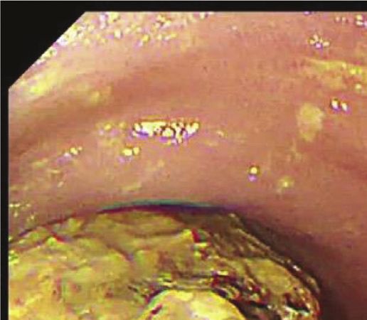
Case Reports in Pediatrics                                                                                                            3

                                         (a)                                                (b)

Figure 4: (a) A colonoscopy revealed a giant brown fecaloma occupying the lumen of the transverse colon. (b) Biopsy forceps splitted the
fecaloma.

                                                                      injection have also been reported in [9, 10]. However, their
                                                                      use requires much time and effort, and one case took more
                                                                      than six hours to treat [9]. Therefore, endoscopic procedures
                                                                      should not be indicated in imminent cases.
                                                                          While the majority of published cases occurred in adults,
                                                                      fecalomas sometimes appear in pediatric patients; however,
                                                                      there are no reports in which the fecalomas were dissolved
                                                                      by endoscopy [7, 8, 11]. We therefore believe this to be the
                                                                      first reported case of pediatric fecalomas successfully treated
                                                                      by endoscopic disimpaction. Endoscopic procedures in
                                                                      pediatric cases require careful maneuvering due to the
                                                                      fragility of these patients’ intestinal tracts. In our present
                                                                      case, we selected the endoscope used for the upper gas-
Figure 5: The pathology of a biopsy sample taken from the             trointestinal tract because of its small diameter and
descending colon wall showed eosinophilic infiltration (arrows)        attempted to reduce insufflation of carbon dioxide during
(hematoxylin and eosin staining, ×400).                               the procedure. Also, treatment should be performed under
                                                                      general anesthesia because pediatric patients are prone to
3. Discussion                                                         poor ventilation conditions due to increased abdominal
                                                                      pressure; there is also a possibility of conversion laparotomy
Although fecal impaction is a common and disturbing                   in an emergency case of perforation.
problem, a fecaloma is a particularly rare form of impaction              There are multiple causes of fecaloma formation, but in
in which a mass separable from the rest of the bowel contents         our case, the cause was not immediately evident as the
is formed. Fecalomas are most frequently seen in the rec-             patient had no reported history of altered bowel habits.
tosigmoid area because the stool becomes firmer, and the               Chronic constipation is a common problem that can lead to
colon diameter is smaller in this area [3, 9]. On the contrary,       fecal impaction and even the development of fecalomas
the proximal colon is a more unusual site for fecalomas, and          [1–4]. Other diseases which reported to result in fecalomas
their presence in this location can cause small bowel ob-             include Hirschsprung’s disease, Chagas disease, psychiatric
struction like seen in our present case.                              disorders, intestinal tuberculosis, and scleroderma [3, 7].
    Regarding the treatment of fecalomas in the rec-                  However, these causes were not apparent in our case, and a
tosigmoid area, conservative procedures such as bowel rest,           gastrointestinal food allergy was ultimately suspected be-
laxatives, enemas, manual evacuation, and colonic lavage are          cause of the pathology of the biopsy specimen taken from
commonly adopted [1–3]. However, especially in the case of            the colon wall. Current recommendations for the diagnosis
a fecaloma in the proximal colon or small intestine, endo-            of gastrointestinal food allergy include biopsies of the
scopic disimpaction or surgical intervention may be re-               intestinal mucosa that can reveal the existence of eosino-
quired [6, 8]. Surgical intervention sometimes becomes                philia at a concentration greater than 20 per HPF. Our
necessary due to difficulty with reaching these locations by            present case demonstrated 22 eosinophils per HPF and
endoscopy. There are some reports in the literature of                fulfilled the diagnostic criteria for a gastrointestinal food
fecalomas successfully dissolved by endoscopic procedures             allergy. Our patient was considered specifically to have
[2–5]. Recently, more creative endoscopic methods of                  food protein-induced enterocolitis syndrome due to barley,
fecaloma disimpaction using jumbo forceps or a cola                   which is a cell-mediated, nonimmunoglobulin E-mediated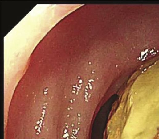
4                                                                                                             Case Reports in Pediatrics

gastrointestinal food allergy [12, 13]. While this allergy                [8] H. Y. Yoo, H. W. Park, S.-H. Chang, and S. H. Bae, “Ileal
sometimes presents together with constipation, it rarely                      fecaloma presenting with small bowel obstruction,” Pediatric
provokes obstruction [14–17]. Some cases of gastrointes-                      Gastroenterology, Hepatology & Nutrition, vol. 18, no. 3,
tinal food allergy have required surgical intervention due to                 p. 193, 2015.
intestinal edema or obstruction [12, 13, 17]. The possible                [9] Y. Matsuo, H. Yasuda, H. Nakano et al., “Successful endo-
                                                                              scopic fragmentation of large hardened fecaloma using jumbo
mechanism of bowel obstruction in this disease is suspected
                                                                              forceps,” World Journal of Gastrointestinal Endoscopy, vol. 9,
to be that inflammatory cytokines such as interferon-c and                     no. 2, p. 91, 2017.
tumor necrosis factor-β induce edema of the gastrointes-                 [10] J. H. Kang and Y. J. Lim, “Can fecaloma be dissolved by cola
tinal mucosa, which causes intestinal peristalsis depression                  injection in a similar way to bezoars?” Intestinal Research,
[18]. In our present case, dehydration due to persistent                      vol. 12, no. 4, p. 333, 2014.
vomiting may also have exacerbated fecal stiffening, which                [11] J. D. Garisto, L. Campillo, E. Edwards, M. Harbour, and
rapidly accelerated the progression to fecaloma develop-                      R. Ermocilla, “Giant fecaloma in a 12-year-old-boy: a case
ment with bowel obstruction rather than simple con-                           report,” Cases Journal, vol. 2, no. 1, p. 127, 2009.
stipation. However, this suggestion is still speculative until           [12] S. Mehr, A. Kakakios, K. Frith, and A. S. Kemp, “Food protein-
an allergy load test is performed, and it also remains un-                    induced enterocolitis syndrome: 16-year experience,” Pedi-
                                                                              atrics, vol. 123, no. 3, pp. e459–e464, 2009.
clear why the patient’s gastrointestinal food allergy ma-
                                                                         [13] S. Jayasooriya, A. T. Fox, and S. H. Murch, “Do not lapa-
terialized mainly in the descending colon.                                    rotomize food protein-induced enterocolitis syndrome,” Pe-
    In conclusion, we herein report a case of fecalomas                       diatric Emergency Care, vol. 23, no. 3, pp. 173–175, 2007.
inducing small and large bowel obstructions in an infant due             [14] K. J. Allen, D. J. Hill, and R. G. Heine, “4. Food allergy in
to a suspected gastrointestinal food allergy. The endoscopic                  childhood,” Medical Journal of Australia, vol. 185, no. 7,
disimpaction of fecalomas is arduous and requires a great                     p. 394, 2006.
deal of time to perform but is an ideal treatment for use in             [15] H. A. Sampson, “Update on food allergy☆,” Journal of Allergy
patients without an imminent condition to avoid the need to                   and Clinical Immunology, vol. 113, no. 5, pp. 805–819, 2004.
progress to surgical intervention.                                       [16] I. Nomura, H. Morita, S. Hosokawa et al., “Four distinct
                                                                              subtypes of non-IgE-mediated gastrointestinal food allergies
                                                                              in neonates and infants, distinguished by their initial symp-
Data Availability                                                             toms,” Journal of Allergy and Clinical Immunology, vol. 127,
                                                                              no. 3, pp. 685–688, 2011.
The clinical data used to support the findings of this study              [17] K. Nakaya, Y. Iinuma, Y. Hirayama, and S. Tsuruhisa, “A case
are included within the article.                                              of suspected gastrointestinal allergy requiring emergent lap-
                                                                              arotomy for fecal ileus,” Journal of the Japanese Society of
Conflicts of Interest                                                         Pediatric Surgeons, vol. 52, no. 5, pp. 1303–1308, 2016.
                                                                         [18] E. Pohjavuori, M. Viljanen, R. Korpela et al., “Lactobacillus
The authors declare no conflicts of interest.                                  GG effect in increasing IFN-c production in infants with
                                                                              cow’s milk allergy,” Journal of Allergy and Clinical Immu-
                                                                              nology, vol. 114, no. 1, pp. 131–136, 2004.
References
 [1] B. T. Wang and S. Y. Lee, “Cecal fecaloma: a rare cause of right
     lower quadrant pain,” European Journal of Radiology Open,
     vol. 6, p. 136, 2019.
 [2] E. Sakai, Y. Inokuchi, M. Inamori et al., “Rectal fecaloma:
     successful treatment using endoscopic removal,” Digestion,
     vol. 75, no. 4, p. 198, 2007.
 [3] S. M. Kim, K. H. Ryu, Y. S. Kim et al., “Cecal fecaloma due to
     intestinal tuberculosis: endoscopic treatment,” Clinical En-
     doscopy, vol. 45, no. 2, p. 174, 2012.
 [4] G. Ghosh, S. Shah, and C. Maltz, “A case of a giant fecaloma,”
     Clinical Gastroenterology and Hepatology, vol. 16, no. 4, p. e48,
     2018.
 [5] R. Sarnoff, B. Girmay, D. Bhakta, R. Mocharla, and
     R. Williams, “An obstructing fecal bezoar in a patient with
     scleroderma with successful colonoscopic disimpaction,”
     ACG Case Report Journal, vol. 6, no. 4, Article ID e00059,
     2019.
 [6] M. Mushtaq, M. A. Shah, A. A. Malik, K. A. Wani, and
     N. Thakur, “Giant fecaloma causing small bowel obstruction:
     case report and review of the literature,” Bulletin of Emergency
     and Trauma, vol. 3, no. 2, pp. 70–72, 2015.
 [7] J. S. Parray, T.-J. Park, J. S. Hwa, J.-H. Seo, C.-H. Park, and
     H.-S. Youn, “Acute urinary retention in a 47-month-old girl
     caused by the giant fecaloma,” Pediatric Gastroenterology,
     Hepatology & Nutrition, vol. 16, no. 3, p. 200, 2013.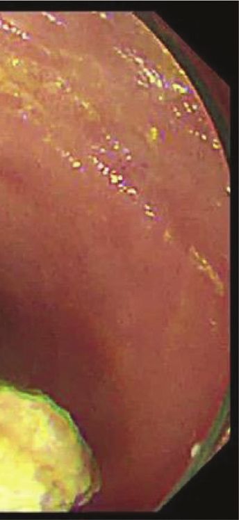
You can also read