Per- pass analysis of acute ischemic stroke clots: impact of stroke etiology on extracted clot area and histological composition - Journal of ...
←
→
Page content transcription
If your browser does not render page correctly, please read the page content below
Ischemic stroke
J NeuroIntervent Surg: first published as 10.1136/neurintsurg-2020-016966 on 9 December 2020. Downloaded from http://jnis.bmj.com/ on July 13, 2021 by guest. Protected by copyright.
Original research
Per-pass analysis of acute ischemic stroke clots:
impact of stroke etiology on extracted clot area and
histological composition
Seán Fitzgerald ,1 Rosanna Rossi ,1,2 Oana Madalina Mereuta ,1,2
Duaa Jabrah,1 Adaobi Okolo,1 Andrew Douglas,1,2 Sara Molina Gil,1,2 Abhay Pandit,2
Ray McCarthy,3 Michael Gilvarry,3 Dennis Dunker,4,5 Annika Nordanstig,6,7
Erik Ceder,4,5 Petra Redfors,6,7 Katarina Jood,6,7 Niclas Dehlfors,4,5 Georgios Magoufis,8
Georgios Tsivgoulis,9 Waleed Brinjikji ,10 David F Kallmes,10 Alan O’Hare,11
Sarah Power,11 Paul Brennan,11 Jack Alderson,11 András Nagy,12 Ágnes Vadász,12
Klearchos Psychogios,8,9 Istvan Szikora ,12 Turgut Tatlisumak,6,7
Alexandros Rentzos,4,5 John Thornton,11 Karen M Doyle 1,2
►► Additional material is ABSTRACT INTRODUCTION
published online only. To view, Background Initial studies investigating correlations A recent study of patients with acute ischemic
please visit the journal online
(http://d x.doi.o rg/10.1136/ between stroke etiology and clot composition are stroke (AIS) from the MR CLEAN clinical trial
neurintsurg-2020-016966). conflicting and do not account for clot size as determined investigators demonstrated that compared with
by area. Radiological studies have shown that cardioembolic strokes, non- cardioembolic strokes
For numbered affiliations see cardioembolic strokes are associated with shorter clot are associated with the presence of a hyperdense
end of article.
lengths and lower clot burden than non-cardioembolic artery sign, longer thrombi, and a more consider-
clots. able clot burden on CT imaging, and a shift towards
Correspondence to
Dr Karen M Doyle, Department Objective To report the relationship between a more proximal thrombus location.1 In agreement,
of Physiology, National stroke etiology, extracted clot area, and histological Dutra et al showed that proximal location, higher
University of Ireland Galway, composition at each procedural pass. clot burden, and longer clot length are associated
Galway, Ireland; k aren.doyle@ Methods As part of the multi-institutional with worse functional outcomes and a longer
nuigalway.ie
RESTORE Registry, the Martius Scarlett Blue stained endovascular procedure.2 These differences in clot
Received 9 October 2020 histological composition and extracted clot area of length and burden affecting procedural and patient
Revised 17 November 2020 612 per-pass clots retrieved from 441 patients during outcomes are probably related to the etiology of
Accepted 20 November 2020 mechanical thrombectomy procedures were quantified. the occlusive clot. Studies investigating the histo-
Correlations with clinical and procedural details were logical composition of AIS clots in relation to stroke
investigated. etiology have proved conflicting to date, limited
Results Clot composition varied significantly with by differing histological staining and analysis
procedural passes; clots retrieved in earlier passes had methods.3–5 In addition to variation of the histo-
higher red blood cell content (H4=11.644, p=0.020) logical clot composition with etiology, it has also
and larger extracted clot area (H4=10.730, p=0.030). been shown that composition varies with increasing
Later passes were associated with significantly higher number of procedural passes.6
fibrin (H4=12.935, p=0.012) and platelets/other In this large multicenter study, the per-pass histo-
(H4=15.977, p=0.003) content and smaller extracted pathological composition and extracted clot area
clot area. Large artery atherosclerotic (LAA) clots were were studied in a series of patients with large vessel
significantly larger in the extracted clot area and more occlusion. The relationship between suspected
red blood cell-rich than other etiologies in passes stroke etiology, histological composition, and
1–3. Cardioembolic and cryptogenic clots had similar extracted clot area at each procedural pass was
histological composition and extracted clot area across investigated.
all procedural passes.
Conclusion LAA clots are larger and associated with a METHODS
© Author(s) (or their large red blood cell-rich extracted clot area, suggesting Patient selection and clinical data
employer(s)) 2021. Re-use soft thrombus material. Cardioembolic clots are smaller This study included patients with AIS from four
permitted under CC BY.
Published by BMJ. in the extracted clot area, consistent in composition and hospitals collected between March 2018 and
area across passes, and have higher fibrin and platelets/ November 2019 as part of the RESTORE Registry.
To cite: Fitzgerald S, Rossi R, other content than LAA clots, making them stiffer clots. This study was approved by the National Univer-
Mereuta OM, et al.
The per-pass histological composition and extracted sity of Ireland Galway research ethics committee
J NeuroIntervent Surg Epub
ahead of print: [please clot area of cryptogenic clots are similar to those of (Study No: 16-SEPT-08) and the regional hospital
include Day Month Year]. cardioembolic clots, suggesting similar formation ethics committees (Beaumont Hospital, Dublin;
doi:10.1136/ mechanisms. Sahlgrenska University Hospital, Gothenburg;
neurintsurg-2020-016966 NICN, Budapest; Metropolitan Hospital, Athens)
Fitzgerald S, et al. J NeuroIntervent Surg 2020;0:1–7. doi:10.1136/neurintsurg-2020-016966 1Ischemic stroke
J NeuroIntervent Surg: first published as 10.1136/neurintsurg-2020-016966 on 9 December 2020. Downloaded from http://jnis.bmj.com/ on July 13, 2021 by guest. Protected by copyright.
following the ethical standards of the Declaration of Helsinki, 2.0 (±1.7) per case) and the clot was successfully retrieved in
and a waiver of informed consent was granted. The inclusion 612 procedural passes (72.5% of all procedural pass attempts).
criteria were patients with a large vessel occlusion, aged ≥18 Median National Institutes of Health Scale score on admission
years, who had undergone mechanical thrombectomy following was 16 (10-20). Forty-eight percent of patients were treated
relevant diagnostic procedures, and with clot material success- with intravenous recombinant tissue plasminogen activator.
fully retrieved in at least one procedural pass. Eighty-eight percent of patients had an occlusion in the ante-
rior circulation, 10% in the posterior circulation, and 2% had
Clot and data collection dual occlusions in both the anterior and posterior circulation.
Endovascular treatment was performed according to the indi- Successful reperfusion (Thrombolysis in Cerebral Infarction
vidual institution’s routine procedures. Clots were collected (TICI) ≥2b) was achieved in 92.3% of patients. Suspected stroke
in a per-pass manner, meaning that where multiple procedural etiologies were LAA (n=115, 26.1%), cardioembolic (n=209,
passes were used to treat the patients, clot material from each 47.4%), cryptogenic (n=101, 22.9%) and other suspected etiol-
pass was collected separately. Data for reperfusion outcome ogies (n=16, 3.6%). The mean number of passes that success-
and suspected stroke etiology were self-reported at the centers fully retrieved a clot were LAA (1.60), cardioembolic (1.26),
and captured on the RESTORE Registry data abstraction form. cryptogenic (1.40), and other suspected etiologies (1.44). Aspi-
Stroke etiology was classified using the Trial of Org 10 172 in ration was used as the first-line treatment approach in 55.3%
Acute Stroke Treatment (TOAST) system: large-artery athero- of patients and stentrievers were used as first-line treatment in
sclerosis (LAA), cardioembolism, a stroke of other determined 44.7%. Clinical and procedural characteristics are shown in
etiology, and cryptogenic.7 online supplemental table 1).
Clot processing, extracted clot area analysis, and histological Clot histological composition and extracted clot area varies
staining with increasing number of procedural passes
Histological analysis of the clot samples was performed as previ- Significant differences in clot composition were found with
ously recommended.8 Following retrieval, clot samples were increasing number of procedural passes (figure 1A, table 1A).
immediately fixed in 10% phosphate-buffered formalin. Samples The proportion of red blood cells was highest in pass 1 (42.8%)
were shipped to the Department of Physiology at the National and was lower in all subsequent passes (H4=11.644, p=0.020).
University of Ireland Galway for analysis. On arrival, each case The proportion of fibrin varied significantly across procedural
and corresponding per- pass procedural data were logged in passes (H4=12.935, p=0.012) and was highest in pass 4 (38.0%;
the RESTORE Registry. Gross photographs of each successful figure 1A, table 1A). The proportion of platelets/other increased
procedural pass were taken using a Canon EOS 1300D camera. significantly with increasing procedural passes (H4=15.977,
ImageJ software (https://imagej.nih.gov/ij/) was used to analyze p=0.003), accounting for 37.4% of the composition in clots
the area of each fragment of clot individually. First the scale removed in passes 5+. The proportion of collagen was also
was set and then the polygon tool was used to draw a region of significantly higher in later procedural passes (H4=10.960,
interest around a fragment of the clot, and the area of that frag- p=0.027), although typically a minor component of clots. The
ment was measured individually. The total extracted clot area proportion of white blood cells did not vary significantly across
for each case is defined as the sum of the clot area from all clot procedural passes (H4=1.988, p=0.738) and was also a minor
fragments within a case. component of most retrieved clots relative to red blood cells,
Per-pass clot material was then processed using a standard fibrin, and platelets/other (figure 1A, table 1A).
tissue processing protocol, embedded in paraffin wax, and cut The per-pass extracted clot area per histological component
into 3 µm sections. Two representative sections were stained showed that extracted clot area removed in earlier passes was
with Martius Scarlett Blue (MSB) to identify the standard clot significantly larger than in later passes (H4=10.730, p=0.030;
components (red blood cells, white blood cells, fibrin, platelets/ figure 1B, table 1B). Specifically, the extracted clot area of
other) as previously described.9 Orbit image analysis (www.orbit. red blood cells (H4=34.418, pIschemic stroke
J NeuroIntervent Surg: first published as 10.1136/neurintsurg-2020-016966 on 9 December 2020. Downloaded from http://jnis.bmj.com/ on July 13, 2021 by guest. Protected by copyright.
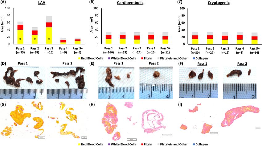
Figure 1 Histological clot composition and extracted clot area per histological component. (A) Mean histological clot composition (%) of each
procedural pass, as determined by MSB staining represented as the percentage of the total. (B) The mean area of each histological component within
a procedural pass is calculated by multiplying each component’s histological composition by the extracted clot area of the corresponding procedural
pass. (C) Mean histological clot composition (%) of each suspected etiology. (D) Mean area of each histological component within each suspected
etiology. ECA, extracted clot area; LAA, large artery atherosclerotic (clots); MSB, Martius Scarlett Blue; RBCs, red blood cells; WBCs, white blood cells.
(33.3%) and platelets/other (28.53%; figure 1C, table 2A). The and collagen (H3=7.577, p=0.056) were not significantly
proportion of white blood cells and collagen did not vary signifi- different across the four etiologies (figure 1D, table 2B).
cantly across the four etiologies (H3=2.218, p=0.528, and
H3=5.533, p=0.137, respectively).
The histological composition of the clots varied consid- Variation of per-pass extracted clot area of histological
erably between suspected etiologies when corrected for the components with etiology
area of the extracted clot material (figure 1D, table 2B). LAA LAA clots had a significantly larger extracted clot area of red
clots had the largest per- pass extracted clot area at (54.96 blood cells in passes 1–3 than in passes 4 and 5+ (H4=14.527,
mm2) and were significantly larger than cardioembolic (33.64 p=0.006; figure 2A, online supplemental table 2). The extracted
mm2), cryptogenic (32.28 mm2), and other (39.60 mm2) clots clot area of white blood cells (H4=7.969, p=0.093), fibrin
(H3=13.810, p=0.003; figure 1D, table 2B). LAA clots had the (H4=7.783, p=0.100), platelets/other (H4=5.872, p=0.209),
largest extracted clot area of red blood cells (32.95 mm2) and and collagen (H4=9.449, p=0.051) were not significantly
white blood cells (1.14 mm2) compared with all other etiologies different across procedural passes in LAA clots (figure 2A and
(H3=27.795, pIschemic stroke
J NeuroIntervent Surg: first published as 10.1136/neurintsurg-2020-016966 on 9 December 2020. Downloaded from http://jnis.bmj.com/ on July 13, 2021 by guest. Protected by copyright.
Table 2 Histological composition per suspected etiology (A) and extracted clot area of each histological component per suspected etiology (B)
Significance
Total number (N) LAA Cardioembolic Cryptogenic Other Test statistic (H) DF (pIschemic stroke
J NeuroIntervent Surg: first published as 10.1136/neurintsurg-2020-016966 on 9 December 2020. Downloaded from http://jnis.bmj.com/ on July 13, 2021 by guest. Protected by copyright.
area of collagen was significantly different in cryptogenic cases staining and analysis techniques.4 5 17–19 More recent studies have
(H4=20.353, pIschemic stroke
J NeuroIntervent Surg: first published as 10.1136/neurintsurg-2020-016966 on 9 December 2020. Downloaded from http://jnis.bmj.com/ on July 13, 2021 by guest. Protected by copyright.
Our study has some limitations: first, only clots that were data interpretation. RR, OMM, DJ, AO, AD, and SMG performed the experiments and
successfully retrieved from the patient were available for analysis described, including measurement of the extracted clot area, histological
staining, and quantification of the cases and statistical analysis. DD, ANo, EC, PR,
histopathological and extracted clot area analysis; however, KJ, ND, GM, AO, SP, PB, JA, ANa, AV, and KP, collected clot samples and extracted
successful reperfusion (TICI ≥2b) was achieved in 92.3% of corresponding clinical data for each patient at each of their respective participating
patients, suggesting that most of the clot was retrieved. Second, hospitals.
the method described for assessing the extracted clot area is an Funding This work was supported by the European Regional Development Fund
extrapolation from a 2D image of a 3D object, which, although and Science Foundation Ireland grant number (13/RC/2073) and by the National
it will lead to some slight inaccuracies in calculating area, gives Institutes of Health grant number (R01 NS105853).
a robust relative estimate. A significant correlation between Competing interests The authors declare competing interests (funding,
extracted clot area as measured by ImageJ and clot weight was employment or personal financial interests) in relation to the work described herein.
found in a subset of samples (n=80, R2=0.898, pIschemic stroke
J NeuroIntervent Surg: first published as 10.1136/neurintsurg-2020-016966 on 9 December 2020. Downloaded from http://jnis.bmj.com/ on July 13, 2021 by guest. Protected by copyright.
12 Johnson S, Chueh J, Gounis MJ, et al. Mechanical behavior of in vitro blood clots 21 Boeckh-Behrens T, Kleine JF, Zimmer C, et al. Thrombus histology suggests
and the implications for acute ischemic stroke treatment. J Neurointerv Surg cardioembolic cause in cryptogenic stroke. Stroke 2016;47:1864–71.
2020;12:853–7. 22 Li Z, Bai Y, Li W, et al. Carotid vulnerable plaques are associated with circulating
13 Fitzgerald S, Ryan D, Thornton J, et al. Preclinical evaluation of Millipede 088 leukocytes in acute ischemic stroke patients: an clinical study based on contrast-
intracranial aspiration catheter in cadaver and in vitro thrombectomy models. J enhanced ultrasound. Sci Rep 2018;8:1–9.
Neurointerv Surg 2020:neurintsurg-2020-016218. 23 Li L, Yiin GS, Geraghty OC, et al. Incidence, outcome, risk factors, and long-term
14 Weafer FM, Duffy S, Machado I, et al. Characterization of strut indentation during prognosis of cryptogenic transient ischaemic attack and ischaemic stroke: a
mechanical thrombectomy in acute ischemic stroke clot analogs. J Neurointerv Surg population-b ased study. Lancet Neurol 2015;14:903–13.
2019;11:891–7. 24 Diener H-C, Sacco RL, Easton JD, et al. Dabigatran for prevention of stroke after
15 Zaidat OO, Castonguay AC, Linfante I, et al. First pass effect. Stroke 2018;49:660–6. embolic stroke of undetermined source. N Engl J Med 2019;380:1906–17.
16 Fennell VS, Setlur Nagesh SV, Meess KM, et al. What to do about fibrin rich ’tough 25 Hart RG, Sharma M, Mundl H, et al. Rivaroxaban for stroke prevention after embolic
clots’? Comparing the Solitaire stent retriever with a novel geometric clot extractor in stroke of undetermined source. N Engl J Med 2018;378:2191–201.
an in vitro stroke model. J Neurointerv Surg 2018;10:907–10. 26 Ntaios G, Wintermark M, Michel P. Supracardiac atherosclerosis in embolic stroke of
17 Liebeskind DS, Sanossian N, Yong WH, et al. CT and MRI early vessel signs reflect clot undetermined source: the underestimated source. Eur Heart J 2020. doi:10.1093/
composition in acute stroke. Stroke 2011;42:1237–43. eurheartj/ehaa218. [Epub ahead of print: 16 Apr 2020].
18 Simons N, Mitchell P, Dowling R, et al. Thrombus composition in acute ischemic 27 Muller C, Roizman M, Wong A. Secondary prevention of ischaemic stroke. Intern Med
stroke: a histopathological study of thrombus extracted by endovascular retrieval. J J 2019;49:1221–8.
Neuroradiol 2015;42:86–92. 28 Hofmeister J, Bernava G, Rosi A, et al. Clot-based radiomics predict a mechanical
19 Boeckh-Behrens T, Schubert M, Förschler A, et al. The impact of histological clot thrombectomy strategy for successful recanalization in acute ischemic stroke. Stroke
composition in embolic stroke. Clin Neuroradiol 2016;26:189–97. 2020;51:2488–94.
20 Sporns PB, Hanning U, Schwindt W, et al. Ischemic stroke: what does the 29 Mohammaden MH, Haussen DC, Perry da Camara C, et al. Hyperdense vessel sign as
histological composition tell us about the origin of the thrombus? Stroke a potential guide for the choice of stent retriever versus contact aspiration as first-line
2017;48:2206–10. thrombectomy strategy. J Neurointerv Surg 2020:neurintsurg-2020-016005.
Fitzgerald S, et al. J NeuroIntervent Surg 2020;0:1–7. doi:10.1136/neurintsurg-2020-016966 7You can also read