Tyler, S., Swales, N., Foster, A., Knowles, T., & Barnard, N. (2019). Otoscopy and aural cytological findings in a population of rescue cats and ...
←
→
Page content transcription
If your browser does not render page correctly, please read the page content below
Tyler, S., Swales, N., Foster, A., Knowles, T., & Barnard, N. (2019).
Otoscopy and aural cytological findings in a population of rescue cats and
cases in a referral small animal hospital in England and Wales. Journal of
Feline Medicine and Surgery. https://doi.org/10.1177/1098612X19834969

Peer reviewed version

Link to published version (if available):
10.1177/1098612X19834969

Link to publication record in Explore Bristol Research
PDF-document

This is the author accepted manuscript (AAM). The final published version (version of record) is available online
via Sage at https://journals.sagepub.com/doi/10.1177/1098612X19834969 . Please refer to any applicable terms
of use of the publisher.

University of Bristol - Explore Bristol Research
General rights

This document is made available in accordance with publisher policies. Please cite only the published
version using the reference above. Full terms of use are available:
http://www.bristol.ac.uk/pure/about/ebr-terms
1   Otoscopy and aural cytological findings in a population of rescue cats and cases in a
 2   referral small animal hospital in England and Wales
 3   S. TYLER*, N. SWALES*, A.P. FOSTER*, T.G. KNOWLES*, N. BARNARD†.
 4
 5   * Bristol Veterinary School, University of Bristol, Langford House, Langford, BS40 5DU, UK
 6   † Highcroft Veterinary Referrals, 615 Wells Road, Whitchurch, Bristol, BS14 9BE , UK
 7
 8   Corresponding author; S. Tyler BVetMed MANZCVS MRCVS sophie.tyler@bristol.ac.uk
 9
10   Acknowledgements
11   Marta Costa for assistance in interpreting the ear cerumen cytology. The rescue centres and
12   owners of cats that allowed sampling of their cat’s ears.
13
14   Funding and Conflict of interest statements
15   Zoetis UK supplied some complementary anti-parasite products. S Tyler and N Barnard have
16   received honorariums and consulted for Zoetis.
17
18
19
20
21
22
23
24
25
26
27
28
29
30
31
32
33
34
35
36
37
38   Abstract
39   Objectives
40   Otitis externa is seen clinically in cats although studies investigating this within the UK are
41   lacking. The objective of this study was to investigate the prevalence of Otodectes cynotis
42   mites and microbial infection in the ear canals of cats in various rescue / charitable centres
43   and a referral hospital.
44
45   Methods
46   Otoscopy was performed in 332 cats from a range of sources. Otoscopic findings were noted,
47   including the gross visualisation of Otodectes. A sample of cerumen was collected for
48   cytological evaluation and a cerumen smear for detection of Otodectes mites if there was a
49   large amount of black or brown aural exudate on otoscopy sufficient exudate for a smear to
50   be mounted in paraffin oil.
51
52   Results
53   Otoscopic evidence of Otodectes cynotis infestation was noted in 3 / 341 cats (0.9 %, 95 %
54   CI = 0.3 - 2.6 %). A total of 129 / 341 (37.8 % CI = 32.7 – 43.0%) cats were found to have
55   Malassezia species within one or both ears. Bacteria were found unilaterally in 9 / 341 (2.6 %
56   CI = 1.4 – 4.9 %) cats. Analysis of the cytological findings showed an increased likelihood for
57   Malassezia to be present in older cats as age increased (Pearson r = 0.204, P < 0.001, n=293).
58   There was also an increased likelihood of finding Malassezia in both ears if found within one
59   ear (r = 0.499, P < 0.001, n = 327). There was a positive correlation between the number of
60   Malassezia organisms and the quantity of aural exudate (r = 0.778, P < 0.001, n = 338). Only
61   10 / 332 cats were found to have no exudate at all upon otoscopy. Cats where Otodectes
62   infestation were noted (n = 3), had moderate or large quantities of cerumen. All cats with
63   bacteria on cytology were found to have small to large quantities of aural exudate present.
64
65   Conclusions and relevance
66   This study shows that there was a low prevalence of O. cynotis in this cohort of cats in the
67   United Kingdom. In normal cats it was not unusual to find Malassezia microorganisms upon
68   aural cytology, bacteria were noted far less frequently and in two cats this was associated with
69   underlying anatomical pathology.
70
71
72   Introduction
 73   Otitis externa is seen more frequently in dogs than cats.1-3 Many studies have investigated the
 74   prevalence of otitis externa in cats, although studies in the United Kingdom are lacking.
 75
 76   Malassezia spp. are known to be part of the normal aural microflora in cats.4-7 Many studies
 77   have used ear swabs for bacterial and fungal culture to investigate the aural microflora of cats,
 78   with and without otitis externa, but fewer studies have used cytology for investigating the
 79   normal feline aural microflora. Malassezia yeasts were cultured from 95.1% and 48.4% of cats
 80   in Iran with and without otitis externa, respectively.8 In a study performed in Brazil, Malassezia
 81   spp. were isolated (also using fungal culture) in 75 % and 28 % of cats with and without otitis
 82   externa, respectively.9 Many studies have taken ear swabs for bacterial and fungal culture to
 83   investigate the aural microflora of cats, with and without otitis externa, but fewer studies have
 84   used cytology for investigating the normal feline aural microflora. A study performed in
 85   Belgium examined a stray population and reported 74 % of cats to have Malassezia spp. in
 86   one or both ears based upon cytological examination alone.10 Fifty-five per cent of cats were
 87   found to have Malassezia upon aural cytological examination and Otodectes cynotis were
 88   found in 29.4 % of cats in an Italian study also examining stray cats.11 In a study performed in
 89   France investigating pet cats, fifteen healthy cats were examined and no Malassezia yeasts
 90   were detected, bacteria were isolated from a single ear.12 In a study performed in the USA,
 91   fifty-two privately owned cats were examined using aural cytology, yeasts were detected in 83
 92   %, and coccoid shaped bacteria in 71 % of cats.6 The median number of microorganisms per
 93   high power dry field was 0.2 and 0.3 for Malassezia and coccoid shaped bacteria respectively.
 94   Far higher numbers of Malassezia and bacteria were found in a study performed in Spain,
 95   where sixteen normal cats were examined; more than or equal to 12 Malassezia and more
 96   than or equal to 15 bacteria per high power dry field were found.5
 97
 98   There is a marked variation in the reported prevalence of O. cynotis in cats, ranging from 0.9
 99   % in Australia13 to 83.7 % in the United Kingdom.14 Many of these studies have examined cats
100   from a feral population which may not be representative of the population seen in primary
101   veterinary care or referral practice. A study from the UK published in 1955 examined 153 cats
102   at post-mortem and the incidence of O. cynotis was reported to be 51 %.15
103
104   The aims of this study were to examine the external ear canal otoscopically and evaluate
105   cytological findings in a large population of cats in a non-feral environment from rescue
106   centres, and in cats presenting to a referral Small Animal Hospital and first opinion practice,
107   from centres in England and Wales.
108
109   Materials and Methods
110   Sampling and data collection
111   Three hundred and forty-one cats were included in this study. Ethical approval was obtained.
112   Cats were recruited from across six rescue centres in the South West of England and South
113   Wales, London and Birmingham (total n= 288, range per centre = 13 to 82). Cats were also
114   recruited from Langford Small Animal Practice and Small Animal Referral Hospital (n=53).
115   Owners of the rescue centres and pet cats gave written or verbal telephone consent for cats
116   to be enrolled on the study. The centre, age, sex, reproductive status, reason for examination,
117   if whether there were ‘in contact’ animals, use of ectoparasite control and frequency, lifestyle
118   (indoor / outdoor) and concurrent medication were recorded for each cat. If treatment was
119   recommended based upon the aural and cytological findings, this was also noted.
120
121   Cytological and microscopic evaluation
122   A clean, non-sterile cotton bud was inserted and rotated into the vertical ear canal to obtain a
123   sample of cerumen for cytological examination. The same person collected the sample and
124   characterised the colour of the cerumen. The sample was rolled onto a clean microscope slide
125   in two lines to distribute the exudate evenly over the slide. The microscope slide was stained
126   with a modified Wright’s stain (Diff-Quik®; Atom Scientific, Manchester, UK), with five one
127   second dips in each of the component three solutions and then the slides were washed and
128   allowed to air dry.
129
130   If there was a sufficient quantity of aural exudate present consistent with that described in O.
131   cynotis infected cats,15,16 an extra sample was taken and mounted in paraffin oil on a
132   microscopy slide and a cover slip was applied. This was examined under a low power using x
133   40 or x 100 magnification and the presence of Otodectes or Demodex adult mites, or their
134   immature life cycle stages (eggs, larvae and nymphs) was noted.
135
136   Each stained microscope slide was examined by the same operator using the same
137   microscope (Olympus, Southend-on-sea, UK), blinded to the previously noted otoscopy
138   findings. Ten fields were examined using immersion oil. Each slide had the total number of
139   Malassezia recorded (the sum of all ten fields) and the average number per oil immersion field
140   (OIF) was calculated.
141
142   The number of bacteria were classified using a previously reported method,17 shown in table
143   1.
144
Classification Description
       0                No bacteria / yeast / inflammatory cells
       1+               Occasional bacteria / yeast / inflammatory cells present, but slide must be scanned
                        carefully for detection
       2+               Bacteria/ yeast / inflammatory cells present in low numbers, but detectable rapidly
                        without difficulties
       3+               Bacteria / yeast / inflammatory cells present in larger numbers and detectable
                        rapidly without any difficulties
       4+               Massive amounts of bacteria / yeast / inflammatory cells present and detectable
                        rapidly without difficulties
145   Table 1 Classification of the quantitative scale used to assess bacteria
146   (based on a previous study17)
147
148   Inflammatory cells, saprophytes, squamous cells and melanin granules were noted as being
149   present or absent for the whole of the slide.
150
151   If otitis (defined as aural discomfort, erythema or abnormal exudate) was noted upon otoscopy
152   whilst examining a cat, cytology samples were evaluated performed on the same day so that
153   medication could be prescribed.
154
155   Otoscopy
156   Each external ear canal was examined using a Heine veterinary hand held otoscope (HEINE
157   Optotechnik, Herrsching, Germany) with a small otoscope head if cerumen sampling was well
158   tolerated. A small number of cats were examined under sedation or general anaesthetic if they
159   were undergoing a procedure at Langford Small Animal Hospital or Small Animal Practice.
160   Table 2 shows the scale used for otoscopic assessment which is an adaptation of a previously
161   reported method of aural clinical scoring.18 The presence of a space occupying lesion such as
162   a polyp or mass, was noted. Assessment also included the gross presence of Otodectes mites
163   (yes / no) and whether it was possible to visualise the tympanic membrane (yes / no). Any
164   other dermatological lesions (ears or whole skin) were noted.
165
166   Data were entered into an Excel (Microsoft) spreadsheet and statistical tests were performed
167   using IBM SPSS Statistics v24 (SPSS, Armonk, NY, USA). Overall prevalences are reported
168   as a percentage of cats, together with a 95 % confidence interval of the estimate calculated
169   using Wilson’s method.
170
171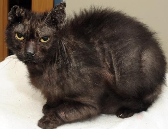
Grade               Quantity of             Degree of              Erythema
                                   cerumen                ulceration
                   0                 None                   None                    None
                   1                 Small                   Mild                   Mild
                   2               Moderate               Moderate                Moderate
                   3                 Large                 Severe                  Severe

172   Table 2 Clinical parameters and scoring system
173
174   Results
175
176   Population
177   Three hundred and forty-one cats were included in this study aged from three weeks to and
178   eighteen years. Two hundred and ninety-one cats were reported to have had contact with
179   other cats or dogs. Two hundred and seventy-five cats had an indoor / outdoor lifestyle, 45
180   cats were indoor only, one cat was outdoor only and for 20 cats their lifestyle was unknown.
181
182   One hundred and forty (41.1 %), cats were male and 198 (58.1 %) were female. One hundred
183   and fifteen (33.7 %) were entire, 224 (65.7 %) cats were neutered with missing data for two
184   cats. Twenty-seven (7.9 %) cats were receiving systemic therapy or topical ear medication at
185   the time of sampling. Fifteen different breeds were sampled (see table S1 in Supplementary
186   material), however, the majority (94.7 %) were classified as domestic long, medium or
187   shorthair, with other breed classifications poorly represented.
188
189   Eight out of 341 (2.3 %) cats, were noted to have focal to generalised signs of dermatological
190   disease including moist and crusting dermatitis, abscessation, pinnal comedones,
191   hypotrichosis of the ventrum, miliary dermatitis, chin acne, pododermatitis, paronychia, and
192   over grooming (see figure 1). and exfoliative dermatitis (see figure 4 in Supplementary
193   material).
194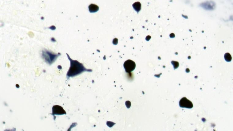
195
196   Figure 1 Concurrent dermatological disease found in some cats
197   a) erythema of the muzzle and chin along with mild feline acne, b) moderate feline acne over
198   the intermandibular region, c) ceruminous cystomatosis
199
200   Otoscopic examination
201   Otoscopy was generally well tolerated although it was not possible in 10 9 / 341 (2.9 %) (2.6
202   %) cats in either one or both ears. The tympanic membrane was visualised partially or
203   completely in 306 / 332 (91.2%) cats in one or both ears. Three cats (0.9%, CI = 0.3-2.5%)
204   were found to have O. cynotis adult mites visible upon otoscopy within one or both ears
205   (confirmed using microscopy).
206
207   Cerumen smear and cytological examination findings
208   An extra sample of aural exudate for low power microscopy (40 x and 100 x) was taken in 13
209   cats (3.8 %), eleven of these cats had excessive aural exudate bilaterally, two had unilateral
210   presentation, therefore twenty-four exudate samples mounted in paraffin oil were examined
211   for microscopic evidence of mites. Cytological findings are shown in table 3. Demodex gatoi
212   was noted unilaterally in one cat. Otodectes cynotis was noted in 3 / 341 (0.9%, CI = 0.3-
213   2.5%) cats using microscopy (see figure S1 in Supplementary material). Two of the three cats
214   had bilateral O. cynotis infestation. One cat with bilateral infestation microscopically only had
215   gross otoscopic evidence in one ear.
216
217
218
219
220
221
222
223
224
Cytological findings
                  Malassezia         Coccoid        Rod shaped          Coccoid and          Otodectes        Demodex            Melanin        Saprophytes
                                     shaped           bacteria          rod shaped             cynotis           gatoi          granules
                                     bacteria                             bacteria

Number of
 cats with        62 (bilateral)   7 (unilateral)   1 (unilateral)      1 (unilateral)      2 (bilateral)    1 (unilateral)   212 (bilateral)   311(bilateral)
cytological      67 (unilateral)                                                            1 (unilateral)                    85 (unilateral)   26 (unilateral)
findings (out
of 341 cats)

     225        Table 3 Cytological findings
     226
     227
     228        Table S2 in Supplementary material shows the otoscopic and cytological findings of four cats
     229        with evidence of Otodectes and / or Demodex. Neither bacteria or inflammatory cells were
     230        noted.
     231
     232        Some of the cytological findings that were noted are shown in figure S2 of the Supplementary
     233        material.
     234
     235        Sixty-two out of 341 cats (18.1 %) were found to have Malassezia bilaterally; sixty-seven cats
     236        had Malassezia unilaterally (19.5 %). There was an increased likelihood for Malassezia to be
     237        present with increasing age as age increases in older cats (Pearson r = 0.204, P < 0.001, n =
     238        293) and an increased likelihood of finding Malassezia in both ears if found within one ear (r
     239        = 0.499, P =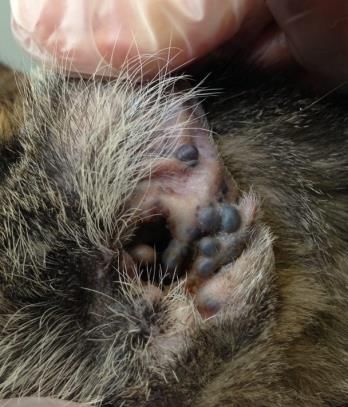
253   Those cats with otitis are shown in table 3 4 with the underlying aetiology of the otitis (if known).
254
                  Demodex     Otodectes     Aural    Allergic   Ceruminous      Generalised    Unknown
                  gatoi       cynotis       mass /   skin       cystomatosis    skin disease
                                            polyp    disease

       Number             1        3          3          4            2               2               24
        of cats

255   Table 3 4 Cats with otitis and the underlying aetiology
256
257   Nine (2.6 %) cats were found to have environmental contaminants (saprophytes) on ear
258   cytology.
259
260   Bacteria were found unilaterally in 9 / 341 (2.6 %) cats. Six of these cats were in the non-otitis
261   group and three were from the otitis group. Seven of these cats had coccoid shaped bacteria
262   only, one cat had both rod and coccoid shaped bacteria and one cat had rod shaped bacteria
263   only. Those cats with higher larger numbers of bacteria (3 or 4+) were within the otitis group.
264   Two of these cats (one with rod shaped bacteria) were found to have a space occupying lesion
265   documented using computed tomography, within the ear where bacterial infection was found.
266   Table 3 in Supplementary material shows the otoscopic and cytological findings of cats where
267   bacteria were found upon cytology. Mites were not detected in any of these cats.
268
269   Melanin granules were noted bilaterally in 212 / 341 (62.2 %) cats, and unilaterally in 85 (24.9
270   %) cats. Squamous cells were noted bilaterally in 311 / 341 (91.2 %) cats and unilaterally in
271   26 (7.6 %) cats.
272
273   Some form of ectoparasite control had been used in 278 / 341 (81.3 %) of cats at the time of
274   enrolment into the study. Nineteen (5.6 %) Thirty eight / 341 (11.14 %) of these cats received
275   regular ectoparasite control at the manufacturers recommended frequency of application.
276
277   Discussion
278   The primary aims of this study were to investigate both the prevalence of O. cynotis in a large
279   cohort of cats and to examine the ear cytology of clinically normal cats from both a rescue
280   centre and veterinary practice setting within the UK. and to examine the ear cytology of
281   clinically normal cats. Those cats presenting for otitis or with disease noted incidentally, were
282   removed when analysing the data for normal ear cytology values. To the best of the authors’
283   knowledge, there have not been any recent studies investigating the prevalence of O. cynotis
284 within a large cohort of cats in the UK, and there have been only three studies that have 285 evaluated the normal external ear cytology in cats.5,6,12 286 287 This study found that the prevalence of O. cynotis was low, recorded as 0.9 %. This result is 288 in agreement with a Belgian study (2%),10 an Australian study (
321 however may be less valuable to veterinary surgeons practicing in the UK who generally treat 322 pet cats receiving regular prophylactic ectoparasite treatments. An alternative method of 323 detecting O. cynotis infection is the use of PCR22 which could be evaluated in future studies. 324 This may however be cost prohibitive in clinical practice and therefore trial treatment may be 325 elected in the first instance. The life style cycle stage of the O. cynotis mite seen upon 326 microscopy was not noted in this study. 327 328 Two out of the three cats were found to have live O. cynotis mites despite having received 329 one application of ectoparasite control (Stronghold®: Selamectin and Broadline®: 330 eprinomectin, fipronil, S-methoprene and praziquantel). One of these cats was a seven-week- 331 old kitten who had received Stronghold within four weeks of enrolment in the study, therefore 332 clinicians should not discount O. cynotis based on previous acaricidal treatment alone. 333 Unfortunately, the exact date of Broadline® application for the other cat was not recorded 334 therefore the acaricidal application may been several weeks to months prior to sampling. One 335 single application of eprinomectin, fipronil, S-methoprene and praziquantel has been shown 336 to be effective in treating otoacariasis where one treatment corresponded to 96% preventive 337 efficacy at day 28 based on ear mite counts.23 A single application of selamectin was found to 338 be 100% effective in resolving infestation 30 days after the treatment application in another 339 study.24 Unfortunately, the date of ectoparasite administration was not recorded in this study. 340 341 Previous studies have found very different values for aural Malassezia counts in normal 342 cats.5,6,12 Two studies used the x 40 objective for examining each high power field.5,6 In our 343 study, similar to a previous study,12 we used the x 100 oil immersion objective. Cytological 344 methods have several limitations when compared to fungal culture. It is a method that is readily 345 available to clinicians and gives semi-quantitative, immediate results. Limitations include 346 inaccuracies in both cellular and microbial counts, operator dependency and reproducibility. 347 Sometimes stain artefact was seen on slides which could easily be misinterpreted as infection 348 if microorganisms were incorrectly noted (see figure S2 in Supplementary material). Some 349 Malassezia organisms did not take up the stain so well therefore appearing as very feint faint 350 structures which could easily be missed (see figure 2 Supplementary material). Seven species 351 of Malassezia have been identified in the cat and of these most are lipid dependent therefore 352 if fungal culture alone is used to detect Malassezia species in feline cerumen, lipid-dependent 353 Malassezia species may go undetected as many laboratories only use mycological culture 354 media without lipids.9 In this instance, cytology may be more sensitive in detecting yeast 355 infection. 356 357 Despite these limitations, Within this cohort of cats, those cats with otitis had five times as
358   many Malassezia per OIF than those with normal ears. The mean number for the otitis group
359   was 0.687 (CI = 0.153 to 1.380) which equates to approximately one Malassezia per two OIFs.
360   The mean number of Malassezia per OIF was 0.169 (CI = 0.114 to 0.228) in the group of cats
361   without otitis, which equates to one Malassezia per six OIFs. It is important to note that some
362   cats without clinical signs or otoscopic evidence of otitis externa had in excess of 10
363   Malassezia per OIF. It is important to note that large numbers of Malassezia (>0.169
364   Malassezia per OIF) were found in some of the cats with normal external ear canals.
365   Therefore, if Malassezia are noted, this should be interpreted along with otoscopy findings
366   and clinical signs of otitis. The finding in this study of the The presence of aural Malassezia in
367   healthy cats in this study corroborated previous studies.4-7
368
369   One cat with O. cynotis and another cat with D. gatoi isolated, were found to have >10 and
370   7.8 Malassezia per OIF respectively, which is not surprising given that it may be an
371   opportunistic microorganism as well as being part of the normal microflora. Interestingly, the
372   ear with D. gatoi cat infestation had previously undergone a pinnectomy of the same ear. One
373   ear with O. cynotis detected however did not have any Malassezia found upon cytology.
374
375   One cat from the otitis group referred to the Langford Small Animal Hospital with various
376   comorbidities along with generalised exfoliative disease (Malassezia exfoliative dermatitis),
377   was found to have very high numbers of aural Malassezia bilaterally (>10 per OIF), see figure
378   S3 in Supplementary material. Unfortunately, this cat presented to the cardiology service at
379   the Small Animal Hospital for congestive heart failure and further investigation including
380   dermatohistopathology was not taken therefore the underling aetiology for the severe
381   exfoliative dermatological disease was unknown. Other than echocardiography, further
382   thoracic imaging was not performed therefore a thymoma could not be excluded. Previous
383   studies have documented increased Malassezia in cats with concurrent illness.12,25
384
385   Two cats with large numbers (4+) of bacteria on cytology (4+) were associated with underlying
386   aural pathology such as otitis media and an aural mass (bilateral otitis media and polyps in
387   one cat and a unilateral aural mass in the other cat) documented using computed tomography
388   (CT). One other cat with large numbers (4+) of bacteria unilaterally (4+) was found to have
389   primary otitis externa and the underlying cause was not found. Only 6 / 341 cats Small
390   numbers of cats (n=6) were found to have low numbers of bacteria (1+ or 2+) which is very
391   different from previous studies where higher numbers of cats were found to have bacteria
392   within the external ear canal.5,6,10,11 These six cats with low bacterial counts were part of the
393   non-otitis group (6 / 302). As bacteria were only noted cytologically in nine cats and two of
394   these had a space occupying lesion present, mean bacterial values were not calculated.
395
396   It is important to note that large numbers of Malassezia (>0.169 Malassezia per OIF) were
397   found in some of the cats with normal external ear canals. Although a mean was calculated
398   for this grup, there was a range from 0 to >10 per oIF. Therefore, if Malassezia are noted, this
399   should be interpreted along with otoscopy findings and clinical signs of otitis. It would also be
400   prudent to take a cerumen smear to check for the presence of ectoparasites even if an
401   acaricidal ectoparasite product is used.
402
403   A link between acne and O. cynotis has been reported.20 The three cats identified as having
404   Otodectes in this study did not have acne like lesions documented.
405
406   Only low numbers of saprophytes were found compared to a previous study,10 most likely
407   because most of the rescue cats were mainly housed indoors at time of sampling. The cats in
408   this study were sampled throughout the spring and summer time. All nine of the cats where
409   saprophytes were detected upon cytology had an indoor / outdoor lifestyle.
410
411   Conclusions
412   Only a small number of cats were found to have O. cynotis in this study. If cats present for
413   otitis, it is important to rule out ectoparasitic disease and to consider other causes of otitis in
414   cats including allergic skin disease (non-flea non-food-induced feline hypersensitivity
415   dermatitis, cutaneous adverse food reaction), space occupying aural lesions such as a polyp,
416   neoplasia and otitis media (especially in cases of bacterial otitis). New mean values of
417   Malassezia counts in the external ear canals of cats were documented in this study which may
418   be a useful benchmark for those clinicians routinely performing ear cytology in cats.
419
420   Acknowledgements
421   Marta Costa for assistance in interpreting the ear cerumen cytology. The rescue centres and
422   owners of cats that allowed sampling of their cat’s ears.
423
424   Funding and Conflict of interest statements
425   Zoetis UK supplied some complementary anti-parasite products. S Tyler and N Barnard have
426   received honorariums and consulted for Zoetis.
427
428   References
429   1.     Hill PB, Lo A, Eden CA et al. Survey of the prevalence, diagnosis and treatment of
430          dermatological conditions in small animals in general practice. Vet Rec 2006; 158: 533–
431          539.

432   2.     Baxter M, Lawler DC. The incidence and microbiology of otitis externa of dogs and cats
433         in New Zealand. N Z Vet J 1972; 20: 29–32.

434   3.    Topală R, Burtan I, Fântânaru M, et al. Epidemiological Studies of Otitis Externa At
435         Carnivores. Lucrări Ştiinłifice Medicină Veterinară, Timişoara 2007; XL: 647–651.

436   4.    Nardoni S, Ebani VV, Fratini F, et al. Malassezia, mites and bacteria in the extrnal canal
437         of dogs and cats with otitis externa. Slov Vet Res 2014; 51: 113-118.

438   5.    Ginel PJ, Lucena R, Rodriguez JC et al. A semiquantitative cytological evaluation of
439         normal and pathological samples from the external ear canal of dogs and cats. Vet
440         Dermatol 2002; 13: 151–156.

441   6.    Tater KC, Scott DW, Miller WH et al. The cytology of the external ear canal in the normal
442         dog and cat. J Vet Med A Physiol Pathol Clin Med. 2003; 50: 370–374.

443   7.    Nardino S, Mancianti F, Rum A et al. Isolation of Malassezia species from healthy cats
444         and cats with otitis. J Feline Med Surg 2005; 7: 141–145.

445   8.    Shokri H, Khosravi A, Rad M et al. Occurrence of Malassezia species in Persian and
446         domestic short hair cats with and without otitis externa. J Vet Med Sci 2010; 72: 293–
447         296.

448   9.    Dizotti CE, Coutinho SDA. Isolation of Malassezia pachydermatis and M. sympodialis
449         from the external ear canal of cats with and without otitis externa. Acta Vet Hung 2007;
450         55: 471–477.

451   10.   Bollez A, de Rooster H, Furcas A et al. Prevalence of external ear disorders in Belgian
452         stray cats. J Feline Med Surg 2018; 20: 149–154.

453   11.   Perego R, Proverbio D, Bagnagatti De Giorgi G et al. Prevalence of otitis externa in
454         stray cats in northern Italy. J Feline Med Surg 2014; 16: 483-490

455   12.   Pressanti C, Drouet C, Cadiergues MC. Comparative study of aural microflora in
456         healthy cats, allergic cats and cats with systemic disease. J Feline Med Surg 2014; 16:
457         992–996.

458   13.   Coman BJ, Jones EH, Driesen MA. Helminth Parasites and Arthropods of Feral Cats.
459         Aust Vet J 1981; 57: 324–327.

460   14.   Pugh KE, Evans JM Hendy PG. Otitis externa in the dog and the cat - an evaluation of
461         a new treatment. J Small Anim Pract 1974; 15: 387–400.

462   15.   Beresford-Jones WP. Observations on the incidence of Otodectes cynotis (Hering) on
463         Dogs and Cats in the London Area. Vet Rec 1955; 67: 716–717.

464   16.   Powell M; Weisbroth SH; Roth L. Reaginic hypersensitivity in Otodectes cynotis
465         infestation of cats and mode of mite feedinge. Am J Vet Res 1980; 41: 877–882.

466   17.   Budach SC, Mueller RS. Reproducibility of a semiquantitative method to assess
467         cutaneous cytology. Vet Dermatol 2012; 23: 426–431.

468   18.   Nuttall T, Bensignor E. A pilot study to develop an objective clinical score for canine
469         otitis externa. Vet Dermatol 2014; 25: 530-e92.

470   19.   Duarte A, Castro I, Pereira da Fonseca IM, et al. Survey of infectious and parasitic
471         diseases in stray cats at the Lisbon Metropolitan Area, Portugal. J Feline Med Surg
472         2010; 12: 441–446.

473   20.   Sotiraki ST, Koutinas AF, Leontides LS, et al. Factors affecting the frequency of ear
474         canal and face infestation by Otodectes cynotis in the cat. Vet Parasitol 2001; 96: 309–
475         315.

476   21.   Akucewich LH, Philman K, Clark A et al. Prevalence of ectoparasites in a population of
477         feral cats from north central Florida during the summer. Vet Parasitol 2002; 109: 129–
478         139.

479   22.   Salib FA, Baraka TA. Diseases I. Epidemiology , genetic divergence and acaricides of
480         Otodectes cynotis in cats and dogs. Vet World 2011; 4: 109–112

481   23.   Beugnet F, Bouhsira E, Halos L et al. Preventive efficacy of a topical combination of
482         fipronil – (S) -methoprene – eprinomectin – praziquantel against ear mite (Otodectes
483         cynotis) infestation of cats through a natural infestation model. Parasite 2014; 21: 40.

484   24.   Shanks DJ, McTier TL, Rowan TG et al. The efficacy of selamectin in the treatment of
485         naturally acquired aural infestations of Otodectes cynotis on dogs and cats. Vet
486         Parasitol 2000; 91: 283–290.

487   25.   Mauldin EA, Morris DO, Goldschmidt MH. Retrospective study: the presence of
488         Malassezia in feline skin biopsies. A clinicopathological study. Vet Dermatol 2002; 13:
489         7-14

490   Supplementary material
491
       Breed                      Number examined
       Bengal                     1
       Birman cross breed         1
       British Short Hair         2
       Burmese                    1
       Devon Rex                  1
       Domestic Long hair         23
       Domestic medium hair       15
       Domestic short hair        285
       Maine coon                 2
       Ragdoll                    1
       Russian Blue               1
       Siamese                    3
       Siamese cross              1
       Snowshoe                   1
       Somali cross               1
Missing data 2
492 Table S1 Breeds of cats examined
493
494
495
496 Figure S1 Microscopic evidence of Otodectes cynotis infestation
497 a) adult mite (100 x) b) one adult mite and three eggs (40 x) c) three nymphs (x 40)
498
Case Signalment Otodectes Otodectes Demodex Ectoparasite Lifestyle Exudate Malassezia
cynotis cynotis gatoi control (quantity) (average
visible visible visible number
upon upon upon per OIF)
otoscopy microscopy microscopy
1 3 years Broadline®*
FN R YES R YES - (Eprinomectin Indoor / R 3+ R0
DSH L YES L YES Fipronil outdoor L 2+ L 0.3
S-
methoprene
Praziquantel)
once
2 4 weeks
ME R NO R NO - - Indoor R 3+ R 2.6
DSH L YES L YES L 3+ L 1.6
3 7 weeks
ME R YES R YES - Stronghold®* Indoor / R 3+ R > 10
DSH L NO L YES (Selamectin) outdoor L 3+ L 7.3
once within 4
weeks prior
to sampling
4 12 years
FN R NO R NO R YES Stronghold®* Indoor / R 3+ R 7.8
DSH L NO L NO L NO (Selamectin) outdoor L 3+ L 0.2
once
499
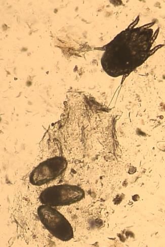
500 Table S2 Otoscopy and aural cytology findings in cats with ear mites (Otodectes cynotis
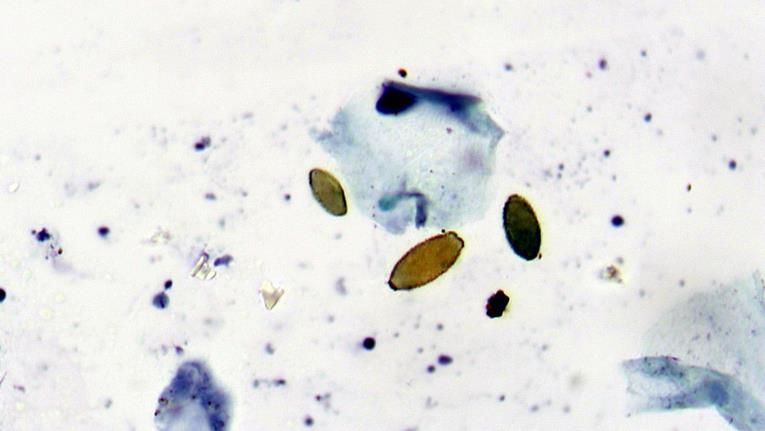
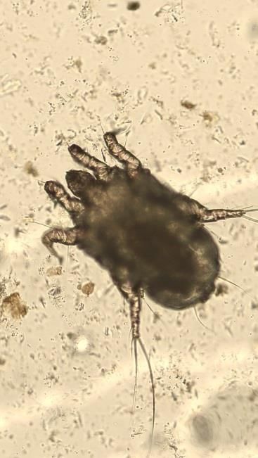
501 or Demodex gatoi)
502503 R = right ear, L = left ear, OIF = oil immersion field, DSH = Domestic short hair, ME = Male 504 entire, FN = Female neutered, * = exact date of application prior to sampling unknown 505
506   a)

507   b)

508   c)

509   d)
510     e)

511     f)

512     g)
513
514
515   Figure S2 Aural cytological findings
516
517   a) Stained Malassezia
518   b) Non-stained Malassezia
519   c) Keratinocytes containing numerous melanin granules
520   d) Degenerate neutrophils and nuclear streaming, with large numbers of coccoid
521   shaped bacteria in a cat with purulent otitis externa in a protein rich background
522   e) Stain artefact
523 f) and g) Environmental likely fungal contaminants
Case Centre Signalment Reason for Lifestyle Otoscopy Exudate Nature of Rod shaped Coccoid shaped Inflammatory Clinical outcome
examination (erythema, (quantity) exudate bacteria bacteria cells
ulceration, (classification) (classification)
oedema)
1 Langford 16 years
SAH FN Study Indoor / Unremarkable R 2+ R dark brown R0 R0 R NO No treatment
DLH (Hyperthyroid outdoor L 3+ L dark brown L0 L 1+ L NO recommended
assessment)
2 Kats and
Kits 17 years Study Indoor / Unremarkable R 3+ R brown R0 R0 R NO Canaural®
MN outdoor L 3+ L cream, purulent L0 L 2+ L NO recommended
DSH
3 Kats and
Kits 10 years Study Indoor / R 1+ R cream R0 R0 R NO No treatment
MN outdoor Unremarkable L 1+ L cream L0 L 2+ L NO recommended
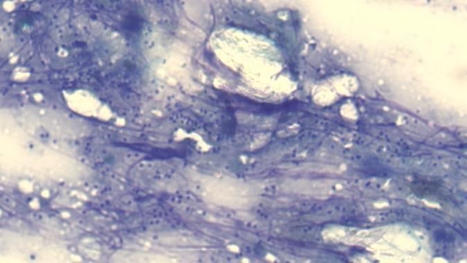
DSH
4 Bridgend 1 years Study Indoor / R 1+ R beige R0 R0 R NO No treatment
CP MN outdoor Unremarkable L 1+ L beige L0 L 2+ L NO recommended
DSH
5 Bridgend Unknown Study Indoor / R 1+ R beige R0 R0 R NO No treatment
CP FN outdoor Unremarkable L 1+ L beige L 2+ L 2+ L NO recommended
DSH
6 Mayhew 2 years Study R 2+ R cream R0 R R NO No treatment
FN Indoor / Unremarkable L cream L0 L 1+ L NO recommended
DSH outdoor L 2+
7 Langford 4 years Otitis externa R beige R0 R0 R NO Resolution with topical
SAH FN (presented to Indoor / Exudate R 1+ L0 L 4+ L YES and treatment and
Siamese the outdoor obscured L 3+ L haemorrhagic, systemic
dermatology vision purulent glucocorticoids
service)
8 Langford 10 months Otitis externa R 1+ R0 R none R0 R0 R NO CT scan and ear flush
SAH MN (presented to Indoor / erythema L 3+ L cream L 4+ L 4+ L YES performed, surgery
Maine coon the outdoor recommended. CT
dermatology L 2+ stenosis, scan revealed bilateral
service) polyp visible otitis media and
post flush bilateral aural polyps
within the middle ear
9 Langford 14 years Otitis externa CT revealed mass at
SAH FN (presented to Indoor / R 2+ R 2+ R brown R0 R 4+ R NO junction of vertical and
DSH the outdoor erythema and L 1+ L yellow L0 L0 L NO horizontal ear canal,
dermatology 2+ ulceration surgery recommended
service)
Table S2 The otoscopic and cytological findings of cats with bacteria found on aural cytology.
R = right ear, L = left ear, DSH = Domestic short hair, ME = Male entire, FN = Female neutered, MN = Male neuteredFigure S3 Cat with generalised exfoliative disease (aetiology unknown) large numbers of
Malassezia noted upon cytology (>10 per oil immersion field)
Page 22 of 22You can also read