Fungal Diversity and Onychomycosis - An Analysis of 8,816 Toenail Samples Using Quantitative PCR and Next-Generation Sequencing - MicrogenDX
←
→
Page content transcription
If your browser does not render page correctly, please read the page content below
ORIGINAL ARTICLES

Fungal Diversity and Onychomycosis
An Analysis of 8,816 Toenail Samples Using Quantitative PCR and Next-
Generation Sequencing
                                                                                Annette Joyce, DPM*
                                                                 Aditya K. Gupta, MD, PhD, FRCP(C)†
                                                                                   Lars Koenig, PhD‡
                                                                                Randall Wolcott, MD§
                                                                                 Jessie Carviel, PhD†

Background: Onychomycosis is a fungal infection of the nail that is often recalcitrant to
treatment and prone to relapse. Traditional potassium hydroxide and culture diagnosis is
costly and time-consuming. Therefore, molecular methods were investigated to
demonstrate effectiveness in diagnosis and to quantify the microbial flora present that
may be contributing to disease.
Methods: A total of 8,816 clinically suspicious toenail samples were collected by
podiatric physicians across the United States from patients aged 0 to 103 years and
compared with a control population (N ¼ 20). Next-generation sequencing and
quantitative polymerase chain reaction were used to identify and quantify dermato-
phytes, nondermatophyte molds, and bacteria.
Results: Approximately 50% of suspicious toenails contained both fungi and bacteria,
with the dermatophyte Trichophyton rubrum contributing the highest relative abundance
and presence in 40% of these samples. Of the remaining 50% of samples, 34% had
bacterial species present and 16% had neither. Fungi only were present in less than 1%
of samples. Nondermatophyte molds contributed to 11.0% of occurrences in fungus-
positive samples. All of the control samples were negative for fungi, with commensal
bacterial species composing most of the flora population.
Conclusions: Molecular methods were successful in efficiently quantifying microbial
and mycologic presence in the nail. Contributions from dermatophytes were lower than
expected, whereas the opposite was true for nondermatophyte molds. The clinical
significance of these results is currently unknown. (J Am Podiatr Med Assoc 109(1): 57-
63, 2019)

Use of genomic techniques has allowed the identi-              and 18S (fungal) ribosomal DNA sequences. Knowl-
fication of fastidious and noncultivable microorgan-           edge of the particular microbe combinations that
isms in nail samples. Consequently, a better                   result in disease can then be applied to the creation
understanding of the role of biodiversity in onycho-           of targeted treatments and improving cure rates.
mycosis and chronic nail infections can be eluci-              Therefore, molecular genetics–based diagnostics
dated. Metagenomics techniques can now deter-                  were used to accurately and rapidly identify
mine the entire genetic composition of a microbial             microbes present in patients with onychomycosis
community. Even organisms in low abundance or                  to improve treatment recommendations.
dormant can be identified through 16S (bacterial)
  *Joyce Podiatry, Westminster, MD. Dr. Joyce is now with      Methods
Dermfoot Educational Ventures, LLC, Pawley’s Island, SC.
  †Mediprobe Research Inc, London, Ontario, Canada.            Patient Population and Sample Collection
  ‡Research and Testing Laboratory, Lubbock, TX.
  §Southwest Regional Wound Care Clinic, Lubbock, TX.
  Corresponding author: Annette Joyce, DPM, 423 Lumbee
                                                               A total of 8,816 toenail samples collected by
Circle, Pawley’s Island, SC 29585. (E-mail: drjoycepodiatry@   podiatric physicians from clinically suspicious
gmail.com)                                                     toenails across the United States were submitted

Journal of the American Podiatric Medical Association  Vol 109  No 1  January/February 2019                    57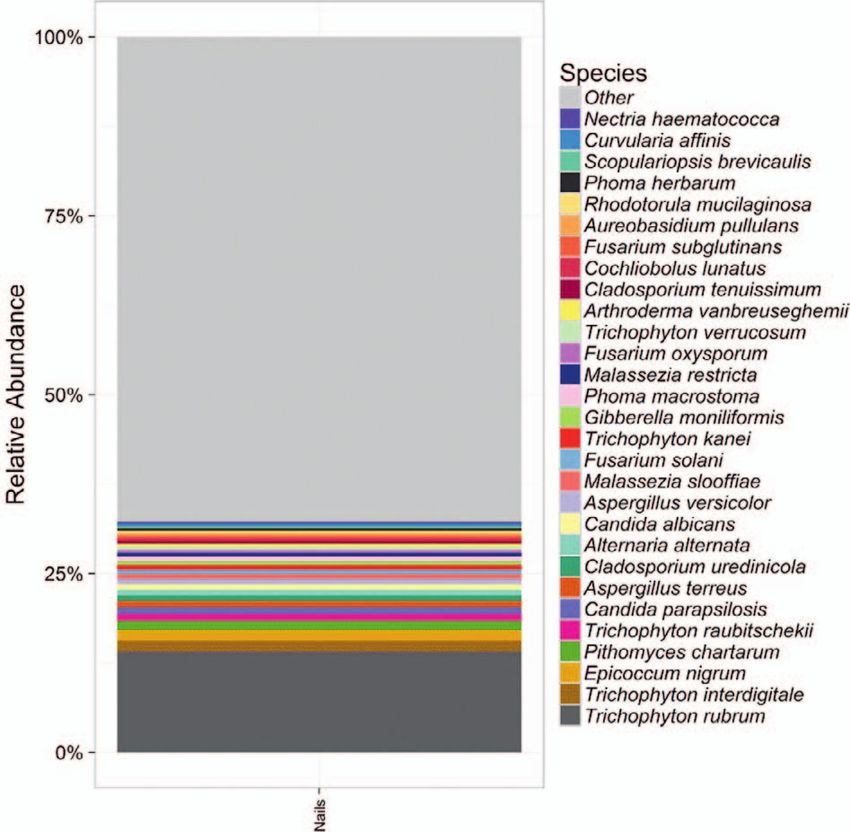
to Pathogenius Laboratories (Lubbock, Texas) for            trimming sequences back using a running average of
laboratory analysis between January 1, 2013, and            Q25. Trimmed sequences were then run through
January 31, 2016. The patient population was                USEARCH1 to cluster the sequences at 4% diver-
composed of 3,487 males and 5,329 females aged 0            gence, perform operational taxonomic unit (OTU)
to 103 years (61 reported no age selected). A second        selection and chimera depletion using the UPARSE2
population (N ¼ 20) of healthy nails was examined           OTU selection algorithm, and map each sequence to
as controls. All of the control nails were investigat-      the corresponding nonchimera OTU. Mapped se-
ed for the presence of bacteria and fungi, and six          quences were then grouped by OTU, and quality
were further examined for species. Fingernail               score–based sequence correction was performed.
samples were excluded from this population as-                 Corrected sequences were run through a taxo-
suming that nail specimens were accurately labeled          nomic analysis pipeline (RTL Genomics, Lubbock,
by physicians at the time of analysis and because           Texas) to determine the taxonomic classifications.
the podiatric physician’s scope of practice is often,       First, quality checking on each corrected sequence
although not always, limited to foot and ankle              was performed to confirm primer removal and a
disease. In addition, the organisms that are found in       minimum sequence length of 250 base pairs.
fingernails are not the same or in the same                 Selection of OTUs was then performed using the
distribution as those found in toenails.                    UPARSE OTU selection pipeline.1,2 Selected OTUs
                                                            were aligned using MUSCLE,3,4 and a phylogenetic
DNA Extraction and Amplification                             tree was generated using FastTree.5,6 The selected
                                                            OTU sequences were globally aligned using
The DNA isolation was performed using the High              USEARCH1 against a database of 16S sequences
Pure polymerase chain reaction (PCR) template               for bacterial assays and internal transcribed spacer
preparation kit (Roche Diagnostics, Indianapolis,           sequences for fungal assays. Sequences were
Indiana) following a modified manufacturer’s pro-           gathered from GenBank and are classified to at
tocol. The extraction procedure was modified to             least genus level with at least partial sequences of
include a beading step for tissue and cell disruption       the target genes. Confidence values were assigned
using 5-mm steel beads, 0.5-mm zirconium oxide              to each OTU classification, and the lowest common
beads, and the TissueLyser II instrument (Qiagen,           ancestor was determined based on these confidence
Germantown, Maryland). The lysate was then run              values. The top hit and lowest common ancestor
through the glass fiber fleece column following the         was reported for each OTU. For the analysis in this
manufacturer’s protocol provided with the Roche             study, we included only the data from microbial
High Pure PCR template preparation kit.                     species that comprised at least two orders of
  DNA was amplified through quantitative PCR                magnitude (two log10 values) of the total bacterial
including a 5-min predenaturation at 958C, followed         population. Therefore, microbial species that repre-
by 35 cycles of denaturation at 948C for 30 seconds,        sented less than 1% of the entire sample are not
annealing at 528C for 40 seconds, extension at 728C         reported in this study.
for 60 seconds, and a final extension at 728C for 10
minutes. DNA was purified using Agencourt AM-               Results
Pure beads (Beckman Coulter, Indianapolis, Indi-
ana) and the Qiagen MinElute PCR purification kit           Of the 8,816 samples, approximately 50% (n ¼ 4,328)
according to the manufacturer’s instructions. DNA           were positive for fungal organisms (as well as
was then pooled based on amplification strength             bacteria), with 19,351 unique fungal occurrences.
and attached to Ion Sphere particles (Thermo Fisher         Therefore, the data are consistent with clinical and
Scientific) through emulsion PCR. Ion Sphere                scientific observations in onychomycosis litera-
particles were enriched and sequenced using next-           ture.7 Of the remaining 50% of samples, 34% (n ¼
generation sequencing technology (Personal Ge-              2,995) had bacterial species present and 16% (n ¼
nome Machine, Life Technologies).                           1,443) had neither. Fungi only were present in less
                                                            than 1% of samples (n ¼ 50). Contradictory to the
Bioinformatic and Biostatistical Analyses                   currently accepted estimate of 90%,8 the dermato-
                                                            phyte Trichophyton rubrum was present in 40% of
Denoising was performed to remove short sequenc-            fungus-positive samples and contributed the highest
es, singleton sequences, and noisy reads, followed          relative abundance (Figs. 1 and 2). Pithomyces
by chimera detection. Correction of sequencing              chartarum was the second most commonly found
errors and chimera removal was performed by first           organism (occurring in 20% of fungus-positive

58                    January/February 2019  Vol 109  No 1  Journal of the American Podiatric Medical Association
Figure 2. Occurrence of the most common derma-
tophytes in fungal (18S) species identification.
species (Fig. 4), including common commensals
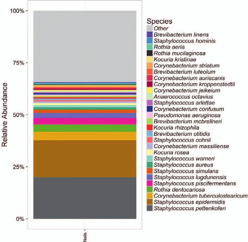
Figure 1. Average relative abundance of the top 30 such as Staphylococcus epidermis (occurring in
fungal species determined by internal transcribed 14% of bacterium-positive samples), Staphylococcus
spacer sequences in 8,816 nail samples. pettenkoferi (occurring in 11% of bacterium-positive
samples), and Staphylococcus lugdenensis (occur-
samples) and has been previously implicated in ring in 5% of bacterium-positive samples), which
onychomycosis.9 Related species Pithomyces sac- were also all present in the control population
chari and Pithomyces maydicus have similarly (Table 2). Not all species isolated were so benign,
been recovered from skin, nails, and respiratory however. Also highly prevalent was Corynebacteri-
tracts.10 um tuberculostearicum, which was present in 8% of
Following these, saprophytic yeasts such as bacterium-positive nail specimens. The clinical
Epicoccum nigrum (occurring in 18% of fungus- relevance of this species has been difficult to assess
positive samples) and Alternaria alternata (occur- because it is commonly present on the skin and
ring in 11% of fungus-positive samples) were overly mucosal surfaces (and was also present in the
abundant. The latter are considered emerging control population [Table 2]) but has been consid-
pathogens in onychomycosis, especially in warm
ered relevant in less than half of the cases where it
and humid environments,11 and the former are
has been identified in patients with chronic dis-
associated with respiratory fungal allergies, includ-
ease.13
ing allergic asthma, rhinitis, hypersensitivity pneu-
monitis, and allergic fungal sinusitis.12
Nondermatophyte molds (NDMs) represented
11.0% of occurrences (2,125 of 19,351) in fungus-
positive samples. The most commonly observed
NDMs included various species of Candida, Asper-
gillus, Fusarium, and Acremonium (Fig. 3). Many
of these species are ubiquitously present in the
environment or are common commensal organisms,
although there is emerging evidence that some may
have pathogenic potential or have been previously
linked to onychomycosis (Table 1). Therefore, the
frequent presence in chronically infected nails (and
lack of presence in the control population [Table 2])
suggests utility in further investigating targeted Figure 3. Occurrence of the most common non-
treatment options. dermatophyte molds in fungal (18S) species iden-
Most bacteria identified were Staphylococcus tification.
Journal of the American Podiatric Medical Association Vol 109 No 1 January/February 2019 59Table 1. Most Commonly Identified Nondermatophyte Molds Observed in Onychomycosis-Suspected Nails
Evidence of Evidence of
Involvement in Virulence/
Nondermatophyte Mold Onychomycosis Pathogenicity Little/No Evidence of Pathogenicity
Candida parapsilosis Yes22,23
Candida albicans Yes22,23
Candida orthopsilosis Yes22
Candida tropicalis Yes23,24
Candida metapsilosis Yes22
Candida sake Noa Yes25-27
Candida glabrata Yes23
Candida zeylanoides Yes28,29
Candida jeffriesii Noa No Xylose-fermenting, associated with rotting wood and the
intestines of wood-boring beetles30
Aspergillus versicolor Yes31-34
Aspergillus terreus Yes35-37
Aspergillus penicillioides/vitricola Noa No Extreme halophile,38 pathogenesis is extremely rare even
in the immunocompromised
Aspergillus tubingenesis Yes36
Aspergillus oryzae Noa No Domesticated, used in industrial fermentation processes39
Aspergillus niger Yes33,36 No
Aspergillus sydowii Yes36
Aspergillus restrictus Noa No Xerophilic, allergen, asthma, found commonly in house
dust,40 pathogenic in immunocompromised, one
anecdotally reported case of onychomycosis
Aspergillus sclerotiorum Noa No Soil fungus, rare reports of onychomycosis in genetically
predisposed (diabetic) individuals
Aspergillus flavus Yes33,35,36
Aspergillus flavipes Noa No Few reports of pathogenicity in immunocompromised
individuals
Aspergillus peyronelii Noa No No evidence
a
In the absence of evidence of previous connections to onychomycosis, presence or absence of reports of pathogenicity are
identified.
Discussion brum in approximately 40% (as opposed to the
expected 90%8) of fungus-positive samples, sug-
Contrary to the current literature, a molecular gesting that the cause of disease may be more
analysis of 8,816 toenail samples suspected of diverse than presently estimated. In comparison, a
onychomycosis revealed the presence of T ru- control population of healthy nails (N ¼ 20)
contained no fungal or yeast species, with com-
Table 2. Average Relative Percentage of Top Microbial mensal bacterial species such as Staphylococcus
Species Determined by 16S Comparison in Six Nail epidermidis composing most of the microbial
Samples flora. Just as importantly, molecular techniques
Bacterial Species Observed (%) were demonstrated as an accurate and efficient
method for diagnosis. Moreover, there have been
Staphylococcus epidermidis 35.83 similar observations of an increased presence of
Corynebacterium tuberculostearicum 15.50
NDMs in other geographic locations reported as
Staphylococcus warneri 13.00
Corynebacterium jeikeium 7.83
well.16,17 Ebihara et al18 also report higher rates of
Staphylococcus lugdunensis 4.17 NDMs and suggest that their observations were
Enterococcus faecalis 2.83 possible due to the sensitivity of the molecular
Staphylococcus pettenkoferi 2.00 methods used for diagnosis and that NDMs may
Anaerococcus tetradius 1.83 currently be underestimated due to the difficulty
Kocuria rosea 1.67 of identifying them by conventional methods.
Brevibacterium mcbrellneri 1.67 Overall these results provide insight into the
Corynebacterium confusum 1.00
chronic nature of the disease as well as implica-
Zimmermannella bifida 1.00
tions for treatment.
60 January/February 2019 Vol 109 No 1 Journal of the American Podiatric Medical Associationconcern for nail infection. Similarly, Shemer et al15
reported that 46% of swimming pool employees had
both tinea pedis and onychomycosis in a recent
study of 169 employees of public pools in Israel,
suggesting that moisture-rich environments such as
public swimming pools may also contribute to the
development of nail disease.
Taken together, evidence from this analysis has
led to the hypothesis that development of onycho-
mycosis may depend on a plethora of microbes
contributing synergistically to disease. Moreover,
there is a possibility of biofilm formation, which
displays similar characteristics (recurrence and
recalcitrance to normally effective treatment) as
observed in patients with onychomycosis.19 This
hypothesis supports the chronic nature of onycho-
mycosis because antifungal therapies are targeted
mainly at T rubrum and NDMs are often resistant to
both oral and topical antifungal agents.20 Thus,
investigations into combining NDM treatment with
Figure 4. Average relative abundance of the top 30
traditional therapy seem worthwhile.
bacterial species determined by 16S comparison in
8,816 nail samples.
Implications for Diagnosis and Treatment
As expected, T rubrum and dermatophytes were
observed most often; however, saprophytic yeasts, Based on the high cost and low accuracy of current
NDMs, and bacteria were also observed on a potassium hydroxide and culture techniques used in
standard diagnosis, use of empirical oral terbinafine
consistent basis. Because the latter organisms are
without the benefit of confirmatory diagnosis has
known to form commensal relationships, a connec-
been suggested, reporting minimal effect on patient
tion to onychomycosis may not be straightforward;
safety with a potential savings of millions of health-
however, there is evidence in the literature to
care dollars annually.21 There are at least two
support this theory of a possible group contribution
drawbacks to this approach, the first being that
to disease (Table 1).
because both oral and topical antifungal therapies
For example, Rothia dentocariosa was found in are specifically focused at T rubrum, we may be
more than 3% of bacterium-positive samples and has actively selecting for alternative organisms by
pathogenic potential as the cause of dental caries targeting the wrong genus and species while
and periodontal disease. As an opportunistic infec- encouraging the development of resistance. Second,
tious organism, pathogenicity has been implicated other similar nail disorders cannot be excluded
in endocarditis, pneumonia, peritonitis, and lung without nail sampling, such as trauma, psoriasis,
infections. Therefore, it is possible that periodontal lichen planus, or other nail disease. Alternatively,
disease and dental procedures may be the first step molecular methods are extremely specific, sensi-
in the inoculation of other distal peripheral tissues tive, and quantitative, which allows not only
such as toenails. diagnosis but is invaluable in assessing response
Likewise, gastrointestinal host colonization with to treatment. In comparison, they are both faster (3-
gram-positive commensal bacteria such as Entero- day turnaround time) and lower in cost than current
coccus faecalis, which was present in almost 1% of methods. Laboratory tests with the required exper-
bacterium-positive samples, is another area of tise are now becoming routinely available at
concern with nail disease. Enterococcus faecalis steadily decreasing costs.
can bind to dentin, alter host responses, and use
serum as a nutritional source, therefore competing Conclusions
with other cells.14 Thus, frequent pedal contamina-
tion with intestinal flora as a result of pooling water Through the evidence provided herein, dermato-
from baths and showers in community-acquired phytes and specifically T rubrum are unsurprisingly
settings such as nursing homes may be a growing a main contributor to onychomycosis; however,
Journal of the American Podiatric Medical Association Vol 109 No 1 January/February 2019 61additional organisms, such as yeasts, NDMs, and enterococci in filled root canals. Int Endod J 42: 277,
possibly bacteria, may also be playing a larger role 2009.
in disease development than previously believed. 15. SHEMER A, GUPTA AK, AMICHAI B, ET AL: Increased risk of
The clinical significance remains unknown, and tinea pedis and onychomycosis among swimming pool
employees in Netanya Area, Israel. Mycopathologia 181:
further confirmatory tests will provide a better
851, 2016.
understanding. To improve treatment outcome, use 16. I OANNIDOU D, M ARAKI S, KRASAGAKIS S, ET AL: The
of new molecular diagnostic techniques are recom- epidemiology of onychomycoses in Crete, Greece,
mended to confirm disease and to target therapy to between 1992 and 2001. J Eur Acad Dermatol Venereol
the presence of specific microbes. 20: 170, 2006.
17. GARCÍA-MARTOS P, DOMÍNGUEZ I, MARÍN P, ET AL: Onycho-
mycoses caused by non-dermatophytic filamentous
Financial Disclosure: None reported. fungi in Cádiz [in Spanish]. Enferm Infecc Microbiol
Conflict of Interest: None reported. Clin 18: 319, 2000.
18. EBIHARA M, MAKIMURA K, SATO K, ET AL: Molecular
References detection of dermatophytes and nondermatophytes in
onychomycosis by nested polymerase chain reaction
1. EDGAR RC: Search and clustering orders of magnitude based on 28S ribosomal RNA gene sequences. Br J
faster than BLAST. Bioinformatics 26: 2460, 2010. Dermatol 161: 1038, 2009.
2. EDGAR RC: UPARSE: highly accurate OTU sequences 19. GUPTA AK, CARVIEL JL, ABRAMOVITS W: Efficacy of
from microbial amplicon reads. Nat Methods 10: 996, tofacitinib in treatment of alopecia universalis in two
2013. patients. J Eur Acad Dermatol Venereol 30: 1373, 2016.
3. EDGAR RC: MUSCLE: multiple sequence alignment with 20. GUPTA AK, DRUMMOND-MAIN C, COOPER EA, ET AL: System-
high accuracy and high throughput. Nucleic Acids Res atic review of nondermatophyte mold onychomycosis:
32: 1792, 2004. diagnosis, clinical types, epidemiology, and treatment. J
4. EDGAR RC: MUSCLE: a multiple sequence alignment Am Acad Dermatol 66: 494, 2012.
method with reduced time and space complexity. BMC 21. MIKAILOV A, COHEN J, JOYCE C, ET AL: Cost-effectiveness of
Bioinformatics 5: 113, 2004. confirmatory testing before treatment of onychomyco-
5. PRICE MN, DEHAL PS, ARKIN AP: FastTree: computing sis. JAMA Dermatol 152: 276, 2016.
large minimum evolution trees with profiles instead of a 22. FENG X, LING B, YANG X, ET AL: Molecular Identification of
distance matrix. Mol Biol Evol 26: 1641, 2009. Candida Species Isolated from Onychomycosis in
6. PRICE MN, DEHAL PS, ARKIN AP: FastTree 2: approximate- Shanghai, China. Mycopathol 180: 365, 2015.
ly maximum-likelihood trees for large alignments. PLoS 23. OTAŠEVI
c S, BARAC A, PEKMEZOVIC M, ET AL: The prevalence
One 5: e9490, 2010. of Candida onychomycosis in Southeastern Serbia from
7. ELEWSKI BE: Onychomycosis: treatment, quality of life, 2011 to 2015. Mycoses 59: 167, 2016.
and economic issues. Am J Clin Dermatol 1: 19, 2000. 24. ARRUA JMM, RODRIGUES LAS, PEREIRA FO, ET AL: Preva-
8. GHANNOUM MA, HAJJEH RA, SCHER R, ET AL: A large-scale lence of Candida tropicalis and Candida krusei in
North American study of fungal isolates from nails: the onychomycosis in João Pessoa, Paraiba, Brazil from
frequency of onychomycosis, fungal distribution, and 1999 to 2010. An Acad Bras Cienc 87: 1819, 2015.
antifungal susceptibility patterns. J Am Acad Dermatol 25. GUCLU E, SOYPACACI Z, YILDIRIM M, ET AL: First case of
43: 641, 2000. continuous ambulatory peritoneal dialysis peritonitis
9. LITZ CE, CAVAGNOLO RZ: Polymerase chain reaction in the due to Candida sake. Mycoses 52: 280, 2009.
diagnosis of onychomycosis: a large, single-institute 26. PRESTERL E, DAXBÖCK F, GRANINGER W, ET AL: Changing
study. Br J Dermatol 163: 511, 2010. pattern of candidaemia 2001-2006 and use of antifungal
10. DA CUNHA KC, SUTTON DA, GENÉ J, ET AL. Pithomyces therapy at the University Hospital of Vienna, Austria.
species (Montagnulaceae) from clinical specimens: Clin Microbiol Infect Off Publ Eur Soc Clin Microbiol
identification and antifungal susceptibility profiles. Infect Dis 13: 1072, 2007.
Med Mycol 52: 748, 2014. 27. PALMISANO A, BENECCHI M, DE FILIPPO M, ET AL: Candida
11. FARWA U, ABBASI SA, MIRZA IA, ET AL: Non-dermatophyte sake as the causative agent of spondylodiscitis in a
moulds as pathogens of onychomycosis. J Coll Physi- hemodialysis patient. Spine J Off J North Am Spine Soc
cians Surg Pak 21: 597, 2011. 11: e12, 2011.
12. GREEN BJ, MITAKAKIS TZ, TOVEY ER: Allergen detection 28. DORKO E, JAUTOVÁ J, TKÁCIKOVÁ L, ET AL: The frequency of
from 11 fungal species before and after germination. J Candida species in onychomycosis. Folia Microbiol
Allergy Clin Immunol 111: 285, 2003. (Praha) 47: 727, 2002.
13. HINIc V, LANG C, WEISSER M, ET AL: Corynebacterium 29. CROZIER WJ: Two cases of onychomycosis due to
tuberculostearicum: a potentially misidentified and Candida zeylanoides. Australas J Dermatol 34: 23, 1993.
multiresistant Corynebacterium species isolated from 30. NGUYEN NH, SUH S-O, MARSHALL CJ, ET AL: Morphological
clinical specimens. J Clin Microbiol 50: 2561, 2012. and ecological similarities: wood-boring beetles associ-
14. ZEHNDER M, GUGGENHEIM B: The mysterious appearance of ated with novel xylose-fermenting yeasts, Spathaspora
62 January/February 2019 Vol 109 No 1 Journal of the American Podiatric Medical Associationpassalidarum gen. sp. nov. and Candida jeffriesii sp. nov. antifungal susceptibility testing. Diagn Microbiol Infect
Mycol Res 110: 1232, 2006. Dis 84: 125, 2016.
31. VERALDI S, CHIARATTI A, HARAK H: Onychomycosis caused 36. NOURIPOUR-SISAKHT S, MIRHENDI H, SHIDFAR MR, ET AL:
by Aspergillus versicolor. Mycoses 53: 363, 2010. Aspergillus species as emerging causative agents of
32. NENOFF P, GINTER-HANSELMAYER G, TIETZ H-J: [Fungal nail onychomycosis. J Mycol Medicale 25: 101, 2015.
infections - an update. Part 2 - From the causative agent 37. FERNÁNDEZ MS, ROJAS FD, CATTANA ME, SOSA M DE LÁ,
to diagnosis - conventional and molecular procedures]. MANGIATERRA ML, GIUSIANO GE: Aspergillus terreus
Hautarzt Z Dermatol Venerol Verwandte Geb 63: 130, complex: an emergent opportunistic agent of Onycho-
mycosis. Mycoses 56: 477, 2013.
2012.
38. NAZARETH SW, GONSALVES V: Halophilic Aspergillus
33. MORENO G, ARENAS R: Other fungi causing onychomyco-
penicillioides from athalassohaline, thalassohaline,
sis. Clin Dermatol 28: 160, 2010.
and polyhaline environments. Front Microbiol 5: 412,
34. TORRES-RODRÍGUEZ JM, MADRENYS-BRUNET N, SIDDAT M, ET 2014.
AL: Aspergillus versicolor as cause of onychomycosis:
39. GIBBONS JG, SALICHOS L, SLOT JC, ET AL: The evolutionary
report of 12 cases and susceptibility testing to antifungal imprint of domestication on genome variation and
drugs. J Eur Acad Dermatol Venereol JEADV 11: 25, function of the filamentous fungus Aspergillus oryzae.
1998. Curr Biol CB 22: 1403, 2012.
35. TSANG C-C, HUI TWS, LEE K-C, ET AL: Genetic diversity of 40. ITABASHI T, HOSOE T, TOYASAKI N, ET AL: [Allergen activity of
Aspergillus species isolated from onychomycosis and xerophilic fungus, Aspergillus restrictus]. Arerugi Aller-
Aspergillus hongkongensis sp. nov., with implications to gy 56: 101, 2007.
Journal of the American Podiatric Medical Association Vol 109 No 1 January/February 2019 63You can also read