Genetic Analysis of Hereditary Polydactyly in Pakistani Families - CUST
←
→
Page content transcription
If your browser does not render page correctly, please read the page content below

CAPITAL UNIVERSITY OF SCIENCE AND
         TECHNOLOGY, ISLAMABAD

 Genetic Analysis of Hereditary
Polydactyly in Pakistani Families
                          by
           Muhammad Sajid Khan
    A thesis submitted in partial fulfillment for the
              degree of Master of Science

                         in the
           Faculty of Health & Life Sciences
              Department of Biosciences

                         2018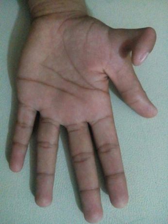
i

            Copyright c 2018 by Muhammad Sajid Khan

All rights reserved. No part of this thesis may be reproduced, distributed, or
transmitted in any form or by any means, including photocopying, recording, or
other electronic or mechanical methods, by any information storage and retrieval
system without the prior written permission of the author.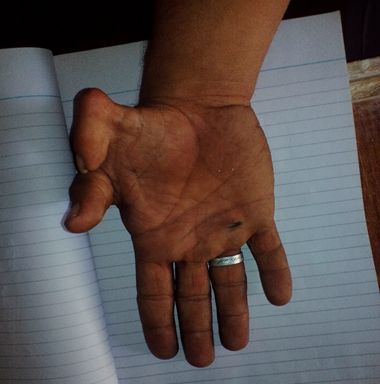
ii

  This thesis is dedicated to my father, who taught me that the best kind of
knowledge to have is that which is learned for its own sake. It is also dedicated
to my mother, who taught me that even the largest task can be accomplished if
                         it is done one step at a time.
CAPITAL UNIVERSITY OF SCIENCE & TECHNOLOGY
                                     ISLAMABAD

               CERTIFICATE OF APPROVAL

Genetic Analysis of Hereditary Polydactyly in Pakistani
                               Families

                                   by
                         Muhammad Sajid Khan
                              MBS161001

                THESIS EXAMINING COMMITTEE

S. No.   Examiner          Name                       Organization
(a)      External Examiner Dr. Sajid Malik            QAU, Islamabad
(b)      Internal Examiner Dr. Syeda Marriam Bakhtiar CUST, Islamabad
(c)      Supervisor        Dr. Shaukat Iqbal Malik    CUST, Islamabad

                         Dr. Shaukat Iqbal Malik
                            Thesis Supervisor
                               April, 2018

  Dr. Sahar Fazal                       Dr. Muhammad Abdul Qadir
  Head                                  Dean
  Dept. of Biosciences                  Faculty of Health & Life Sciences
  April, 2018                           April, 2018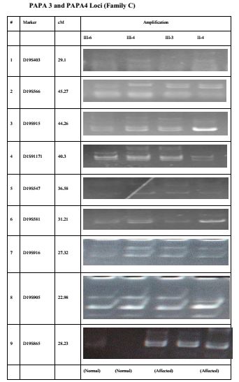
iv

                  Author’s Declaration
I, Muhammad Sajid Khan hereby state that my MS thesis titled “Genetic
Analysis of Hereditary Polydactyly in Pakistani Families” is my own work
and has not been submitted previously by me for taking any degree from Capital
University of Science and Technology, Islamabad or anywhere else in the coun-
try/abroad.

At any time if my statement is found to be incorrect even after my graduation,
the University has the right to withdraw my MS Degree.

(Muhammad Sajid Khan)

Registration No: MBS161001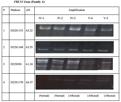
v

                 Plagiarism Undertaking
I solemnly declare that research work presented in this thesis titled “Genetic Anal-
ysis of Hereditary Polydactyly in Pakistani Families” is solely my research work
with no significant contribution from any other person. Small contribution/help
wherever taken has been dully acknowledged and that complete thesis has been
written by me.

I understand the zero tolerance policy of the HEC and Capital University of Science
and Technology towards plagiarism. Therefore, I as an author of the above titled
thesis declare that no portion of my thesis has been plagiarized and any material
used as reference is properly referred/cited.

I undertake that if I am found guilty of any formal plagiarism in the above titled
thesis even after award of MS Degree, the University reserves the right to with-
draw/revoke my MS degree and that HEC and the University have the right to
publish my name on the HEC/University website on which names of students are
placed who submitted plagiarized work.

(Muhammad Sajid Khan)

Registration No: MBS161001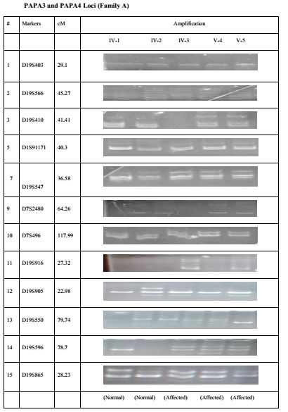
vi

List of Publications
vii

                     Acknowledgements
In the name of Allah, the most Merciful, the most Gracious. All praise is due
to Allah; we praise Him, seek His help, and ask for His forgiveness. I am thank-
ful to Allah, who supplied me with the courage, the guidance, and the love to
complete this research. Also, I cannot forget the ideal man of the world and
most respectable personality for whom Allah created the whole universe, Prophet
Mohammed (Peace Be upon Him).

I would like to thank my thesis supervisor Dr. Shaukat Iqbal Malik Assistant
Professor, Department of Biosciences. I am very grateful for his since he gave me
the chance to work on an interesting topic.

I am thankful to Dr.Sahar Fazal, Head of Department of Biosciences, Capi-
tal University of Science & Technology, Islamabad, for Provision of all possible
facilities.

My deepest thanks to Shabir Hussain, PhD student, Department of Biochem-
istry QAU Islamabad who helped me in lab work. Some of my friends had direct
participation in this study. They helped in wet lab during wet lab work. I am
proud to have such fellows who deserve my high appreciation. My sincere and
profound gratitude is due to my Parents. I cannot forget their kind care and their
interest in my success. Their prayers and moral support will always boost my
progress. During the course of this thesis, my sisters, and family members looked
closely at my progress and kept encouraging me towards success. I cannot express
my deepest feeling and high appreciation through this acknowledgement.
viii
Abstract
In vertebrates, the development of limb is a field of active research in developmen-
tal as well as in evolutionary biology. It initiates during fourth week of gestation
period in which several genes and molecular factors are involved via different bi-
ological pathways. Congenital limb abnormalities may occur if these signaling
pathways are disturbed. These limb abnormalities may be present as an isolated
entity of upper as well as lower limbs or may present in association with any other
syndrome. Polydactyly is widely reported congenital hand abnormality. It has
prevalence of 519/10000. Isolated form of polydactyly is categorized into postax-
ial, central and preaxial polydactyly. Postaxial polydactyly (PAPA) is classified
into two main types i.e. type A and type B. Postaxial polydactyly type A has been
further classified into seven types as PAPA1, PAPA2, PAPA3, PAPA4, PAPA5,
PAPA6 and PAPA7. Postaxial type B is further classified into two types. Preaxial
polydactyly has been classified into four types i.e. PPDI, PPDII, PPDIII and
PPDIV. Polydactyly is frequently associated with syndactyly, a digit disorder in
which adjacent fingers are webbed. This is due to failure of separation of digits
during the period of development.
In current study, three families one with postaxial polydactyly and syndactyly of
4th and 5th fingers (A) and two (B, C) with preaxial polydactyly belonging to var-
ious areas of Pakistan were selected for linkage analysis. It indicated autosomal
dominant mode of inheritance on basis of pedigree. Linkage analysis for already
reported loci/genes of these families was done by homozygosity mapping having
highly polymorphic microsatellite markers. The family A with postaxial poly-
dactyly in addition to syndactyly of 4th and 5th finger was mapped to known loci
and genes GLI3, SHH, LMBR1 (ZRS ), ZNF141 and IQCE, HOXD13 ). Genotyp-
ing analysis showed that markers D7S2541, D7S2548, and D7S691 showed some
evidence of linkage to GLI3 gene. Homozygosity mapping of family B showed no
linkage with any of the known gene. Exclusion of linkage indicates that an un-
known gene is responsible for preaxial polydactyly in this family. Family C with
affected individuals having preaxial polydactyly was tested for linkage to severalix genes as mentioned for family A. By genotyping analysis marker D17S54 showed homozygous pattern for both affected members of the family despite absence of immediate consanguinity. The marker lies midway between LMBRI and WDR60. The presence of mild brachydactyly and mild short stature in one of the patients indicated the possible involvement of WDR60 that causes short rib thoracic dys- plasia. No other gene showed linkage to the phenotype.
Contents

Author’s Declaration                                                                                        iv

Plagiarism Undertaking                                                                                       v

List of Publications                                                                                        vi

Acknowledgements                                                                                           vii

Abstract                                                                                                   viii

List of Figures                                                                                            xii

Abbreviations                                                                                              xvi

1 Introduction                                                                                               1
  1.1 Introduction . . . . . . . . . . . . . . . . . . .   .   .   .   .   .   .   .   .   .   .   .   .     1
       1.1.1 Pre-Axial Polydactyly . . . . . . . . .       .   .   .   .   .   .   .   .   .   .   .   .     2
             1.1.1.1 Pre-Axial Type I . . . . . . .        .   .   .   .   .   .   .   .   .   .   .   .     2
             1.1.1.2 Pre-Axial Type II . . . . . .         .   .   .   .   .   .   .   .   .   .   .   .     3
             1.1.1.3 Pre-Axial Type III . . . . . .        .   .   .   .   .   .   .   .   .   .   .   .     3
             1.1.1.4 Pre-Axial Type IV . . . . . .         .   .   .   .   .   .   .   .   .   .   .   .     4
  1.2 Post-Axial Polydactyly . . . . . . . . . . . . .     .   .   .   .   .   .   .   .   .   .   .   .     4
       1.2.1 PAPA . . . . . . . . . . . . . . . . . .      .   .   .   .   .   .   .   .   .   .   .   .     5
       1.2.2 PAPB . . . . . . . . . . . . . . . . . .      .   .   .   .   .   .   .   .   .   .   .   .     5
  1.3 Other Polydactylies . . . . . . . . . . . . . . .    .   .   .   .   .   .   .   .   .   .   .   .     6
       1.3.1 Mirror-Image Polydactyly . . . . . . .        .   .   .   .   .   .   .   .   .   .   .   .     6
       1.3.2 Haas Type Polydactyly . . . . . . . . .       .   .   .   .   .   .   .   .   .   .   .   .     7
       1.3.3 Central Polydactyly . . . . . . . . . . .     .   .   .   .   .   .   .   .   .   .   .   .     7
       1.3.4 Dorsal and Palmer/Ventral Polydactyly         .   .   .   .   .   .   .   .   .   .   .   .     7
       1.3.5 Syndactyly . . . . . . . . . . . . . . .      .   .   .   .   .   .   .   .   .   .   .   .     8
  1.4 Limb Development . . . . . . . . . . . . . . .       .   .   .   .   .   .   .   .   .   .   .   .     8
       1.4.1 Digit Development . . . . . . . . . . .       .   .   .   .   .   .   .   .   .   .   .   .     8
       1.4.2 Digit Specification . . . . . . . . . . .     .   .   .   .   .   .   .   .   .   .   .   .     9
  1.5 Molecular Pathways in Limb Development . .           .   .   .   .   .   .   .   .   .   .   .   .     9

                                        x
xi
1.6 Purpose . . . . . . . . . . . . . . . . . . . . . . . . . . . . . . . . . 11
1.7 Objectives . . . . . . . . . . . . . . . . . . . . . . . . . . . . . . . . 11
2 Literature Review 12
2.1 Molecular Genetics of Polydactyly . . . . . . . . . . . . . . . . . . . 12
2.1.1 PAPA1 . . . . . . . . . . . . . . . . . . . . . . . . . . . . . . 12
2.1.2 PAPA2 . . . . . . . . . . . . . . . . . . . . . . . . . . . . . . 12
2.1.3 PAPA3 . . . . . . . . . . . . . . . . . . . . . . . . . . . . . . 13
2.1.4 PAPA4 . . . . . . . . . . . . . . . . . . . . . . . . . . . . . . 13
2.1.5 PAPA5 . . . . . . . . . . . . . . . . . . . . . . . . . . . . . . 14
2.1.6 PAPA6 . . . . . . . . . . . . . . . . . . . . . . . . . . . . . . 14
2.1.7 PAPA7 . . . . . . . . . . . . . . . . . . . . . . . . . . . . . . 14
2.1.8 PPD1 . . . . . . . . . . . . . . . . . . . . . . . . . . . . . . 14
2.1.9 PPD2 . . . . . . . . . . . . . . . . . . . . . . . . . . . . . . 15
2.1.10 PPD3 . . . . . . . . . . . . . . . . . . . . . . . . . . . . . . 17
2.1.11 PPD4 . . . . . . . . . . . . . . . . . . . . . . . . . . . . . . 17
3 Methodology 18
3.1 Study Subjects . . . . . . . . . . . . . . . . . . . . . . . . . . . . . 18
3.2 Pedigree Sketch . . . . . . . . . . . . . . . . . . . . . . . . . . . . . 18
3.3 Collection of Blood . . . . . . . . . . . . . . . . . . . . . . . . . . . 19
3.4 DNA Extraction . . . . . . . . . . . . . . . . . . . . . . . . . . . . 19
3.5 Agarose Gel Electrophoresis . . . . . . . . . . . . . . . . . . . . . . 21
3.6 Homozygosity Mapping . . . . . . . . . . . . . . . . . . . . . . . . . 22
3.7 PCR . . . . . . . . . . . . . . . . . . . . . . . . . . . . . . . . . . . 22
3.8 Polyacrylamide Gel Electrophoresis . . . . . . . . . . . . . . . . . . 23
4 Results 27
4.1 Description of families . . . . . . . . . . . . . . . . . . . . . . . . . 27
4.1.1 Family A . . . . . . . . . . . . . . . . . . . . . . . . . . . . 27
4.1.2 Family B . . . . . . . . . . . . . . . . . . . . . . . . . . . . . 29
4.1.3 Family C . . . . . . . . . . . . . . . . . . . . . . . . . . . . 30
5 Conclusion and Future work 53
5.1 Conclusion and Future work . . . . . . . . . . . . . . . . . . . . . . 53
Bibliography 56List of Figures
3.1 List of Solutions . . . . . . . . . . . . . . . . . . . . . . . . . . . . . 21
3.2 List of Highly Polymorphic Microsatellite Markers . . . . . . . . . . 26
4.1 Pedigree design of family A with hereditary Post-axial polydactyly.
Circles and squares represent females and males respectively, A
filled symbol represents affected individual while unfilled symbol
represents unaffected individual. Consanguineous marriages indi-
cated by double lines. The number of generations indicates by Ro-
man numerals. . . . . . . . . . . . . . . . . . . . . . . . . . . . . . 28
4.2 Photograph of member V-2 of family A: (a) showing post-axial poly-
dactyly in both hands with syndactyly of digits 3 and 4; (b) showing
post-axial polydactyly of both feet. . . . . . . . . . . . . . . . . . . 28
4.3 Pedigree design of family B with hereditary Pre-axial polydactyly.
Circles and squares represent females and males respectively. A
filled symbol represents affected individual while unfilled symbol
represents unaffected individual. Consanguineous marriages indi-
cated by double lines . The number of generations indicates by
Roman numerals . . . . . . . . . . . . . . . . . . . . . . . . . . . . . 29
4.4 Photograph of member IV-1 of family B, showing pre axial poly-
dactyly in one hand only. . . . . . . . . . . . . . . . . . . . . . . . . 30
4.5 Pedigree design of family C with hereditary Pre-axial polydactyly.
Circles and squares represent females and males respectively. A
filled symbol represents affected individual while unfilled symbol
represents unaffected individual. Consanguineous marriages indi-
cated by double lines . The number of generations indicates by
Roman numerals . . . . . . . . . . . . . . . . . . . . . . . . . . . . 32
4.6 Photograph of member III-3 of family C, showing pre axial poly-
dactyly in one hand only. . . . . . . . . . . . . . . . . . . . . . . . . 32
4.7 Electropherogram of 8% non-denaturing polyacrylamide gel obtained
by staining with ethidium bromide. Banding pattern illustrates the
genotype of alleles amplified with respective markers within GLI3
gene at chromosome 7p14.1 Numbers indicates the family members
of the pedigree. . . . . . . . . . . . . . . . . . . . . . . . . . . . . . 33
xiixiii
4.8 Electropherogram of 8% non-denaturing polyacrylamide gel obtained
by staining with ethidium bromide. Banding pattern illustrates
the genotype of alleles amplified with respective markers within
SHH/ZRS gene at chromosome 7q36. Numbers indicates the fam-
ily members of the pedigree . . . . . . . . . . . . . . . . . . . . . . 34
4.9 Electropherogram of 8% non-denaturing polyacrylamide gel obtained
by staining with ethidium bromide. Banding pattern illustrates the
genotype of alleles amplified with respective markers within ZNF141
gene at chromosome 4pl6.3. Numbers indicates the family members
of the pedigree . . . . . . . . . . . . . . . . . . . . . . . . . . . . . 35
4.10 Electropherogram of 8% non-denaturing polyacrylamide gel obtained
by staining with ethidium bromide. Banding pattern illustrates the
genotype of alleles amplified with respective markers within IQCE
gene at chromosome 7p22.3. Numbers indicates the family members
of the pedigree . . . . . . . . . . . . . . . . . . . . . . . . . . . . . 36
4.11 Electropherogram of 8% non-denaturing polyacrylamide gel obtained
by staining with ethidium bromide. Banding pattern illustrates the
genotype of alleles amplified with respective markers within FBLNI
gene at chromosome 22q13.31. Numbers indicates the family mem-
bers of the pedigree . . . . . . . . . . . . . . . . . . . . . . . . . . 37
4.12 Electropherogram of 8% non-denaturing polyacrylamide gel obtained
by staining with ethidium bromide. Banding pattern illustrates
the genotype of alleles amplified with respective markers within
HOXD13 gene at chromosome 7p22.3. Numbers indicates the fam-
ily members of the pedigree. . . . . . . . . . . . . . . . . . . . . . . 38
4.13 Electropherogram of 8% non-denaturing polyacrylamide gel obtained
by staining with ethidium bromide. Banding pattern illustrates the
genotype of alleles amplified with respective markers within PAPA2
and PAPA5 gene at chromosome 13q21-32 & 13q13.3-21. Numbers
indicates the family members of the pedigree. . . . . . . . . . . . . 39
4.14 Electropherogram of 8% non-denaturing polyacrylamide gel obtained
by staining with ethidium bromide. Banding pattern illustrates the
genotype of alleles amplified with respective markers within PAPA3
and PAPA4 gene at chromosome 19p13.1-13.2. & 7q21-34. Num-
bers indicates the family members of the pedigree . . . . . . . . . . 40
4.15 Electropherogram of 8% non-denaturing polyacrylamide gel obtained
by staining with ethidium bromide. Banding pattern illustrates the
genotype of alleles amplified with respective markers within GLI3
gene at chromosome 7p14.1 Numbers indicates the family members
of the pedigree. . . . . . . . . . . . . . . . . . . . . . . . . . . . . . 41
4.16 Electropherogram of 8% non-denaturing polyacrylamide gel obtained
by staining with ethidium bromide. Banding pattern illustrates
the genotype of alleles amplified with respective markers within
SHH/ZRS gene at chromosome 7q36. Numbers indicates the fam-
ily members of the pedigree. . . . . . . . . . . . . . . . . . . . . . . 42xiv
4.17 Electropherogram of 8% non-denaturing polyacrylamide gel obtained
by staining with ethidium bromide. Banding pattern illustrates the
genotype of alleles amplified with respective markers within ZNF141
gene at chromosome 4pl6.3. Numbers indicates the family members
of the pedigree. . . . . . . . . . . . . . . . . . . . . . . . . . . . . . 43
4.18 Electropherogram of 8% non-denaturing polyacrylamide gel obtained
by staining with ethidium bromide. Banding pattern illustrates the
genotype of alleles amplified with respective markers within IQCE
gene at chromosome 7p22.3 Numbers indicates the family members
of the pedigree . . . . . . . . . . . . . . . . . . . . . . . . . . . . . 44
4.19 : Electropherogram of 8% non-denaturing polyacrylamide gel ob-
tained by staining with ethidium bromide. Banding pattern il-
lustrates the genotype of alleles amplified with respective markers
within PAPA2 and PAPA5 at chromosome 13q21-32 & 13q13.1-21
Numbers indicates the family members of the pedigree . . . . . . . 45
4.20 Electropherogram of 8% non-denaturing polyacrylamide gel obtained
by staining with ethidium bromide. Banding pattern illustrates the
genotype of alleles amplified with respective markers within PAPA3
and PAPA4 at chromosome Numbe7q21-34. Numbers indicates the
family members of the pedigree . . . . . . . . . . . . . . . . . . . . 46
4.21 Electropherogram of 8% non-denaturing polyacrylamide gel obtained
by staining with ethidium bromide. Banding pattern illustrates the
genotype of alleles amplified with respective markers within GLI3
gene at chromosome 7p14.1 Numbers indicates the family members
of the pedigree. . . . . . . . . . . . . . . . . . . . . . . . . . . . . . 47
4.22 Electropherogram of 8% non-denaturing polyacrylamide gel obtained
by staining with ethidium bromide. Banding pattern illustrates the
genotype of alleles amplified with respective markers within SHH/
ZRS gene at chromosome 7q36. Numbers indicates the family mem-
bers of the pedigree . . . . . . . . . . . . . . . . . . . . . . . . . . 48
4.23 Electropherogram of 8% non-denaturing polyacrylamide gel obtained
by staining with ethidium bromide. Banding pattern illustrates the
genotype of alleles amplified with respective markers within ZNF141
gene at chromosome 4pl6.3. Numbers indicates the family members
of the pedigree. . . . . . . . . . . . . . . . . . . . . . . . . . . . . . 49
4.24 Electropherogram of 8% non-denaturing polyacrylamide gel obtained
by staining with ethidium bromide. Banding pattern illustrates the
genotype of alleles amplified with respective markers within IQCE
gene at chromosome 7q22.3. Numbers indicates the family members
of the pedigree. . . . . . . . . . . . . . . . . . . . . . . . . . . . . . 50
4.25 Electropherogram of 8% non-denaturing polyacrylamide gel obtained
by staining with ethidium bromide. Banding pattern illustrates the
genotype of alleles amplified with respective markers within PAPA2
and PAPA5 at chromosome 13q21-32 & 13q13.3-21 Numbers indi-
cates the family members of the pedigree . . . . . . . . . . . . . . 51xv
4.26 Electropherogram of 8% non-denaturing polyacrylamide gel obtained
by staining with ethidium bromide. Banding pattern illustrates the
genotype of alleles amplified with respective markers within PAPA3
and PAPA4 gene at chromosome 19p13.1-13. & 7q21-34. Numbers
indicates the family members of the pedigree. . . . . . . . . . . . . 52Abbreviations
PAP Post-axial polydactyly
PPD Pre-axial polydactyly
SNPs Single nucleotide polymorphism
PCR Polymerase chain reaction
SHH Sonic hedgehog
CNV Copy number variation
TPT-PS Triphalangeal thumb-polysyndactyly syndrome
ZPA Zone of polarizing activity
PPD Pre axial polydactyly
PAPA Post axial polydactyly type A
PAPB Post axial polydactyly type B
STS Short tandem sequences
WES Whole exome sequencing
EDTA Ethylene diamine tetraacetic acid
ZRS ZPA regulatory sequence
OMIM Online Mendelian Inheritance in Man
MIPOL Mirror image polydactyly
TGFb Transforming growth factor, beta-1
SDS Sodium dodecyl sulphate
EtBr Ethidium bromide
cM Centimorgan
xviChapter 1
Introduction
1.1 Introduction
Polydactyly is a congenital abnormality which is primarily indicated as superficial
toe/fingers that is completely grown in addition to the bony component or an addi-
tional softhearted material with no bone component. This disorder is a hereditary
limb abnormality having prevalence of 519/10 000 babies which are alive (Schwabe
and Mundlos, 2004; Christensen, 2011; Malik, 2014). Association of the feet have
been identified to be less frequent as compare to the upper limbs, right hand more
familiar as compared to left hand and the right foot is less prevalent as compared
to left foot (Malik et al., 2014). It may be present in addition to other phenotypes
called as syndromic or it may be present as isolated entity without having any ab-
normality beyond this disorder so called as non-syndromic polydactyly (Biesecker,
2011). The main classes of non- syndromic polydactyly are pre-axial polydactyl,
mesoaxial polydactyl and post- axial polydactyly (Deng, 2014; Malik, 2014). Pre-
axial polydactyly has been further classified into four types PPD1, PPD2, PPD3
and PPD4. On the other hand post-axial polydactyly further classified into type A
and type B. Type A has seven types named as PAPA1, PAPA2, PAPA3, PAPA4,
PAPA5, PAPA6 and PAPA7(Malik, 2013). These different types of non-syndromic
polydactyly are caused by nine genes and about 10 loci reported up till now. These
1Introduction 2 reported genes are GLI3, HOXD13, ZNF141, IQCE, MIPLO1, PITXI, ZRS/SHH, GJA1 and FBLN1. A locus has been reported for autosomal recessive PAP-type A5 and (Umm-e-Kalsoom, 2012a; Umm-e-Kalsoom, 2012b). Four PPD types were identified by Temtamy and McKusick as mentioned above. 1.1.1 Pre-Axial Polydactyly 1.1.1.1 Pre-Axial Type I The type of polydactyly in which the components of biphalangeal thumb are re- peated one or more times. It is reported in a study, hands are favorably involved as compare to feet and in case of hands mostly cases are bilateral. It is also re- ported that mostly right hand is involved as compare to left hand (Malik S. et al., 2012). There is high prevalence of this type in males that are affected than females (Malik et al., 2012; Orioli et al., 1999). In Pre-axial Type I, the appearance diversity is variable which includes recurrence of ends of bones of fingers also known as distal phalanx, the extra incompletely developed thumb and the ends of bones of fingers may show bifurcation and these end might also be broad, two extra phalanges may be present on the same in- completely developed thumb or there may be the repetition of the whole thumb (Temtamy et al., 1978). The name of triplicated thumb may also be given to this type that results in total of seven digits (heptadactyly) (Zuidam et al., 2008). It has also been reported that hereditary type I pre-axial polydactyly which is commonly segregates in a dominant way (autosomal) having decreases extent of expression (Castilla et al.,, 1978; Orioli et al., 1999). It is a heterogeneous disor- der. Variations in the gene sonic hedgehog are responsible to produce this type of abnormality (Wieczorek et al., 2010; Lettice et al., 2002). This disorder of thumb polydactyly may possibly come with doubling of hallux in spite of this, hallux polydactyly also present as a separated element or a prevalent appearance in families (Castilla et al., 1973). Hallux duplication is infrequent than polydactyly of thumb (0.025/1000 vs 0.154/1000 present in South American
Introduction                                                                     3

country. Hallux doubling showed a meaningful overabundance in men, associated
mostly the limb of right side. Mostly cases reported are those in which disorder
is on one side only (Orioli et al., 1999). Hallux polydactyly still has unknown
molecular basis.

1.1.1.2   Pre-Axial Type II

It is a type of polydactyly which exhibits the existence of generally opposable
triphalangeal thumb. It may be with or without extra duplication of thumb (Tem-
tamy et al., 1978). It is also known as triphalangeal thumb polydactyly (TPT).
The disorder may possibly be present in lower appendages as well and opposable
in most cases and exhibit toe doubling (Temtamy et al., 1978). TPT is often bi-
lateral and symmetrical (Swanson et al., 1962). It has also been reported that this
disorder segregates autosomal dominantly having incomplete penetrance (Tem-
tamy et al., 1978). This disorder type showed a reduced prevalence in women.
Triphalangeal thumb polydactyly is a somewhat infrequent type. It has been ob-
served that variations in ZRS/SHH locus on chromosome 7q36 are responsible to
cause separate triphalangeal thumb polydactyly, many other abnormalities and
several of the triphalangeal appendage anomalies (Lettice LA et al., 2002). In
spite of this, ZRS/SHH alterations are not responsible for whole polydactyly of
lonely triphalangeal thumb relatives not various varieties of triphalangeal thumb
polydactyly reflecting other processes may be the source of this type of pre-axial
polydactyly.

1.1.1.3   Pre-Axial Type III

In this type of pre-axial polydactyly which is uncommon, there is doubling of index
finger. It is considered as autosomal dominant. One or two triphalangeal digits
take the place of thumb. In this type epiphysis has been shown for the metacarpal
of the additional finger, so this unit become distinct than other types (Swanson et
al., 1962). Ist and 2nd toes may be affected. As it is considered a modified form
Introduction                                                                     4

of thumb repetition but sometimes it is added in the type of polydactyly called as
central polydactyly (Wood, 1970). The radial-sided doubling normally is smaller
and the stage of repetition may possibly at the metacarpal or more distal. The
deviation of the superfluous finger occur here radially or deviation of ulna occur
in normal finger (Graham and Ress, 1998).

1.1.1.4   Pre-Axial Type IV

It is also called as polysyndactyly in which repetition of thumb occurs to some
extent along with deviation of the phalanx in outward direction. The broadness
in the thumb also occur. Mostly 3rd and 4th finger show the sign of syndactyly
(Temtamy et al., 1978). There is divergence of 1st metacarpal having abnormality
in former toe. Digits of the whole foot may show syndactyly or some time 2nd
and 3rd toe exhibit syndactyly. Pre-axial type 4 is different from synpolydactyly.
In synpolydactyly extra finger exhibit syndactyly (Malik and Grzeschik, 2008). A
word crossed polydactyly is normally in use on behalf of existence of PAPA and
PPD in addition to variation in angle of the extra fingers present among the feet
as well as hand. Cross polydactyly shows a form of polydactyly that is pre-axial in
feet while post-axial in hands. In cross polydactyly type II post-axial polydactyly
exist in feet along with pre-axial polydactyly present in hands (Temtamy et al.,
1978). It is reported that type 1 is allelic to PAP (A/B) and variation in ZRS/SHH
and GLI3 are directly involved in this disorder. (Radhakrishna et al., 1999).

1.2       Post-Axial Polydactyly

The type of polydactyly in which one or more additional fibular or ulnar digits
are present. There may be only a part of ulnar or digits exist. Particularly for
the ulnar polydactyly, two different units A and B have been documented. Both
these two types of post-axial polydactyly show marked differences of seriousness,
heritage model as well as penetration assessments (Temtamy et al., 1978).
Introduction                                                                     5

1.2.1    PAPA

The kind of polydactyly which exhibit complete development of additional digit
which is functional. The articulation of additional 5th digit or additional carpel
(Temtamy et al., 1978). 1 to 3 bony elements present in additional digit along with
the nail which is formed completely. It indicates the mode of inheritance which is
autosomal dominant and show 70% penetrance rate (Kucheria et al., 1981).

It has been reported that it exist as autosomal recessive type (OMIM-263450)
(Briard and Kaplan, 1982). In an epidemiological study, enrolled 6586 people
having PAP (A/ B). By complicated segregated analysis, these writers found large
heritability assessment for this feature and a main recessive gene involvement.
(Castilla et al., 1998). It was reported that chromosome 13q13.3-q21.2 as well
as 4p16.3 (ZNF141 ) had been linked to 2 types of recessive PAP respectively
(Kalsoom et al., 2012). On the basis of studies of mapping different types of PAP
have been observed having phenotypes of A and B. These are PAPA-1, 2, 3, 4,5,6,7.
It was reported that mutated ZRS/SHH as well as GLI3 are responsible for PAPA-
1 (Radhakrishna et al., 1999). However on genetic bases the heterogeneity of both
these types of polydactyly was not confirmed.

1.2.2    PAPB

It is one of the most frequent type of polydactyly in different inhabitants. There
is incomplete development of additional digit. There may be an existence of small
lump on ulnar feature with 5th finger or the length may lead up to four centimeter
like a nubbin having an element of bone and normally nail may also be present
(Castilla et al., 1973). The site of articulation of this lump with the 5th finger
show alteration. It was indicated that this type show more complication geneti-
cally. Reported penetrated rate of this type is 42% (Castilla et al., 1973). The
disturbance in hands and left hand is mostly reported (Malik, 2013). The genetics
of PAP-A and PAP-B shows that there is heterogeneity and diversity in both these
types. The reason behind that is their array of segregation is different and they
Introduction                                                                     6

occur independently (Castilla et al., 1973). Although it was reported occurrence
of PAP-A and B in same person (Kucheria et al., 1981).

1.3     Other Polydactylies

Beyond PPD and PAP phenotypes other phenotypes are also familiar which are
different from both these types.

1.3.1    Mirror-Image Polydactyly

The type of polydactyly in which digits on the posterior side are duplicated. There
is replacement of digits of anterior one by posterior one while the sequence is
reversed. The sequence of additional fingers from index finger 5-4-3-2-3-4-5 while
hallux or thumb is not present (Temtamy et al., 1978). The pattern of segregation
is autosomal dominant. Non syndromic cases are very infrequent, mostly it exist
in addition with other abnormalities i.e. syndromic cases are frequent (OMIM-
135750) where ulna and fibula are duplicated, thumb is not present while rest
fingers are duplicated. Hands and feet tubular bones show repetition and on the
basis of this various types of MIP are suggested. One of them is infrequent in
which tibia is faulty in addition to pre-axial polydactyly. This type is indicated
as autosomal dominant with different penetration and expression. The second
type is that in which tibial development remain incomplete and mirror feet. The
third type shows duplication in ulna fibula (Temtamy et al., 1978). The region in
chromosome 14q13 harboring MIPOL1 mutated to cause a kind of mirror image
polydactyly (Kondoh et al., 2002). It was also reported that variation in PITX1 is
also responsible to cause mirror image polydactyly in feet (Klopocki et al., 2012).
Introduction                                                                         7

1.3.2     Haas Type Polydactyly

It is the type of polydactyly whole digits are merged showed a complete fusion. The
syndactyly is present along with additional ray of digit. The additional ray may be
post axial or pre axial (Haas SL 1940). The shape of hand looks like a cup because
of presence of severe syndactyly. This form is regarded as syndactyly of type 4
(Malik S. 2012). It was reported that the variations in ZRS/SHH gene and GLI3
gene are responsible for causing this type of polysyndactyly on the other hand this
form of polydactyly is observed as heterogeneous genetically (Radhakrishna et al.,
1999).

1.3.3     Central Polydactyly

This type of polydactyly is mostly bilateral while abnormalities of various kinds
are also observed. There is presence of lump of tissue in middle of upper limb in
addition to syndactyly and nail are fused. Repetitions may considered as hidden
but not all of its forms. Index digit is duplicated. As compare to index digit the
repetition of ring digit is mostly observed (Temtamy et al., 1978).

1.3.4     Dorsal and Palmer/Ventral Polydactyly

This type of polydactyly is very infrequent and uncommon in population. In this
type additional raised near dorsal or ventral side of auto pod. There is partial
development of digit ray or it shows complete development but nail is absent. The
additional digit may be moveable as well as functional. It was reported that the
basis of this moveable digit is present in hand at ventral or palmar side (Nair et al.,
2001). Similarly it was also reported in lower limb as well at dorsum site (Hussain
et al., 2007).
Introduction                                                                      8

1.3.5     Syndactyly

It is the form of polydactyly some digits attain a design like a web. This is due
to failure of separation during development of limb. Although the association
between syndactyly and polydactyly is very close so that it become difficult to
classify them. However it may be regarded as part of kind 4 of pre-axial poly-
dactyly (Temtamy et al., 1978). Its incidence rate is 3 to 10 in 10000 births.
(Malik et al., 2012). Mostly 1st to 2nd or 3rd to 5th digit may accompanied in
triphalangeal type. I t was observed that extra toe in foot may fused with other
digit in non-syndromic form of pre-axial polydactyly. It was reported that 4th to
5th digits of hands and feet are fused when PAP and PPD both are present along
with syndactyly (Goldstein et al., 1994). It is indicated that this abnormality is
inherited recessively (Briard et al., 1982). The hand looks like a shape of a cup
in case of MIP when whole syndactyly is present (Temtamy et al., 1978). There
may be heterogeneity in the same person and he attain asymmetrical phenotypes
hands and feet and right as well as left appendages. Syndactyly may be complete
or incomplete, phalanges only or metatarsal and metacarpals are also involved
(Malik et al., 2012).

Various classes of syndactyly have been observed. These are syndactyly type 1
to IX on bases of involvement of digits/toe. The syndactyly type 1 is further
classified into type 1-a, type 1-b, type 1-c and type 1-d (Malik et al., 2012).

1.4      Limb Development

1.4.1     Digit Development

For learning organ development the limb has long considered as a model. A re-
peated set of fingers and toes shows distal feature of appendage of vertebrate.
These form a consecutive pattern on the other hand there is variety in shape, size
Introduction                                                                     9

and structure of these digits and toes. It was reported that the process of devel-
opment of fingers and toe is different from the process to give them identification
(Zwilling, 1964).

1.4.2    Digit Specification

The idea of positional information gives specificity of digit identification. Mor-
phogens which are called variety of signals are involved in establishing the posi-
tional values. It was proposed that this information is interpreted by the tissues
in the next step. Digits are formed as a result of these values (Saunders et al.,
1957). The conclusions of the experiments of inserting of cells at ZPA towards
the anterior distal margin from the posterior one are according to the informa-
tion provided by morphogens (Tickle et al., 1975). This the way that hint to
duplication of fingers and toes (Saunders and Gasseling, 1968). Zone of polarizing
activity provide the strength of morphogens. In this view, the concentration of
a morphogen produced by the ZPA. The digit primordia inferred this strength of
morphogens in limb region as positional information help in digits identification.
Although this morphogens exemplary gives only the morphology of digits and not
position or spaces pattern between digits.

1.5     Molecular Pathways in Limb Development

In a publishing (Raspopovicet al., 2014) molecules were reported which control
the space pattern of establishing centers. The molecules that are associated with
are WNT and BMP. WNT was observed to be associated with interdigit field
and Bmp with digit field. The action of Wnt hinders the chondrification so that
SOX9 is not expressed. This help in specification of inter digit. On the other hand
the action of BMP enhances chondrification so that SOX9 expression is enhanced
beyond the approach of WNT activity so specification of digits occur. The arrival
of BMP and WNT suppressed by SOX9 in digit. It becomes the origin of a model
called as three-node Turing model. (Raspopovic et al., 2014).
Introduction                                                                     10

This model not properly explain the initiation of digit pattern so there is require-
ment of more study for understanding the mechanism of digit pattern initiation.
The Wnt/ planer cells polarity and TGF-b are also involved in digit initiation
pattern. Preventing of origination of extra PFR/DCs digit controllers during the
expansion of auto pod is necessary. Wnt is possible nominee responsible to cause
anti-chondrogenesis in appendages (Hartmann and Tabin, 2001; ten Berge et al.,
2008) and is known to be the part of turning like models suggested for specificity
of digits by Raspopovic et al., (2014). When the controlling hubs are created
near the metacarpal origin the extra digit does not usually created. Due to the
delivery of Bmp at abnormal place at the edge of developing digit, there may be
the bifurcation of rays of the digits (Ganan et al., 1996; Duprez et al., 1996).
If the controlling hub is not split one ray of cartilage may arranged proximally.
On the other hand when the controlling hub is split results in bifurcation into
more outlets. If the ectoderm is removed or TGF-b is applied, the ectopic dig-
its which are completely independent could be provoked among endogenously ray
of the digits (Hurle and Ganan, 1987; Ganan et al., 1996; Macias et al., 1993).
It was suggested that this digit is started just like one particular pimple away
from the origin having TGF-b and due to the distal development of auto pod this
pimple lengthens to develop into a shape like digit (Lorda-Diez et al., 2011). So
that ectopic digit managing hubs are induced via these processes. Then these
hubs perform as endogenous managing hubs in the course of development of hand
plate. A cluster of cells of mesenchyme produced at the posterior margin of the
limb bud called ZPA. This is the signaling hub are proposed to involved in digit
development. Mirror image digit repetition may be result of polarizing actions of
these cells. SHH is responsible to intervene its activity (Saunders et al., 1968).
The antagonistic factors are SHH and GLI3. These are responsible to control not
only number of digit and identification of digits as well.
Introduction                                                                    11

1.6     Purpose

More than a hundred genes have been discovered in human to be involved in
polydactyly (Xiang et al., 2016). Most of these genes show syndromic polydactyly.
Very few genes are reported in non-syndromic polydactyly. The purpose of this
study is to identify the genes in the selected families of Pakistani origin showing
inherited non-syndromic polydactyly.

1.7     Objectives

   • To perform genotyping of known genes of polydactyly.

   • To check heterozygosity in family with dominant inheritance.

   • To check homozygosity in family with consanguanity.
Chapter 2

Literature Review

2.1     Molecular Genetics of Polydactyly

2.1.1    PAPA1

Heterozygous mutation in GLI3 gene was observed by Radhakrishna et al.,. (1997)
in family of India having post axial polydactyly type A-1 and the linkage was found
to chromosome 7p. A variation in GLI3 gene in an individual having PAP- type
B. The variation is heterozygous in nature and this variation is because of decay of
nonsense mediated mRNA (Furniss et al., 2007). A family of Saudi Arabia having
PAP. The patients of family have broadness in thumb and additionally syndactyly
was also present in upper limbs as well as in lower limbs. A deletion of 2bp of
GLI3 gene was observed (Al-Qattan 2012).

2.1.2    PAPA2

A family having PAPA selected by Akarsu et al., (1997) for linkage analysis.
The family showed a total of 18 informative meioses of which 11 were phase-
known. He omitted association to 7p15-q11.23, where a form of PAPA (PAPA1;
174200) had been mapped in a large Indian kindred. They demonstrated linkage

                                        12
Literature Review 13 to markers from 13q21-q32. An extreme lod score 2.34 at 7 cM was observed with D13S1230. They observed no recombination in affected members between centromeric D13S800 and telomeric D13S154 in a 23-cM interval. One unaffected member of the family received the ’affected haplotype’ from his affected parent. Coding this individual as of ’unknown’ status, they obtained the highest lod score with D13S1230 (maximum lod = 3.612 with no recombination). A karyotyping was performed by (van der Zwaag et al., 2010) in an individual having PAP in upper limbs. The disorder was present in both of upper limbs. He indicated heterozygous condition. He found that chromosome 13 has de novo inverted repetition in its large arm. 2.1.3 PAPA3 A family in china having PAP studied by Zhao et al., (2002). The family showed autosomal dominant mode of inheritance. A negative lod score was observed by him with already known forms PAPA locus on 7p (PAPA1; 174200) plus 13q (PAPA2; 602085). But for STS on chromosome 19p a positive lod score was also observed. This was founded after performing genome screen. An extreme 5.86 lod score was observed with D19S221. 2.1.4 PAPA4 A Dutch family having 32 people along with 11 polydactyly patients was studied by Galjaard et al., (2003). The family has post-axial polydactyly with various types i.e. post-axial type A, type B or both in addition to syndactyly in some individuals. 4th locus was observed in this family. An association was observed between the polydactyly and markers on 7q chromosome after performing the whole genome analysis. Between D7S1799 and D7S500 a maximum lod score was observed i.e. 3.33.
Literature Review                                                              14

2.1.5    PAPA5

Umm-e-Kalsoom et al., (2012) studied a family in Pakistan having PAP. The
family have autosomal recessive mode of inheritance and have a consanguineousity
as well. By utilizing highly polymorphic microsatellite markers by performing
genome wide search. She observed association to markers on 7q. A highest lod
score of 3.85 was observed with phenotype. Among markers D13S1288 (32.39
cM) and D13S632 (57.16 cM) on chromosome 13q with 17.87 cM critical field was
demarcated by recombinations (NCBI36).

2.1.6    PAPA6

Kalsoom et al.,. (2013) studied a family in Pakistan with PAP-A. The family ex-
hibited recessive mode of inheritance and have consanguineousity as well. Mutated
ZNF141 was found to be responsible by using a technique of WES. Genotyping
was performed by using 26 markers of above mentioned gene. Linkage was found
to be 6.52-Mb on 4p16.3-p16.2 chromosome. For markers D4S412 highest 3.38 lod
score was observed.

2.1.7    PAPA7

Umair et al., (2017) studied a family in Pakistan having PAP in foot. The family
exhibited recessive mode and have consanguineousity as well Mutated IQCE gene
was found to be responsible for this abnormality. Homozygous splice site variation
in the IQCE (617631.0001) was observed after performing WES.

2.1.8    PPD1

Kelly (1982) identified a family with polydactyly. He found that thumb and toe
was duplicated symmetrically. He observed that the abnormality was transmitted
men to men over many generations. Graham et al.,. (1985, 1987) was studied
Literature Review                                                              15

the pre-axial polydactyly. He proposed a polydactyly type in which thumb is
incompletely developed. Mostly it may inherited with autosomal dominant mode
of inheritance. As a French biologist (Fromont, 1895) defined this previously so
they called this slight variation as Fromont anomaly.

Ray (1987) founded an Indian family with PPD. The family comprising 15 men
and 5 women in several generations. Repetition of thumbs were not as much stable
property. There was variability in expression and repetition of large toe in both
feet exhibited condensed penetration. Orioli and Castilla (1999) performed a study
based on epidemiology. They obtained a data from a Latin American department.
They selected a total of 3,444,374 newborns from 1967 to 1995 in which 921 were
born with 1st digit repetition. The incidence of non-syndromic cases was found to
be 714 (2.08/ 10,000). These are further divided into 5 classes. Thumb repetition
(568), hallux repetition (82), and polysyndactyly (37), TPT (24), Thumb/hallux
repetition (4). Men were mostly affected, right limbs were more involved, and
mostly cases were unilateral. It was also founded in this study that incidence was
more in Bolivia 3.4 per ten thousand as compared to ten Latin states of America.

2.1.9    PPD2

The variations in SHH regulatory unit was responsible to cause PPD-2. This
element was located on chromosome 7q36. Variations in ZRS which is present
in between intron five of LIMBR1 gene showed segregation with abnormality in
families of PPD-2(605522.0002, 605522.0004, and 605522.0005) (Lettuce et al.,
2003).

Four families were examined by Gurnett et al., (2007) having phenotypes of PPD-2
and TPT. The families were already studied by Dobbs et al., (2000). He pointed
out two variations in three families (605522.0007 and 605522.0008, respectively)
while 4th family had just TPT phenotype.
Literature Review                                                               16

A large family having four generations in which nine individuals were involved
in disorder of TPT plus syndactyly (Klopocki et al., (2008). He pointed out a
heterozygous condition for identified 589-kb repetition.

Sun et al., (2008) studied five families having TPT PS and few individuals had
syndactyly-4. He pointed out repetitions involves ZRS having a range of 130-
397kb. He used polymerase chain reaction technique and identified a common
coinciding section of 32757 base pair. This section possessed ZRS enhancer. It was
found co-segregation of the repetitions with phenotypes in five family individuals.
The normal individuals did not occupy these duplications.

Wang et al., (2007) examined any of these families. Point mutation was observed
in LIMBR1 gene and the location was intron five. The variation could denote
polymorphism which was infrequent proposed by Sun et al., (2008).

Furniss et al., (2008) studied three families in England having TPT polydactyly
in both hands and one of them had unilateral PPD. He pointed out variation in
ZRS in LIMBR1. There was observed heterozygosity in mutation in all disturbed
members. In England TPT was commonly caused by variation in 295T-C which
was dominant mutation. Balci et al., (1999), Wieczorek et al., (2010) studied
a family in Turkey having TPT-PS. Mutation in ZRS/ LMBR1 was observed.
The mutation was found in those individuals of the family which were abnormal
while normal individuals did not had such mutation. The doubling of 276-kb
was nominated arr7q36.3. Both of them explained that repetition region was
impossible to be identified because of its repetitive nature.

Albuisson et al., (2011) studied two families in France having PPD-2 phenotype.
He observed two dissimilar variations showing heterozygosity in ZRS area of LIM-
BRI (297G-A; 605522.0013 and 334T-G; 605522.0014). These were present in
expected attachment locations for SOX9 and PAX3 which were transcription fac-
tors. The two families had the variations completely penetrated. It was observed
that SOX9 as well as PAX3 have a role pattern of digit formation during the
development of embryo in mouse. Therefore it was proposed that SHH might be
regulated from ZRS by SOX9 as well as PAX3.
Literature Review                                                              17

Vander Meer et al., (2014) examined two families in Mexica having TPT as well
as PPD phenotypes which were segregated in an autosomal dominant way. The
abnormal members of these two families showed heterozygosity for variations in
ZRS field of LIMBR1 gene (297G-A; 605522.0013 and 334T-G; 605522.0014).

2.1.10     PPD3

Manoiloff, 1931 observed the transmission of abnormality in a Scipio kindred. He
stated that the transmission might be for about 2000 years in this family (Swanson
and Brown, 1962) stated that triphalangeal digits substituted the thumb. The
feature of opposability could or could not be present (Manoiloff, 1931; James and
Lamb, 1963) stated that PPD of first and second toes showed by lower limbs in few
circumstances (Swanson and Brown, 1962) found that the extra digit metacarpal
showed distal epiphysis.

2.1.11     PPD4

Radhakrishna et al., (1999) examined a family having PPD-4. He observed a
frameshift insertion of one nucleotide. It was resulted in abnormal protein con-
sisting of 1245 amino acids. Fujioka et al., (2005) studied a family having PPD-4.
A nonsense variation was observed in GLI3 (R290X; 165240.0014). which was
heterozygous in nature. It was observed that left upper limb of the baby showed
syndactyly of 3rd as well as 4th digits while dad possessed no irregularities with
upper limbs. It indicated that different expressions might be observed between
PPD cases with same variation in GLI3.
Chapter 3

Methodology

3.1      Study Subjects

Three families from different areas of Rawalpindi including family A with post-
axial polydactyly along with syndactyly of 4th and 5th finger while family B and
C both have pre-axial polydactyly were examined in the current study. Historical
and clinical information of families was collected after visiting at their residencies.
After getting the informed consent the blood samples of abnormal and unaffected
members were drawn at local clinic. After the approval from the supervisor and
higher authorities of CUST Islamabad Pakistan the study was performed.

3.2      Pedigree Sketch

Different questions were asked regarding the history of family and then pedigree
was drew using method provided by Bennet et al., (1995). Circles are used to
denote females while squares are used to denote the males in this pedigree. Filled
circles and squares are used to represent abnormal males and females respectively.
On the other hand unaffected males and females are represented by empty squares
and circles respectively. Doubles lines are used to represent consanguineousity

                                          18
Methodology 19
in the families. Every generation is represented by roman numerals. Deceased
members are denoted by crossed circles and squares.
3.3 Collection of Blood
Sterilized syringes were used to take the 5 ml blood samples of affected and normal
individuals. The samples were then shifted to Vacutainers containing potassium
EDTA. Then samples in the tubes were stored in human molecular genetics labo-
ratory, Department of Biochemistry, CUST, Islamabad.
3.4 DNA Extraction
DNA was then extracted from the blood by the manual method named as Phenol-
chloroform DNA extraction method (Sambrook et al., 1989).
• In the first step 750 microliter of blood and equal amount of solution A
was added in an eppendorf tube (1.5 ml), and mixed gently by inverting
the eppendorf tube 4 to 6 times. After mixing the blood with solution A,
eppendorf tube was incubated for 20-30 minutes at room temperature.
• In second step, the eppendorf tube was spun for one minute at l3, 000
rpm (Revolutions per minutes) in a centrifuge (eppendorf Microfuge, 5415D;
USA), after incubation at room temperature.
• Pellet was separated after the removal of supernatant present in 460ul of
solution A
• Centrifugation of mixture was repeated for 60 seconds at 13000 rpm.
• Again pellet was separated after removal of supernatant and again it was
suspended in 450 microliter of B solution, 10 microliter of proteinase K
(10mg/ ml) and 15 ul of SDS (2W.4) and l0 microliter of proteinase K
(10mg/ ml).Methodology 20
• Sample was placed for overnight incubation at 37o C.
• Solution C and D having equimolar volume was mixed with sample coming
day.
• Centrifugation of mixture was performed for 15 minutes at 13000 rpm after
mixing
• Three different layers were appeared. Micropipette was used to separate the
DNA which was present in top layer in eppendorf.
• Thereafter solution D was added about 0.5 ml to the new eppendorf tube
containing DNA sample.
• The centrifugation was repeated for 15 minutes at 13000 rpm and top layer
was separated containing DNA in new eppendorf tubes. The precipitation of
DNA was done after mixing 66 microliter of three molar CH3COONa along
with 455 microliter of cold isopropanol. DNA was precipitated after gentle
inversion of tubes.
• To get the pellet of precipitated DNA again centrifugation was performed
for 10 minutes at 13000 rpm, supernatant was discarded carefully.
• 200 µI of chilled 70% ethanol was added to the tubes having the DNA pellet
followed by centrifugation for 7 minutes at 13000 rpm.
• Again carefully removal of ethanol was done so that DNA pellet was not
disturbed. Vacuum concentrator was used to dry the pellet at 460 C for 15
minutes.
• Finally DNA pellet was dissolved in 120-200 ul (depending on the quantity
of pellet) of Tris-EDTA (TE) buffer by incubating whole night at 37o C.
• DNA was stored at 40 C after complete dissolution of DNA pellet in TE buffer
after 24 hours.Methodology 21
Figure 3.1: List of Solutions
3.5 Agarose Gel Electrophoresis
To analyze the magnitude and quality of obtained DNA, 1% agarose gel was
used. It was prepared (100 ml) by addition of 1 gram agarose into flask. Then
100 milliliter of IX TBE (0.90 molar Tris, 0.026 molar Borate, and 0.033 molar
EDTA) buffer was added. For proper miscibility the mixture containing agarose
was heated in oven for about one or two minutes. After fully dissolution of agarose
2-6 microliter of EtBr (0.5 g/ml) was poured into the solution and shacked. Then
it was shifted to gel tank and solution was left at 260 C for an hour to solidify.
Before further process combs were separated to load the sample in the well. Then
2 microliter of DNA after mixing with the dye was introduced into the well. The
dye used for this purpose was bromophenol blue 0.26% along with 39% sucrose.
Then Electrophoresis was done four half an hour at a voltage of 140 volts carefully
in IX TBE buffer. After that gel was visualized in UV illuminator to analyze the
DNA and the image was got with the help of camera EDAS 280 (Kodak, USA).Methodology 22 3.6 Homozygosity Mapping The family A with post axial polydactyly plus syndactyly while family B and C both having pre axial polydactyly were undergo homozygosity mapping for loci which were already known by using polymorphic microsatellite markers. The distance of these loci were determined with the help of UCSC genome browser followed by finding the genetic distance of markers nearby these loci. Many markers of distinct cM were utilized to map any locus. The purpose of this mapping was to point out the heterozygous nature in unaffected members and homozygous nature in abnormal members. When these markers were linked with a specific locus in all abnormal individuals, the same pattern were observed by descendant in abnormal members at a specific locus and more likely there will be disturbance in gene occupy that specific locus. To find out the mutation the abnormal gene will be suggested to perform sequencing. For homozygosity mapping the STS markers utilized are noted in Figure. 3.2. 3.7 PCR The STS markers used for mapping were undergo amplification by PCR. PCR tubes of about 200 microliter were used to prepare the PCR mixture. The whole volume of this blend was about 26 microliter comprising the following ingredients i.e. 1.5µl of 25 mM MgCl2, 0.5µl of forward and reverse primers, 2.5µl l0X buffer (750 mM Tris HCl pH 8.8, 200 mM (NH4)2so4, 0.1 % Tween 20), 0.5 l of 10 mM dNTPs, 0.2µl of Taq polymerase, 17.5- µl of PCR water, 2µl (20 ng/µl) DNA dilution. For shaking and mixing of the whole material, it was rotated for I minute at 4500 rpm. After that PCR tubes containing mixture were shifted to thermocycler. Thermocycler possessed the following conditions 1. DNA denaturation at 950 C for 480 seconds.
Methodology                                                                    23

  2. Amplification having forty cycles and every cycle comprising of following sub
      stages.

       (a) DNA denaturation (increased product) at 950 C for 60 seconds

      (b) Primers annealing designed for amplification of STS markers to their
           corresponding DNA bp at 54-650 C for 60 seconds,

       (c) Elongation of complementary DNA strands by Taq DNA polymerase
           at 720 C for I minute.

  3. Residual partial corresponding DNA filaments undergo last elongation by
      Taq DNA polymerase at 720 C for about 400-600 seconds.

3.8      Polyacrylamide Gel Electrophoresis

8% polyacrylamide gel was used to analyze the amplified DNA for genotyping
of banding pattern. To prepare the gel for 1 plate having a volume of about 50
ml in a cylinder the following ingredients were used i.e. 13.6 ml of 30 % acryl
amide solution (N, N Methylene-bisacrylamide plus 28: l ratio of acryl amide), 5-6
ml of 10X Tris-Borate-EDTA. Then distilled water was poured in solution in 500
ml measuring cylinder to make the whole volume about 50ml. Then 10% APS
(Ammonium per sulphate) of about 355 microliter was added along with TEMED
and shacked the whole mixture for proper mixing.

The two glass plates were assembled together using metal clips and the distance
between plates were 1.7 mm. Several plates were prepared according to the re-
quirement and then the mixture was added in between the plates. Then combs
were introduced in between plates in gel to create the wells and then left the
solution for 1 hour at room temperature to polymerize acrylamide. After that,
for electrophoresis these plates were hanged into gel tanks which were filled with
buffer IX-TBE. Then 5 ul loading dye (0.25% bromo- phenol blue with 40% su-
crose) and the amplified DNA were mixed with each other. The dye gave blue
Methodology 24 colour to the mixture. After that mixture was added into wells and left the solu- tion for 2.5 hours at 140 volts for electrophoresis. At the end of 2.5 hours the gels were separated from the tanks and stained with ethidium bromide (10mg/ml). To visualize these gels UV transilluminator was used and camera was used to take the images.
Methodology 25
Methodology                                                              26

         Figure 3.2: List of Highly Polymorphic Microsatellite Markers
Chapter 4
Results
4.1 Description of families
4.1.1 Family A
Family A is native of Rawalpindi, province of Punjab, Pakistan. The information
regarding family history and pedigree sketch was taken from family members after
their consent. Affected members were present in many generations and each af-
fected member had an affected parent. It indicated an autosomal dominant mode
of inheritance. Due to the presence of consanguinity in the family, autosomal
recessive mode cannot be ruled out. Clinical examination of affected members
showed post-axial polydactyly of both hands and feet. Syndactyly of 4th and 5th
finger was present in both hands. The pedigree sketch (Figure 4.1) showed five
generations, consisting of twenty one members with six affected members in sec-
ond, third, fourth and fifth generations. Blood samples were collected in a local
medical clinic from normal individuals and affected individuals.
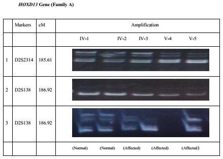
Six genes GLI3, SHH, LMBR1 (ZRS ), ZNF141, HOXD13 and IQCE were checked
for their possible involvement in the present case of polydactyly. Highly polymor-
phic microsatellite markers (Figure 3.2) close to each genes were used in genotyp-
ing. Markers D7S2541, D7S2548 and D7S691 showed linkage to GLI3 gene. No
27You can also read