Genetically Targeted Connectivity Tracing Excludes Dopaminergic Inputs to the Interpeduncular Nucleus from the Ventral Tegmentum and Substantia ...
←
→
Page content transcription
If your browser does not render page correctly, please read the page content below

Research Article: New Research

Integrative Systems

Genetically Targeted Connectivity Tracing Excludes
Dopaminergic Inputs to the Interpeduncular
Nucleus from the Ventral Tegmentum and
Substantia Nigra
Nailyam Nasirova,1,p Lely A. Quina,1,p Shoshana Novik,1 and                             Eric E. Turner1,2

https://doi.org/10.1523/ENEURO.0127-21.2021
1
 Center for Integrative Brain Research, Seattle Children’s Research Institute, 1900 Ninth Avenue, Seattle, WA 98101
and 2Department of Psychiatry and Behavioral Sciences, University of Washington, Seattle, WA 98195

Abstract
The “habenulopeduncular system” consists of the medial habenula (MHb) and its principal target of innervation, the
interpeduncular nucleus (IP). Neurons in the ventral MHb (MHbV) express acetylcholine along with glutamate, and
both the MHb and IP are rich in nicotinic acetylcholine receptors. Much of the work on this system has focused on
nicotinic mechanisms and their clinical implications for nicotine use, particularly because the IP expresses the a5 nic-
otinic receptor subunit, encoded by the CHRNA5 gene, which is genetically linked to smoking risk. A working model
has emerged in which nicotine use may be determined by the balance of reinforcement mediated in part by nicotine
effects on dopamine reward pathways, and an aversive “brake” on nicotine consumption encoded in the MHb-IP
pathway. However, recent work has proposed that the IP also receives direct dopaminergic input from the ventral
tegmental area (VTA). If correct, this would significantly impact the prevailing model of IP function. Here, we have
used Chrna5Cre mice to perform rabies virus-mediated retrograde tracing of global inputs to the IP. We have also
used Cre-dependent adeno-associated virus (AAV) anterograde tracing using Slc6a3Cre (DATCre) mice to map VTA
dopaminergic efferents, and we have examined tract-tracing data using other transgenic models for dopaminergic
neurons available in a public database. Consistent with the existing literature using non-genetic tracing methods,
none of these experiments show a significant anatomic connection from the VTA or substantia nigra (SN) to the IP,
and thus do not support a model of direct dopaminergic input to the habenulopeduncular system.
Key words: dopamine; habenula; interpeduncular nucleus; nicotine; substantia nigra; ventral tegmental area

    Significance Statement
    The interpeduncular nucleus (IP) is the central node in a descending brain pathway linking the medial habenula
    (MHb) of the epithalamus to the mesopontine tegmentum. Neurons in this “habenulopeduncular pathway” express
    unusual nicotinic acetylcholine receptors and are thought to play a role in behavioral responses to nicotine. In par-
    ticular, the IP expresses the a5 nicotinic receptor, human variants of which confer increased risk of smoking.
    Recently it has been reported that inputs from midbrain dopaminergic neurons to the IP also influence behavior.
    Here, we have used a mouse genetic model to map specific inputs to the a5-expressing neurons in the IP.
    Contrary to the published reports, we do not identify dopaminergic inputs to the IP.

Received March 25, 2021; accepted April 21, 2021; First published June 4,     Author contributions: E.E.T. designed research; N.N., L.A.Q., and S.N.
  2021.                                                                     performed research; E.E.T., N.N., L.A.Q., and S.N. analyzed data; E.E.T. wrote
  The authors declare no competing financial interests.                     the paper.

May/June 2021, 8(3) ENEURO.0127-21.2021 1–10
Research Article: New Research      2 of 10

Introduction                                                                     cluster, and have been used to show that optogenetic acti-
   The habenula is a bilateral epithalamic structure that                        vation of a5-expressing neurons in the IP is aversive if mice
provides a major link between the limbic forebrain and nu-                       have been previously exposed to nicotine (Morton et al.,
clei in the mesopontine tegmentum, including the ventral                         2018).
tegmental area (VTA), the rostromedial tegmental nucleus                            In contrast, the best characterized outputs of the LHb are
(RMTg), the interpeduncular nucleus (IP), and the raphe                          projections to the VTA, both directly and via GABAergic in-
nuclei. The two major divisions of the habenula, the medi-                       termediaries in the RMTg (Petzel et al., 2017; Li et al., 2019),
al habenula (MHb) and lateral habenula (LHb), appear to                          and projections to the mesopontine raphe (Sego et al.,
serve largely distinct, parallel pathways (Fakhoury, 2018;                       2014; Tchenio et al., 2016; Quina et al., 2017, 2020).
Metzger et al., 2021). Although the MHb and LHb effer-                           Reciprocal connections between the LHb and both dopami-
ents initially travel via a single prominent tract, the fasci-                   nergic and non-dopaminergic neurons in the VTA have been
culus retroflexus, their outputs terminate in distinct                           demonstrated (Root et al., 2014; Zahm and Root, 2017).
tegmental areas. The MHb predominantly innervates the                            Thus, in terms of neurotransmitter systems, work on the
IP, with glutamatergic/cholinergic fibers from the ventral                       LHb has emphasized regulation of dopamine and serotonin
MHb (MHbV), and glutamatergic/peptidergic fibers from                            (Metzger et al., 2021).
the dorsal MHb (MHbD), connecting to specific IP subnu-                             Given these largely parallel pathways, little attention
clei to form the “habenulopeduncular system” (Quina et                           has been paid to the possibility of a direct interaction be-
al., 2017). Although the precise function of the IP is not                       tween midbrain DA neurons and the MHb-IP pathway.
well understood, much of the interest in MHb-IP pathway                          Classic tract-tracing studies using histochemical techni-
has been driven by the expression of specific nicotinic ac-                      ques have shown that the IP integrates inputs from many
etylcholine receptors in the MHbV and IP (Quina et al.,                          brain regions, including tegmental afferents from the me-
2009; Hsu et al., 2013), the dual glutamatergic/cholinergic                      dian raphe, nucleus incertus (NI), and laterodorsal teg-
nature of MHbV neurons that project to the IP (Qin and                           mentum, but have not identified inputs from the VTA or
Luo, 2009; Hsu et al., 2013; Frahm et al., 2015), the effect                     substantia nigra (SN; Marchand et al., 1980; Hamill and
of nicotine on signaling in and between the MHbV and IP                          Lenn, 1984; Groenewegen et al., 1986; Shibata et al.,
(Ables et al., 2017; Morton et al., 2018; Wolfman et al.,                        1986; Lima et al., 2017; Bueno et al., 2019). Despite these
2018; Arvin et al., 2019), and its possible relevance to the                     studies, it has been recently proposed that the IP receives
balance of nicotine reward and aversion that regulates                           functional dopaminergic inputs from the VTA, which mod-
smoking (Fowler and Kenny, 2014; McLaughlin et al.,                              ulate nicotine withdrawal-induced anxiety and novelty
2017; Lee et al., 2019).                                                         signaling in behavioral tests of social familiarity (Zhao-
   The IP expresses particularly high levels of the a5                           Shea et al., 2015; Molas et al., 2017). Such a direct inter-
nicotinic receptor subunit, product of the Chrna5 gene                           action between DA neurons and the MHb-IP pathway, if
(Boulter et al., 1990; Hsu et al., 2013). The strongest                          confirmed, could shift the paradigm for how this pathway
known genetic risk for increased tobacco consumption                             functions in reinforcement and other behaviors. Here,
is associated with certain haplotypes that occur at                              however, we have used rabies-virus mediated retrograde
high frequency in European populations, and contain                              tracing in Chrna5Cre mice, and virally-mediated antero-
a nonsynonymous CHRNA5 polymorphism, CHRNA5                                      grade tracing in multiple transgenic models, and find no
(D398N) (Berrettini and Doyle, 2012; Lassi et al., 2016),                        evidence for an anatomic connection between midbrain
which appears to reduce the function of nicotinic re-                            DA neurons and the IP.
ceptors into which this variant a5 subunit is incorporated
(Kuryatov et al., 2011; Sciaccaluga et al., 2015). Although in                   Materials and Methods
humans and rodents the Chrna5 gene locus is part of a con-
served cluster with two other nicotinic receptor genes,                          Mouse strains
Chrna3 and Chrnb4, transgenic mice have been devised for                            Transgenic targeting of the IP was achieved with a Chrna5-
expression of Cre recombinase regulated by the Chrna5                            BAC-Cre transgenic line (Chrna5Cre; Morton et al., 2018).
locus without over-expression of the other receptors in this                     Transgenic reporting of Cre-expression was achieved by
                                                                                 crossing Chrna5Cre mice with the Cre-dependent ZsGreen re-
  This work was supported by the National Institute of Mental Health Grant       porter strain Gt(ROSA).26Sortm6(CAG-ZsGreen1)Hze/J (Ai6, Jax
R01MH093667 and the National Institute on Drug Abuse Grant R01DA035838.          #007906; Madisen et al., 2010). Viral reporters were tar-
  *N.N. and L.A.Q. contributed equally to this work.
  Acknowledgements: We thank Julie Harris, Karla Hirokawa, Ali Cetin,
                                                                                 geted to tegmental DA neurons using the transgenic line
Shenqin Yao, and Marty Mortrud of the Allen Institute for Brain Research for     Slc6a3tm1.1(cre)Bkmn/J (DATCre, Jax #006660; Bäckman et
the gift of tract-tracing adeno-associated and rabies viruses; Hongkui Zeng of   al., 2006). All strains were maintained on a C57BL/6NCrl
the Allen Institute for information about the Allen Connectivity Atlas; and      genetic background (Charles River). Adult mice of both
Kamiliam Nasirova for technical assistance.
                                                                                 sexes were used in the experiments. Data for three other
  Correspondence should be addressed to Eric E. Turner at eric.turner@
seattlechildrens.org.                                                            transgenic lines were obtained from the Allen Mouse
  https://doi.org/10.1523/ENEURO.0127-21.2021                                    Brain Connectivity Atlas, as described below.
Copyright © 2021 Nasirova et al.
This is an open-access article distributed under the terms of the Creative
Commons Attribution 4.0 International license, which permits unrestricted use,
                                                                                 Immunofluorescence and in situ hybridization
distribution and reproduction in any medium provided that the original work is     Mouse brain tissue was prepared by fixation via trans-
properly attributed.                                                             cardial perfusion with 4% paraformaldehyde. Brains were

May/June 2021, 8(3) ENEURO.0127-21.2021                                                                                                 eNeuro.org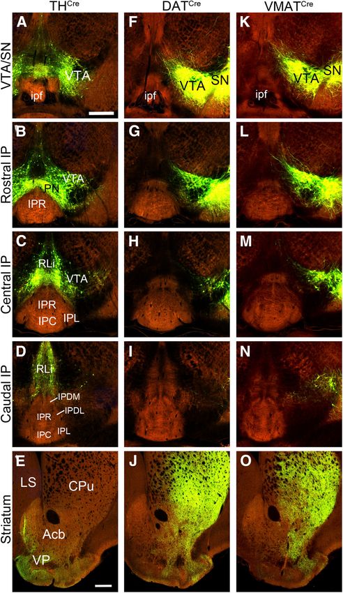
Research Article: New Research      3 of 10

then removed and equilibrated in graded sucrose solu-           Retrograde tracing
tions, frozen at 8°C in OCT solution, and cryosectioned           For retrograde tracing from a genetically defined cell
at 25 mm for fluorescence/immunofluorescence imaging.           population, the helper virus AAV1-Syn-DIO-TVA66T-
Tissue processed in this way was suitable for imaging of        dTom-CVS N2cG, (AAV1-N2cG) a tricistronic virus which
endogenous protein fluorescence, immunofluorescence,            expresses the pseudotyping receptor TVA, tdTomato,
and fluorescence in situ hybridization (FISH). Tyrosine hy-     and the rabies glycoprotein G (Lo et al., 2019) was in-
droxylase (TH) immunoreactivity was detected using rab-         jected by pressure injection into the IP of Chrna5Cre mice,
bit anti-TH (AB152, EMD Millipore, RRID:AB_390204).             followed by the rabies virus EnvA CVS-N2cDG-histone-
Multi-channel FISH was performed with the RNAscope              eGFP (RV-GFP), injected 21 d later into the same location.
Multiplex Fluorescent V2 kit, according to the manufac-         The injection coordinates were: AP 3.4, ML 0.0, DV 5.0,
turer’s instructions (Advanced Cell Diagnostics). The           and the injected volume was 200 nl for the helper virus
probes used included: EGFP, #400281-C2 (channel 2)              and 300 nl for the rabies virus. AAV1 and RV for retro-
and Mm-Rln3-C1 (channel 1).                                     grade tracing were the gifts of Shenqin Yao and Ali Cetin
                                                                (Allen Institute for Brain Science). Further details regard-
                                                                ing rabies reagents are available on request from Shenqin
Anterograde tracing                                             Yao and Ali Cetin. Mice were euthanized 10 d later and
   The targeted coordinates for each anterograde or retro-      the brains were processed as described above to visual-
grade tracing injection were based on a standard atlas          ize the nuclear GFP signal, or virally expressed GFP
(Paxinos and Franklin, 2001). For anterograde tracing of        mRNA using FISH, in presynaptic neurons.
VTA efferents, 100 nl of viral stock was pressure injected
at coordinates: AP 3.4, ML 0.5, DV 4.5. Anterograde             Results
tract tracing data derived from the Allen Mouse Brain             RV-mediated retrograde tracing is usually targeted by
Connectivity Atlas were generated using iontophoretic in-       the specific expression of Cre-recombinase in the postsy-
jection of AAV, and detailed methods have been pub-             naptic neurons of interest. In order to trace specific inputs
lished in conjunction with the Atlas (Harris et al., 2012; Oh   to the IP, we used a well-characterized mouse BAC trans-
et al., 2014). Animals were fixed by transcardial perfusion     genic strain, Chrna5Cre, which expresses Cre recombi-
with 4% paraformaldehyde at 14–21 d after injection and         nase in most neurons of the IP, and in a small population
processed as described above. Anterograde tracing was           of GABAergic neurons in the adjacent median raphe
performed using Cre-activated (FLEX) adeno-associated           (Morton et al., 2018). Although a5 mRNA is expressed in
virus (AAV; capsid strain 1). Enhanced labeling of presyn-      some neurons of the VTA, the regulatory sequences in
aptic areas was performed by expression of a synapto-           this BAC transgene do not target VTA neurons, leading to
physin-EGFP fusion protein (sypGFP). The plasmid                high local specificity of Cre expression in the IP. This
pCAG.Flex.sypEGFP.WPRE (“FLEX-sypGFP”) was con-                 specificity of Cre expression was verified by crossing
structed by replacing the EGFP moiety of pCAG-FLEX-             Chrna5Cre mice with a genetic reporter strain, Ai6 (Fig. 1A;
EGFP-WPRE (Addgene #51 502) with the sypEGFP con-               Materials and Methods), which allows Cre-dependent ex-
struct from phSyn1(S)-FLEX-tdTomato-T2A-SypEGFP-                pression of the reporter ZsGreen in targeted cells. The
WPRE (Addgene #51509) by Julie Harris, Karla Hirokawa           ZsGreen reporter was expressed abundantly in all of the
and Hong Gu of the Allen Institute for Brain Science (gift      IP subnuclei, except the lateral subnucleus, which
of Julie Harris).                                               showed sparse labeling (Fig. 1B–D), whereas labeling was
                                                                not observed in the VTA (Fig. 1B).
                                                                  A cohort of five Chrna5Cre mice were used for retro-
Anterograde tract tracing: database information                 grade tracing. These mice were first injected in the IP with
   The Allen Mouse Brain Connectivity Atlas provides a          the “helper” virus AAV1-N2cG, which is a tricistronic vec-
searchable database of brain-wide AAV tract-tracing da-         tor expressing pseudotyping receptor TVA, tdTomato,
tasets using wild-type and Cre-recombinase expressing           and the rabies glycoprotein G (Materials and Methods).
mouse strains (https://connectivity.brain-map.org/). A          Three weeks later the same site was injected with RV ex-
source structure database search was performed for the          pressing nuclear GFP (nGFP), and after 10 d the mice
VTA, and a target structure search was performed for the        were killed, and brains were processed for localization of
IP. Three informative cases (experiments) were identified       RV-nGFP and other markers. The two cases with the
with the VTA as the fiber source, using three different Cre-    most comprehensive labeling of the targeted IP neurons
drivers related to monoaminergic transmission to specifically   were chosen for detailed characterization (Cases r1, r2;
target the VTA: (1) TH-IRES-CreER (JAX #008532, THCre;          Fig. 1F–K). As expected, tdTomato from AAV1-N2cG and
Rotolo et al., 2008), experiment 156314762, published online    nGFP-labeled “starter cells” capable of infecting IP affer-
10/04/2012; (2) Slc6a3-Cre (DATCre; Zhuang et al., 2005), ex-   ents were observed in all IP subnuclei, but were sparse in
periment 160539283, published 03/07/2013; (3) Slc18a2-          IPL. In order to determine the efficiency of RV-mediated
Cre_OZ14 (GENSAT BAC-Cre, VMAT2Cre; Gong et al., 2007),         retrograde labeling, we examined RV-nGFP expression in
experiment 292958638, published 03/06/2014.                     two areas known to have strong inputs to the IP, the ha-
   The online publication dates of the Allen Connectivity       benula (Fig. 1L,M, ref) and the NI (Fig. 1N; Nasirova et al.,
Atlas datasets were provided by Hongkui Zeng, Allen             2020). RV-Case r1 resulted in strong labeling of the
Institute for Brain Science, Seattle, WA.                       MHbV, and partial labeling of the MHbD and LHb. RV-

May/June 2021, 8(3) ENEURO.0127-21.2021                                                                               eNeuro.org
Research Article: New Research      4 of 10

Figure 1. Genetic and viral strategy for transsynaptic labeling of IP afferents. A, A genetic strategy for labeling Cre-expressing neu-
rons in the IP of Chrna5Cre mice. Chrna5Cre mice were interbred with the mouse strain Ai6, which conditionally expresses the fluoro-
phore ZsGreen (Materials and Methods). B–D, ZsGreen expression in the rostral, central, and caudal IP of Chrna5Cre, Ai6
compound heterozygous mice, generated as shown in A. E, RV transsynaptic labeling strategy. A helper virus AAV1-N2cG was in-
jected into the IP of Chrna5Cre mice, followed by RV three weeks later (Materials and Methods). The habenula and IP were examined
for the expression of nuclear GFP (nGFP) expressed by RV. Blue lines indicate the planes of section for the habenula, IP, and NI
(rostral to caudal). F–K, Imaging of nGFP expressed by RV and cytoplasmic tdTomato (tdT) expressed by the AAV helper virus in
the rostral (F, I), central (G, J), and caudal (H, K) IP of two injected cases, r1 and r2 (retrograde 1 and 2). Insets in G, J show higher
magnification of the boxed area. L, M, Expression of RV nGFP transsynaptic label in the habenula. Coronal sections correspond to
bregma 1.7 in a standard atlas (Paxinos and Franklin, 2001). N, Dual-label FISH for RV-expressed GFP mRNA and Rln3 mRNA in
the NI of injected Case r1. IP, interpeduncular nucleus; IPC, central part; IPDL, dorsolateral part; IPDM, dorsomedial part; IPI, inter-
mediate part; IPL, lateral part; IPR, rostral part; LHbL, lateral habenula, lateral part; LHbM, lateral habenula, medial part; MHbD, me-
dial habenula, dorsal part; MHbV, medial habenula, ventral part; NI, nucleus incertus; RMTg, rostromedial tegmental nucleus; VTA,
ventral tegmental area. Scale bar: 200 mm (B, L, N).

Case r2 resulted in strong labeling of both MHbV and                   (Quina et al., 2017). The NI of Case r1 was examined
MHbD, as well as partial labeling of LHb. More intense la-             for expression of RV-GFP mRNA together with mRNA for
beling of MHbD in Cases r2 is expected, because the in-                the characteristic NI neuropeptide, relaxin-3 (Rln3). Con-
jected area is more caudally positioned in the IP, where               sistent with prior studies using anterograde tracing of
MHbD fibers are known to cross the midline and terminate               Rln3-positive and Rln3-negative NI neurons (Nasirova et

May/June 2021, 8(3) ENEURO.0127-21.2021                                                                                        eNeuro.org
Research Article: New Research      5 of 10

                                                                    al., 2020), most of the RV-nGFP labeled neurons in the NI
                                                                    did not express Rln3. Together these two RV-injected
                                                                    cases appear to give nearly complete coverage of the a5-
                                                                    expressing cell population in the IP.
                                                                       To assess for dopaminergic afferents to the IP, we then
                                                                    examined the tegmental dopamine system of the
                                                                    Chrna5Cre RV-traced mice, including the entire rostrocau-
                                                                    dal extent of the SN and VTA, for the expression of RV-
                                                                    nGFP. TH immunostaining was used to identify dopami-
                                                                    nergic neurons. Since TH is cytoplasmic, and the RV-ex-
                                                                    pressed GFP is nuclear, we looked for neurons that
                                                                    showed a circular or semicircular pattern of TH staining
                                                                    with a nuclear “hole” indicating that the cell nucleus was
                                                                    in the plane of section. Dopaminergic neurons projecting
                                                                    to the IP should exhibit a “fried egg” appearance with a
                                                                    nGFP-positive nucleus and a TH-immunoreactive periph-
                                                                    ery. Overall, RV-nGFP labeled neurons were sparse in the
                                                                    VTA (Fig. 2A,B). They appeared at a somewhat higher fre-
                                                                    quency in the caudal part of the SN (Fig. 2B). Few, if any,
                                                                    of the nGFP-labeled neurons appeared to be dopaminer-
                                                                    gic. Using Z-stacked confocal images, we counted RV-
                                                                    nGFP labeled neurons and TH-immunoreactive neurons
                                                                    in one hemisphere of the tegmentum from bregma 2.8
                                                                    to bregma 3.6 (in Cases r1 and r1), encompassing the
                                                                    entire extent of the VTA1SN a standard atlas (Paxinos
                                                                    and Franklin, 2001). RV-nGFP neurons were counted
                                                                    within a manually outlined area of interest encompassing
                                                                    all of the VTA1SN TH-immunoreactive cell bodies (Fig.
                                                                    2A,B). TH immunostaining that did not incorporate a nu-
                                                                    clear profile was assumed to be contributed by fibers of
                                                                    passage or by neurons with nuclei out of the plane of sec-
                                                                    tion and was ignored in cell counts. Figure 2C shows an
                                                                    example of a rarely encountered TH-positive neuron that
                                                                    may have a GFP-positive nucleus, and an example of a
                                                                    GFP-positive nucleus that overlaps TH signal in the image
                                                                    but does not appear to be the nucleus of a TH-labeled
                                                                    cell. In Case r1 we counted 1104 TH-immunoreactive
                                                                    neurons in the series, and 373 nGFP labeled nuclei within
                                                                    the area of interest defined by the TH staining (Fig. 2D). In
                                                                    1099/1104 TH-immunoreactive neurons, a GFP-labeled
                                                                    nucleus could be excluded; only 5 cells showed sufficient
                                                                    overlap to suggest possible co-localization of the
Figure 2. RV-mediated retrograde tracing of projections from
the VTA and SN to the IP. Inputs to the IP were retrogradely la-
                                                                    markers. Results for Case r2 were similar, with only one
beled with RV as described in Figure 1 (Case r1), producing a       cell showing possible co-localization (Table 1). We con-
nuclear GFP label in presynaptic neurons. Serial 25-mm sec-         clude that DA neurons in the SN/VTA very rarely make
tions were examined at 100-mm intervals through the VTA and         contact with a5-expressing neurons in the IP. Labeling
SN, at levels from bregma 2.8 to bregma 3.6 in a standard           data for Cases r1 and r2 across the rostrocaudal extent of
atlas (Paxinos and Franklin, 2001), and imaged as Z-stacks. DA      the VTA/SN appear in Table 1.
neurons were labeled by immunostaining for TH, and all cells
were counterstained for nucleic acids with DAPI. TH-positive
neurons were counted if the TH immunostaining formed a cyto-
plasmic circle or semicircle around a nuclear hole, indicating      continued
that the labeled neuron was in the plane of section. Areas of in-   indicates an example of an nGFP-labeled nucleus which may
terest were drawn manually around the area containing the           be in a TH-labeled neuron. The arrowhead indicates an nGFP
VTA1SN (outlines), and all of the nGFP and TH-immunoreactive        nucleus which overlies TH-labeling but does not appear to be
cells in the left hemisphere of these structures were counted. A,   the nucleus of the TH-expressing cell. D, Count of nGFP la-
RV nGFP and TH labeling in the rostral VTA/SN. In the region of     beled, TH-positive, and dual-labeled neurons in the left VTA/SN
interest outlined, 9/1606 DAPI-labeled cells were labeled with      in sections at the designated coordinates in Case r1. IP, inter-
GFP (0.56%). B, C, RV nGFP and TH labeling in the caudal            peduncular nucleus; ml, medial lemniscus; MM, medial mam-
VTA/SN. In the region of interest outlined, 83/2595 DAPI-la-        millary nucleus; PN, paranigral nucleus; SNC, substantia nigra,
beled cells were labeled with GFP (3.2%). Co-localization of        pars compacta; SNR, substantia nigra, pars reticulata; VTA,
nGFP and TH was very rarely observed. In C, the arrow               ventral tegmental area. Scale bar: 200 mm (A).

May/June 2021, 8(3) ENEURO.0127-21.2021                                                                                    eNeuro.org
Research Article: New Research 6 of 10
Table 1: Cell counts for RV-labeling and TH immunoreactiv- together, these three cases gave good coverage of DA
ity in Cases r1 and r2 neurons in the tegmentum and their striatal targets of in-
Bregma Case r1 cell count Case r2 cell count nervation. No case showed evidence of dopaminergic in-
Coordinate nGFP TH Both nGFP TH Both nervation of the IP (Fig. 3B–D,G–I,L–N).
2.8 12 43 0 2 28 0 To complement these tract-tracing cases available in a
2.9 9 116 0 2 65 0 public database, we performed independent injections in
3.0 24 134 1 15 112 0 DATCre mice targeting the central to caudal VTA, which
3.1 31 117 0 19 102 0 were relatively under-labeled in the Allen DATCre cases
3.2 52 192 1 39 110 0
(Fig. 4). In order to enhance detection of any projections
3.3 54 156 1 38 121 0
3.4 83 158 1 38 145 1
from tegmental DA neurons to the IP, we used a Cre-de-
3.5 59 101 1 36 138 0 pendent AAV expressing synaptically targeted GFP
3.6 49 87 0 33 128 0 (sypGFP), which produces intense punctate signal in pre-
Sum 373 1104 5 222 949 1 synaptic areas. Serial sections through the tegmentum
of the injected cases were immunostained for TH to visu-
alize the DA cell bodies. Case a1 was a unilateral injection
Although most IP neurons express Chrna5Cre, we also in the central/caudal VTA, also labeling the PN, and
considered the possibility that DA neurons in the VTA largely sparing the SN (Fig. 4A–D). Case a2 was a midline
could project exclusively to a subset of IP neurons that injection labeling both VTA hemispheres, the PN, and the
are a5-negative, and thus would not be labeled in the ret- RLi (Fig. 4E–H). Intensely labeled DA neuron cell bodies
rograde tracing experiments. In addition, it is possible were observed in all of the injected areas. Rare GFP-la-
that RV does not efficiently label all kinds of presynaptic beled cell bodies were also detected in the caudal IP, indi-
neurons (Rogers and Beier, 2021). For these reasons, we cating that a few IP cells express DATCre (Fig. 4G). These
examined data available in a public database addressing cells were not TH immunoreactive and their identity is un-
the projections of tegmental DA neurons, and performed clear. However, no sypGFP labeling was detected in the
further Cre-mediated anterograde tracing experiments. IP indicative of dopaminergic afferents.
The Allen Connectivity Atlas is a large, searchable data-
base of cases in which the mouse brain has been injected Discussion
with AAV tracers, then imaged by serial two-photon to- Two recent papers have proposed a novel and behav-
mography through the entire neural axis (Oh et al., 2014). iorally significant connection between tegmental DA neu-
Many of the Allen Connectivity Atlas experiments use rons and the IP (Zhao-Shea et al., 2015; Molas et al.,
Cre-dependent AAV combined with mouse strains ex- 2017). Certainly, new pathways may be discovered linking
pressing Cre recombinase in specific populations of neu- well-studied brain regions. However, the projections of
rons. Allen cases mapping the efferents of the SN/VTA are the midbrain DA system have been extensively studied in
available using three Cre-drivers for catecholaminergic rats, mice, primates, and other species, so it is surprising
neurons: TH-IRESCreER (THCre), Slc6a3Cre (DATCre), and that such a pathway, if present, has not been previously
Slc18a2Cre (VMAT2Cre). We thus examined AAV-GFP ex- reported. Indeed, the mesolimbic and nigrostriatal DA
pression in the SN/VTA and IP of each of these transgenic pathways may be the most-studied subcortical systems.
models (Fig. 3). The use of multiple transgenic models A comprehensive review of the first ;170 studies of the
helps to overcome possible heterogeneity in the labeling VTA efferent system in birds, rodents, and primates
of DA neurons in different transgenic systems (Lammel et makes no mention of a VTA-IP projection (Oades and
al., 2015; Stuber et al., 2015). As a positive control for the Halliday, 1987), nor do subsequent reviews of studies
successful labeling of efferents from the tegmental DA using classical methods (Bentivoglio and Morelli, 2005).
system, we examined labeled fibers in the striatum in Further studies of this system using a wide range of ge-
each case. The available THCre case, injected near the netic, viral, and optogenetic methods, and reviews of
midline, labeled DA neurons predominantly in the caudal those studies, also do not mention projections of tegmen-
VTA, as well as DA neurons of the paranigral nucleus (PN), tal DA neurons to the IP (Yetnikoff et al., 2014; Aransay et
and the rostral linear nucleus raphe (RLi; Fig. 3A–D). As al., 2015; Beier et al., 2015; Wise and McDevitt, 2018).
expected in a case that did not label the SN, fibers in the Likewise, a viral anterograde study of GABAergic and glu-
striatum terminated mainly in the ventral pallidum (VP), tamatergic VTA neurons did not identify IP projections
rather than the caudate/putamen (CPu; Fig. 3E). Two (Taylor et al., 2014).
DATCre mediated VTA-labeling cases are available in the Although the existing IP literature is much sparser, prior
Allen database, and the more extensively labeled case is studies of global inputs to the IP also have not described
shown (Fig. 3F–J). In this case, injected unilaterally in the a VTA-IP dopaminergic connection. Brain-wide studies of
right tegmentum, there are numerous labeled cell bodies projections to the IP using classical methods in the rat
in the rostral and central parts of the SN and VTA (Fig. 3F– have not identified VTA or SN neurons projecting to the IP
H). Terminal fibers are seen in both the ventral (VP, Acb) (Marchand et al., 1980; Groenewegen et al., 1986;
and dorsal (CPu) striatum. A reported case using a Shibata et al., 1986). A recent study using retrograde CTB
VMATCre mouse strain shows labeling of cell bodies in the tracing from the IP in rats showed rare labeling in the VTA,
SN/VTA and terminal fibers in the striatum that is very sim- similar to that observed here using RV, but these neurons
ilar to that obtained with DATCre (Fig. 3K–O). Taken were not identified as dopaminergic (Lima et al., 2017). A
May/June 2021, 8(3) ENEURO.0127-21.2021 eNeuro.orgResearch Article: New Research 7 of 10 Figure 3. Cre-mediated tract-tracing of tegmental DA neurons in the Allen Connectivity Atlas. Three cases of Cre-mediated AAV tract tracing from the Allen Connectivity Atlas are shown. A–E, Labeling mediated by THCre, Allen experiment 156314762, with la- beled neurons in the bilateral VTA, PN, and RLi, and sparing the SN. F–J, Labeling mediated by Slc6a3Cre (DATCre), Allen experiment 160539283, with signal in the right VTA and SN. K–O, Labeling using a VMAT2Cre driver, Allen experiment 292958638, with expres- sion in the right VTA and SN. The planes of section from top to bottom are: (A, F, K) VTA/SN, incorporating the injected area, close to bregma 3.1 as designated in a standard atlas (Paxinos and Franklin, 2001); (B, G, L) rostral IP, bregma 3.3; (C, H, M) central IP, bregma 3.5; (D, I, N) caudal IP, bregma 3.8; (E, J, O) striatum, bregma 0.7. The reported target coordinates are (bregma, AP, ML, DV): experiment 156314762 ( 3.28, 0.36, 4.13); experiment 160539283 ( 3.08, 1.25, 4.08); experiment 292958638 ( 3.08, 1.25, 4.15). Acb, accumbens nucleus; CPu, caudate/putamen; IP, interpeduncular nucleus; IPC, central part; IPDL, dorsolateral part; IPDM, dorsomedial part; IPL, lateral part; IPR, rostral part; ipf, interpeduncular fossa; LS, lateral septum; PN, paranigral nu- cleus; RLi, rostral linear nucleus raphe; SN, substantia nigra; VP, ventral pallidum; VTA, ventral tegmental area. Scale bar: 400 mm (A, E). prior Cre-mediated RV retrograde tracing study of the present study, also did not report VTA labeling from the IP specific inputs to populations of IP neurons expressing (Ables et al., 2017). Amigo1 and Epyc, each of which identifies a subset of the In the present study, we have used two different Cre- a5 nicotinic receptor-expressing neurons labeled in the driven transgenic strategies in mice to search for May/June 2021, 8(3) ENEURO.0127-21.2021 eNeuro.org
Research Article: New Research      8 of 10

Figure 4. Cre-mediated tract tracing of tegmental DA neurons using synaptically-targeted GFP. DATCre mice were injected with a
Cre-dependent AAV expressing synaptically targeted GFP. Sections were stained for TH by immunofluorescence (red), and with the
nuclear marker DAPI (blue). The intended target of the injection was at the level shown in B, F. The targeted coordinates for both
cases were: AP 3.4, ML 0.5, DV 4.5. A–D, Case a1 (anterograde 1), injected in the central part of the right VTA, with some spread
laterally into SN and RRF. E–H, Case a2, injected nearer the midline, labeling the medial part of the VTA bilaterally, as well as the
PN and RLi. Arrows in G indicate rare cell bodies in the IP labeled by AAV, indicating that a few IP cells may express enough Cre re-
combinase to activate the Cre-dependent AAV. The planes of section from top to bottom are: (A, E) injected area, bregma 3.1; (B,
F) rostral IP, bregma 3.3; (C, G) central IP, bregma 3.5; (D, H) caudal IP, bregma 3.8. IF, interfascicular nucleus; IP, interpedun-
cular nucleus; IPC, central part; IPDL, dorsolateral part; IPDM, dorsomedial part; IPL, lateral part; IPR, rostral part; ipf, interpeduncu-
lar fossa; ml, medial lemniscus; PN, paranigral nucleus; RLi, rostral linear nucleus raphe; RRF, retrorubral field; SN, substantia nigra;
VTA, ventral tegmental area. Scale bar: 200 mm (A).

connections between tegmental DA neurons and the IP.                   the GFP label in presynaptic areas, essentially no dopami-
First, we used a well described BAC-Cre transgenic line,               nergic fibers were identified in the IP.
Chrna5Cre (Morton et al., 2018), to express RV as a spe-                  Given these findings, we also searched for supporting
cific retrograde tracer in the habenula-recipient subnu-               data from other transgenic models in the Allen Connectivity
clei of the IP. No significant RV labeling from the IP was             Atlas. Useful experiments were found in the Atlas using
observed in tegmental DA neurons, although other neu-                  three other mouse strains used to identify DA neurons:
rons in the VTA were sparsely labeled. The habenula, in                THCre, VMAT2Cre, and a different DATCre transgenic line
contrast, was heavily labeled by RV via the well-known                 from that used in our own experiments. AAV injections into
habenulopeduncular pathway. Second, Cre-mediated                       the tegmentum in these experiments labeled DA neurons in
anterograde tracing was performed by injection of a                    the VTA, as well as other tegmental DA neurons in the SN,
Cre-dependent AAV expressing a synaptically-targeted                   PN, and RLi. Although none of these cases, taken alone,
GFP marker into the VTA of a DATCre transgenic line.                   completely labeled all of these structures, together they
Despite the enhanced sensitivity obtained by concentrating             gave extensive coverage of the tegmental DA system. In

May/June 2021, 8(3) ENEURO.0127-21.2021                                                                                         eNeuro.org
Research Article: New Research        9 of 10

each case, as expected, dopaminergic projections to                       neurophysiology of habenulo-interpeduncular circuitry. J Neurosci
the striatum were heavily labeled, but none of these                      39:4268–4281.
cases showed significant fiber labeling in the IP. These               Bäckman CM, Malik N, Zhang Y, Shan L, Grinberg A, Hoffer BJ,
                                                                          Westphal H, Tomac AC (2006) Characterization of a mouse strain
experiments were published online as part of the Allen                    expressing Cre recombinase from the 3’ untranslated region of the
Connectivity Atlas between October 2012 and March                         dopamine transporter locus. Genesis 44:383–390.
2014, so they were publicly available at the time the re-              Beier KT, Steinberg EE, DeLoach KE, Xie S, Miyamichi K, Schwarz L,
sults of Molas et al. (2017) were reviewed and                            Gao XJ, Kremer EJ, Malenka RC, Luo L (2015) Circuit architecture
published.                                                                of VTA dopamine neurons revealed by systematic input-output
   Surprising new findings in a well-studied system should                mapping. Cell 162:622–634.
                                                                       Bentivoglio M, Morelli M (2005) The organization and circuits of mes-
be supported by strong evidence. However, in the pub-
                                                                          encephalic dopaminergic neurons and the distribution of dopa-
lished work reporting a VTA-IP pathway (Molas et al.,                     mine receptors in the brain. Handb Chem Neuroanat 21:1–107.
2017), the evidence for dopaminergic projections to the IP             Berrettini WH, Doyle GA (2012) The CHRNA5-A3-B4 gene cluster in
is quite limited. In these experiments, Cre-dependent AAV                 nicotine addiction. Mol Psychiatry 17:856–866.
encoding ChR2-eYFP was injected into the VTA of the                    Boulter J, O’Shea-Greenfield A, Duvoisin RM, Connolly JG, Wada E,
same strain of DATCre mice used to generate the data                      Jensen A, Gardner PD, Ballivet M, Deneris ES, McKinnon D (1990)
shown in Figure 2. Only a single VTA injected case is                     Alpha 3, alpha 5, and beta 4: three members of the rat neuronal
                                                                          nicotinic acetylcholine receptor-related gene family form a gene
shown, in which a few labeled fibers appear in the periph-                cluster. J Biol Chem 265:4472–4482.
ery of the IP (Molas et al., 2017; see their Supplementary             Bueno D, Lima LB, Souza R, Gonçalves L, Leite F, Souza S, Furigo
Fig. 10). The origin of these fibers is not well defined, be-             IC, Donato J Jr, Metzger M (2019) Connections of the laterodorsal
cause no data were shown to delineate the extent of viral                 tegmental nucleus with the habenular-interpeduncular-raphe sys-
infection. The VTA injections performed in this report em-                tem. J Comp Neurol 527:3046–3072.
ployed 800 nl of AAV, a large volume relative to subcorti-             Fakhoury M (2018) The dorsal diencephalic conduction system in re-
                                                                          ward processing: spotlight on the anatomy and functions of the
cal structures in the mouse CNS. This is in contrast to                   habenular complex. Behav Brain Res 348:115–126.
the 100-nl injection volume used in the present study, and             Fowler CD, Kenny PJ (2014) Nicotine aversion: neurobiological
the focused iontophoretic injections used in the Allen                    mechanisms and relevance to tobacco dependence vulnerability.
Connectivity Atlas experiments. Thus, it is unclear whether               Neuropharmacology 76:533–544.
the injection was restricted to the VTA, might have spread to          Frahm S, Antolin-Fontes B, Görlich A, Zander J-F, Ahnert-Hilger G,
other DAT-expressing areas, or could have infected the TH-                Ibañez-Tallon I (2015) An essential role of acetylcholine-glutamate
                                                                          synergy at habenular synapses in nicotine dependence. Elife 4:
negative DATCre labeled cells of unknown phenotype that
                                                                          e11396.
are intrinsic to the IP (Fig. 4G). These authors also cite prior       Gong S, Doughty M, Harbaugh CR, Cummins A, Hatten ME, Heintz
work from the same laboratory as support for a VTA-IP pro-                N, Gerfen CR (2007) Targeting Cre recombinase to specific neuron
jection (Zhao-Shea et al., 2015). In that paper, supported by             populations with bacterial artificial chromosome constructs. J
a single image (see their Supplementary Fig. 9), the authors              Neurosci 27:9817–9823.
state that the VTA projects to the centrally located IP subnu-         Groenewegen HJ, Ahlenius S, Haber SN, Kowall NW, Nauta WJ
clei, IPI and IPC, rather than the periphery of the rostral IP,           (1986) Cytoarchitecture, fiber connections, and some histochemi-
                                                                          cal aspects of the interpeduncular nucleus in the rat. J Comp
an entirely different pattern of innervation from that de-                Neurol 249:65–102.
scribed in the 2017 paper.                                             Hamill GS, Lenn NJ (1984) The subnuclear organization of the rat in-
   In summary, we are unable to reconcile the current find-               terpeduncular nucleus: a light and electron microscopic study. J
ings with the hypothesis that VTA DA neurons have direct                  Comp Neurol 222:396–408.
input to the IP, and thus mediate behavioral responses to              Harris JA, Wook OS, Zeng H (2012) Adeno-associated viral vectors
social novelty by this pathway, as proposed. Likewise,                    for anterograde axonal tracing with fluorescent proteins in non-
                                                                          transgenic and cre driver mice. Curr Protoc Neurosci Chapter 1:
there do not appear to be IP inputs from the other next-
                                                                          Unit 1.20.1-18.
closest tegmental DA cell groups in the SN, PN, or RLi.                Hsu YW, Tempest L, Quina LA, Wei AD, Zeng H, Turner EE (2013)
Thus, the conclusion that there is a direct functional inter-             Medial habenula output circuit mediated by a5 nicotinic receptor-
section of the DA reinforcement pathway and the habenu-                   expressing GABAergic neurons in the interpeduncular nucleus. J
lopeduncular pathway at the level of the IP should be                     Neurosci 33:18022–18035.
reconsidered unless better evidence for such a connec-                 Kuryatov A, Berrettini W, Lindstrom J (2011) Acetylcholine receptor
                                                                          (AChR) a5 subunit variant associated with risk for nicotine de-
tion can be found.
                                                                          pendence and lung cancer reduces (a4 b 2)(2)a5 AChR function.
                                                                          Mol Pharmacol 79:119–125.
References                                                             Lammel S, Steinberg EE, Földy C, Wall NR, Beier K, Luo L, Malenka
                                                                          RC (2015) Diversity of transgenic mouse models for selective tar-
Ables JL, Görlich A, Antolin-Fontes B, Wang C, Lipford SM, Riad           geting of midbrain dopamine neurons. Neuron 85:429–438.
  MH, Ren J, Hu F, Luo M, Kenny PJ, Heintz N, Ibañez-Tallon I          Lassi G, Taylor AE, Timpson NJ, Kenny PJ, Mather RJ, Eisen T,
  (2017) Retrograde inhibition by a specific subset of interpeduncu-      Munafò MR (2016) The CHRNA5-A3-B4 gene cluster and smoking:
  lar alpha5 nicotinic neurons regulates nicotine preference. Proc        from discovery to therapeutics. Trends Neurosci 39:851–861.
  Natl Acad Sci USA 114:13012–13017.                                   Lee HW, Yang SH, Kim JY, Kim H (2019) The role of the medial habe-
Aransay A, Rodríguez-López C, García-Amado M, Clascá F, Prensa            nula cholinergic system in addiction and emotion-associated be-
  L (2015) Long-range projection neurons of the mouse ventral teg-        haviors. Front Psychiatry 10:100.
  mental area: a single-cell axon tracing analysis. Front Neuroanat    Li H, Vento PJ, Parrilla-Carrero J, Pullmann D, Chao YS, Eid M, Jhou
  9:59.                                                                   TC (2019) Three rostromedial tegmental afferents drive triply dis-
Arvin MC, Jin XT, Yan Y, Wang Y, Ramsey MD, Kim VJ, Beckley NA,           sociable aspects of punishment learning and aversive valence en-
  Henry BA, Drenan RM (2019) Chronic nicotine exposure alters the         coding. Neuron 104:987–999.e4.

May/June 2021, 8(3) ENEURO.0127-21.2021                                                                                          eNeuro.org
Research Article: New Research 10 of 10
Lima LB, Bueno D, Leite F, Souza S, Gonçalves L, Furigo IC, Donato Quina LA, Walker A, Morton G, Han V, Turner EE (2020) GAD2 ex-
J Jr, Metzger M (2017) Afferent and efferent connections of the in- pression defines a class of excitatory lateral habenula neurons in
terpeduncular nucleus with special reference to circuits involving mice that project to the raphe and pontine tegmentum. eNeuro 7:
the habenula and raphe nuclei. J Comp Neurol 525:2411–2442. ENEURO.0527-19.2020.
Lo L, Yao S, Kim DW, Cetin A, Harris J, Zeng H, Anderson DJ, Rogers A, Beier KT (2021) Can transsynaptic viral strategies be used
Weissbourd B (2019) Connectional architecture of a mouse hypo- to reveal functional aspects of neural circuitry? J Neurosci
thalamic circuit node controlling social behavior. Proc Natl Acad Methods 348:109005.
Sci USA 116:7503–7512. Root DH, Mejias-Aponte CA, Zhang S, Wang HL, Hoffman AF,
Madisen L, Zwingman TA, Sunkin SM, Oh SW, Zariwala HA, Gu H, Lupica CR, Morales M (2014) Single rodent mesohabenular axons
Ng LL, Palmiter RD, Hawrylycz MJ, Jones AR, Lein ES, Zeng H release glutamate and GABA. Nat Neurosci 17:1543–1551.
(2010) A robust and high-throughput Cre reporting and characteri- Rotolo T, Smallwood PM, Williams J, Nathans J (2008) Genetically-
zation system for the whole mouse brain. Nat Neurosci 13:133– directed, cell type-specific sparse labeling for the analysis of neu-
140. ronal morphology. PLoS One 3:e4099.
Marchand ER, Riley JN, Moore RY (1980) Interpeduncular nucleus Sciaccaluga M, Moriconi C, Martinello K, Catalano M, Bermudez I,
afferents in the rat. Brain Res 193:339–352. Stitzel JA, Maskos U, Fucile S (2015) Crucial role of nicotinic a5
McLaughlin I, Dani JA, De Biasi M (2017) The medial habenula and subunit variants for Ca21 fluxes in ventral midbrain neurons.
interpeduncular nucleus circuitry is critical in addiction, anxiety, FASEB J 29:3389–3398.
and mood regulation. J Neurochem 142:130–143. Sego C, Gonçalves L, Lima L, Furigo IC, Donato J Jr, Metzger M
Metzger M, Souza R, Lima LB, Bueno D, Gonçalves L, Sego C, (2014) Lateral habenula and the rostromedial tegmental nucleus
Donato J Jr, Shammah-Lagnado SJ (2021) Habenular connections innervate neurochemically distinct subdivisions of the dorsal raphe
with the dopaminergic and serotonergic system and their role in nucleus in the rat. J Comp Neurol 522:1454–1484.
stress-related psychiatric disorders. Eur J Neurosci 53:65–88. Shibata H, Suzuki T, Matsushita M (1986) Afferent projections to the
Molas S, Zhao-Shea R, Liu L, DeGroot SR, Gardner PD, Tapper AR interpeduncular nucleus in the rat, as studied by retrograde and
(2017) A circuit-based mechanism underlying familiarity signaling
anterograde transport of wheat germ agglutinin conjugated to
and the preference for novelty. Nat Neurosci 20:1260–1268.
horseradish peroxidase. J Comp Neurol 248:272–284.
Morton G, Nasirova N, Sparks DW, Brodsky M, Sivakumaran S,
Stuber GD, Stamatakis AM, Kantak PA (2015) Considerations when
Lambe EK, Turner EE (2018) Chrna5-expressing neurons in the in-
using cre-driver rodent lines for studying ventral tegmental area
terpeduncular nucleus mediate aversion primed by prior stimula-
circuitry. Neuron 85:439–445.
tion or nicotine exposure. J Neurosci 38:6900–6920.
Taylor SR, Badurek S, Dileone RJ, Nashmi R, Minichiello L, Picciotto
Nasirova N, Quina LA, Morton G, Walker A, Turner EE (2020)
MR (2014) GABAergic and glutamatergic efferents of the mouse
Mapping cell types and efferent pathways in the ascending relax-
ventral tegmental area. J Comp Neurol 522:3308–3334.
in-3 system of the nucleus incertus. eNeuro 7:ENEURO.0272-
20.2020. Tchenio A, Valentinova K, Mameli M (2016) Can the lateral habenula
Oades RD, Halliday GM (1987) Ventral tegmental (A10) system: neu- crack the serotonin code? Front Synaptic Neurosci 8:34.
robiology. 1. Anatomy and connectivity. Brain Res 434:117–165. Wise RA, McDevitt RA (2018) Drive and reinforcement circuitry in the
Oh SW, Harris JA, Ng L, Winslow B, Cain N, Mihalas S, Wang Q, Lau brain: origins, neurotransmitters, and projection fields.
C, Kuan L, Henry AM, Mortrud MT, Ouellette B, Nguyen TN, Neuropsychopharmacology 43:680–689.
Sorensen SA, Slaughterbeck CR, Wakeman W, Li Y, Feng D, Ho A, Wolfman SL, Gill DF, Bogdanic F, Long K, Al-Hasani R, McCall JG,
Nicholas E, et al. (2014) A mesoscale connectome of the mouse Bruchas MR, McGehee DS (2018) Nicotine aversion is mediated
brain. Nature 508:207–214. by GABAergic interpeduncular nucleus inputs to laterodorsal teg-
Paxinos G, Franklin KBJ (2001) The mouse brain in stereotaxic coor- mentum. Nat Commun 9:2710.
dinates, Ed 2. San Diego; London: Academic. Yetnikoff L, Lavezzi HN, Reichard RA, Zahm DS (2014) An update on
Petzel A, Bernard R, Poller WC, Veh RW (2017) Anterior and posterior the connections of the ventral mesencephalic dopaminergic com-
parts of the rat ventral tegmental area and the rostromedial teg- plex. Neuroscience 282:23–48.
mental nucleus receive topographically distinct afferents from the Zahm DS, Root DH (2017) Review of the cytology and connections of
lateral habenular complex. J Comp Neurol 525:2310–2327. the lateral habenula, an avatar of adaptive behaving. Pharmacol
Qin C, Luo M (2009) Neurochemical phenotypes of the afferent and Biochem Behav 162:3–21.
efferent projections of the mouse medial habenula. Neuroscience Zhao-Shea R, DeGroot SR, Liu L, Vallaster M, Pang X, Su Q, Gao G,
161:827–837. Rando OJ, Martin GE, George O, Gardner PD, Tapper AR (2015)
Quina LA, Wang S, Ng L, Turner EE (2009) Brn3a and Nurr1 mediate Increased CRF signalling in a ventral tegmental area-interpedun-
a gene regulatory pathway for habenula development. J Neurosci cular nucleus-medial habenula circuit induces anxiety during nico-
29:14309–14322. tine withdrawal. Nat Commun 6:6770.
Quina LA, Harris J, Zeng H, Turner EE (2017) Specific connections of Zhuang X, Masson J, Gingrich JA, Rayport S, Hen R (2005) Targeted
the interpeduncular subnuclei reveal distinct components of the gene expression in dopamine and serotonin neurons of the mouse
habenulopeduncular pathway. J Comp Neurol 525:2632–2656. brain. J Neurosci Methods 143:27–32.
May/June 2021, 8(3) ENEURO.0127-21.2021 eNeuro.orgYou can also read