Giant Lipoma of the Scalp in Aba, South East Nigeria: A Case Report
←
→
Page content transcription
If your browser does not render page correctly, please read the page content below

Open Access Library Journal
                                                                                                            2021, Volume 8, e7898
                                                                                                           ISSN Online: 2333-9721
                                                                                                             ISSN Print: 2333-9705

Giant Lipoma of the Scalp in Aba, South East
Nigeria: A Case Report

Ndukauba Eleweke

Department of Surgery, Abia State University, Uturu, Nigeria

How to cite this paper: Eleweke, N. (2021)      Abstract
Giant Lipoma of the Scalp in Aba, South
East Nigeria: A Case Report. Open Access        Lipomas are ubiquitous soft tissue swellings that can be found in any part of
Library Journal, 8: e7898.                      the body. They are the commonest non-malignant tumours of the fatty tissue
https://doi.org/10.4236/oalib.1107898
                                                with varying sizes. When they are more than 10 cm in size along any dimen-
Received: August 27, 2021                       sion or weigh more than 1000 g, they are described as giant lipomas. I present
Accepted: October 9, 2021                       a case of giant lipoma of the scalp, managed in Jonex Hospital, Aba, Nigeria.
Published: October 12, 2021                     After clinical and relevant radiological investigations to characterize and as-
                                                certain the extent of the scalp lesion, and ensure patients suitability for sur-
Copyright © 2021 by author(s) and Open
Access Library Inc.
                                                gery, the mass was excised surgically. The specimen underwent histology to
This work is licensed under the Creative        confirm the diagnosis. From available literature search, this is the first case of
Commons Attribution International               giant Lipoma of the Scalp reported from this area. Conclusion: giant lipoma
License (CC BY 4.0).
                                                of the scalp in Aba, Abia State of Nigeria has been diagnosed and adequately
http://creativecommons.org/licenses/by/4.0/
                                                treated by surgical excision.
               Open Access

                                                Subject Areas
                                                Oncology

                                                Keywords
                                                Giant Lipoma, Scalp, Subcutaneous Tissue, Aba

                                              1. Introduction
                                              Lipomas are benign fatty tumours that can occur anywhere in the body where
                                              there is fat [1]. They are majorly found in the subcutaneous layers, occurring in
                                              all ages, but they are more commonly seen in adults aged between 40 and 60
                                              years [2]. They originate from primordial fat cells, so they increase in size with
                                              accumulation of adipose tissue, but weight loss does not affect the size [3] [4].
                                                They are mostly solitary but may be multiple. For solitary lipomas the male:
                                              female ratio is about the same, but females tend to have higher incidence of mul-

DOI: 10.4236/oalib.1107898       Oct. 12, 2021                      1                                   Open Access Library Journal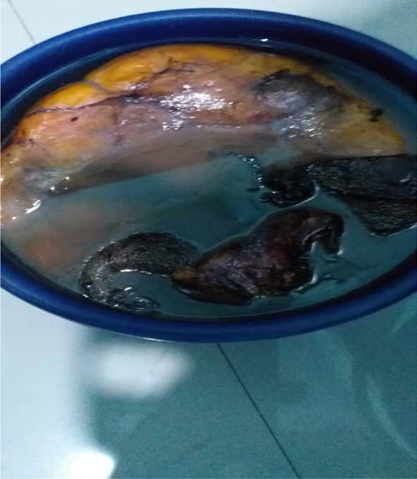
N. Eleweke

                             tiple lipomas [5].
                                Lipomas could be superficial when they can be encapsulated or non encapsu-
                             lated, or deep [6]. Superficial lipomas are more common than deep lipomas [6].
                             Deep lipomas are generally larger than superficial lipomas and could be inter-
                             muscular, intramuscular, interosseous or visceral, displacing adjacent structures
                             [7].
                                Lipomas are described as giant when they weigh more than 1 kg or measure
                             more than 10cm in any dimension [8]. Giant Lipomas can get infected, have ad-
                             verse cosmetic effects, limit movement, cause pain, lymphoedema, pressure
                             symptoms and may undergo malignant transformation [9].
                                Giant lipomas of various parts of the body have been reported. Emegoakor et
                             al. in Awka Nigeria, reported giant lipomas of the gluteal region and lower limb
                             [10]. Nakamura Y et al. [11], Chatterton BD et al. [12], Danzi M et al. [13] have
                             also reported Giant Lipomas of various anatomical regions. Brandler [4] in 1894
                             reported a lipoma of the left scapular region in a 26-year-old man which
                             weighed about 22.7 kg. This is the largest reported lipoma in English literature.
                                I present giant lipoma of the scalp in a 23-year-old female Nigerian in Aba,
                             South East Nigeria. From available literature, this is the first case of such a li-
                             poma to be reported from this area.

                             2. Case Report
                             A 23 years old female presented to us with progressively increasing swelling on
                             the head of about 6 years duration. The swelling has remained painless with no
                             constitutional disturbance. She however cannot expose her head in public be-
                             cause of the embarrassing size and shape of her head.
                                There was no history of trauma and no similar swelling elsewhere in the body.
                             There was no family member with a similar swelling.
                                The past medical and surgical histories were unremarkable.
                                Physical examination showed a healthy-looking young girl. The vital signs
                             were normal. Her body mass index was 19.4 kg/m2. Significant findings were on
                             the head where an elliptical mass measuring 15 × 12 × 10 cm was located. It ex-
                             tended from the frontal region to the occiput and laterally to the parietal regions
                             (Figure 1).
                                The mass was freely mobile in all directions, firm and non-tender. It was
                             non-pulsatile and non-compressible. The overlying skin was normal with nor-
                             mal hair growth over the swelling (Figure 2). There was no palpable cervical
                             lymphadenopathy.
                                We made a diagnosis of giant lipoma of the scalp. Blood work including lipid
                             profile was normal. Plain x-ray of the skull revealed a well-circumscribed soft
                             tissue mass with no attachment to bone and no bony defect (Figure 3).
                               The patient was worked up for excision biopsy. In theatre under general an-
                             aesthesia, with patient in prone position, an elliptical skin incision was made
                             over the swelling. An encapsulated fatty mass under the subcutaneous layer was
                             shelled out (Figure 4). The wound was closed over an active drain.

DOI: 10.4236/oalib.1107898                         2                                  Open Access Library Journal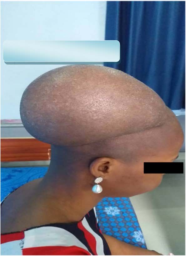
N. Eleweke

                                 Figure 1. The scalp swelling at pres-
                                 entation.

                                 Figure 2. Normal hair growth over
                                 the scalp swelling.

                                 Figure 3. Plain skull x-ray showing
                                 soft tissue swelling of the scalp with
                                 no bone involvement.

DOI: 10.4236/oalib.1107898   3                                      Open Access Library Journal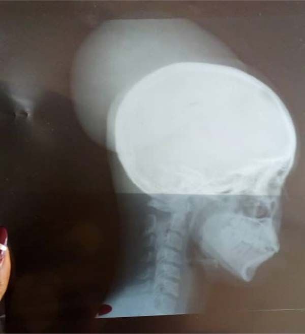
N. Eleweke

                                                       Figure 4. The specimen excised from
                                                       the scalp of the patient with giant li-
                                                       poma of the scalp.

                               The mass which weighed 3.7 kg was sent for histology. The result of histology
                             showed the mass to be fibrolipoma. The post-operative period was unremarkable
                             and she was discharged home after three days. Eighteen months after the sur-
                             gery, she has remained in good clinical condition and is happy with the treat-
                             ment.

                             3. Discussion
                             Lipomas are slow-growing benign tumours of the fatty tissue, with a few attain-
                             ing gigantic sizes [1] [4]. Although the exact cause of lipoma is not known, it has
                             been associated with genetic abnormalities, familial tendencies [2] obesity, hy-
                             percholesterolaemia, and trauma [1] [11].
                                The reasons for lipomas attaining gigantic sizes have not been ascertained but
                             theories abound including the role of trauma. Trauma is said to induce release of
                             cytokines which cause differentiation and maturation of pre-adipocytes [8] [9].
                             Trauma is also postulated to lead to separation of fibrous septa and connections
                             between the skin and deep fascia making the fat cells proliferate rapidly [9].
                             Trauma may also lead to herniation of the lipoma through fascia planes causing
                             pseudo lipomas [9]. Familial and genetic abnormalities and hypercholesterolae-
                             mia have also been postulated as possible aetiological factors of giant lipoma [1].
                             In the index patient, there were none of the above predisposing conditions to li-
                             poma.
                                Lipomas rarely undergo malignant transformation into liposarcomas—the
                             most common soft tissue malignancy in long-standing lipomas [2]. Malignant
                             transformations occur more when there is delayed diagnosis as in retroperito-
                             neal lipomas. [12]. When lipoma increases in size, becomes painful, deep-seated,
                             intramuscular in position and irregular in shape, the likelihood of malignant
                             transformation is high [2]. The malignant nature or otherwise of the lipoma can
                             be ascertained by ultrasonography, CT Scan, MRI, and needle biopsy [14].
                                Open surgical excision as in the index case is the treatment of choice. The en-

DOI: 10.4236/oalib.1107898                         4                                       Open Access Library Journal
N. Eleweke
capsulation of lipomas makes the surgery relatively easy. There is a need for
careful dissection and control of bleeding to prevent damage to surrounding
structures and formation of haematoma and seroma postoperatively.
The excised specimen should be subjected to histopathological examination to
rule out malignancy [15]. In this index case, the result came out as fibrolipoma.
4. Conclusion
We have presented the first case of giant lipoma of the scalp seen and success-
fully managed in Aba, South East Nigeria. From literature search, no such giant
scalp lipoma has been seen and managed in Aba, South East Nigeria.
Conflicts of Interest
The author declares no conflicts of interest.
References
[1] Singh, M., Saxena, A., Kumar, L., Karande, L.K. and Kolhe, Y. (2014) Giant Lipoma
of Posterior Cervical Region. Case Reports in Surgery, 2014, Article ID: 289383.
https://doi.org/10.1155/2014/289383
[2] Rydholm, A. and Berg, N.O. (1983) Site, Size and Clinical Incidence of Lipoma.
Factors in the Differential Diagnosis of Lipoma and Sarcoma. Acta Orthopaedica
Scandinavica, 54, 929-934. https://doi.org/10.3109/17453678308992936
[3] Allen, B., Radar, C. and Babigian, A. (2007) Giant Lipoma of the Upper Extremity.
The Canadian Journal of Plastic Surgery, 15, 141-144.
https://doi.org/10.1177/229255030701500308
[4] Brandler, T.I. (1894) Large Fibrolipoma. British Medical Journal, 1, 574.
[5] Nigri, G., Dente, M., Valabrega, S., Beccaria, G., Aurelio, P.D., Angelo, F., et al.
(2008) Giant Inframuscular Lipoma Disclosed 14 Years after a Blunt Trauma: A
Case Report. Journal of Medical Case Reports, 2, Article No. 318.
https://doi.org/10.1186/1752-1947-2-318
[6] Di, B., Enedetto, G., Aquinati, A., Astolffi, M. and Bertani, A. (2004) Giant Com-
pressing Lipoma of the Thigh. Plastic and Reconstructive Surgery, 114, 1983-1985.
https://doi.org/10.1097/01.PRS.0000143929.24240.9A
[7] Roberts, C.C., Liu, P.T. and Colby, T.V. (2003) Encapsulated versus Non-Encapsulated
Superficial Fatty Masses; a Proposed MR Imaging Classification. American Journal
of Roentgenology, 180, 1419-1422. https://doi.org/10.2214/ajr.180.5.1801419
[8] Sanchez, M.R., Golomb, F.M., Moy, J.A. and Potozkin, J.R. (1993) Giant Lipoma:
Case Report and Review of Literature. Journal of the American Academy of Der-
matology, 28, 266-268. https://doi.org/10.1016/S0190-9622(08)81151-6
[9] Kshirsagar, A.Y., Nangare, N.R., Gupta, V., Vekeriya, M.A., Patankar, R., Mahna,
A., et al. (2014) Multiple Giant Intra Abdominal Lipomas: A Rare Presentation. In-
ternational Journal of Surgery Case Reports, 5, 399-402.
https://doi.org/10.1016/j.ijscr.2014.04.002
[10] Emegoakor, C.D., Echezona, C.N., Onwukamuche, M.E. and Nzeakor, H.O. (2017)
Giant Lipomas. A Report of Two Cases. Nigerian Journal of General Practice, 15,
46-49. https://doi.org/10.4103/NJGP.NJGP_17_16
[11] Nakamura, Y., Teramato, Y., Sato, S., Yamada, K., Fujisada, Y., Fujumoto, M., et al.
DOI: 10.4236/oalib.1107898 5 Open Access Library JournalN. Eleweke
(2014) Axillary Giant Lipoma: A Report of 2 Cases and Published Review. The
Journal of Dermatology, 41, 841-844. https://doi.org/10.1111/1346-8138.12598
[12] Chatterton, B.D., et al. (2013) An Exceptionally Large Giant Lipoma of the Hand.
BMJ Case Reports, 2013, bcr2013200206. https://doi.org/10.1136/bcr-2013-200206
[13] Danzi, M., Grimaldi, L., Reggio, S. and Danzi, R. (2010) Giant Atypical Lipoma of
the Thigh. Case Report and Literature Review. Il Giornale di Chirurgia, 31, 108-111.
[14] Yakubu, A.A., Edino, S.T., Mohammed, A.Z., Sheshe, A.A. and Alhassan, S.U.
(2008) Giant and Complicated Subcutaneous Lipoma of the Neck. West African
Journal of Medicine, 27, 44-46.
[15] Guler, O. (2015) Giant Lipoma of the Back Affecting Quality of Life. Annals of
Medicine and Surgery, 4, 279-282. https://doi.org/10.1016/j.amsu.2015.08.001
DOI: 10.4236/oalib.1107898 6 Open Access Library JournalYou can also read