GOAT DISEASES AND FARM HERD-HEALTH SAFETY - Compiled by Charlotte Clifford-Rathert, Veterinarian, Small Ruminant Extension Specialist Cooperative ...
←
→
Page content transcription
If your browser does not render page correctly, please read the page content below
GOAT DISEASES
                       AND
             FARM HERD-HEALTH SAFETY

Compiled by Charlotte Clifford-Rathert,
            Veterinarian, Small Ruminant Extension Specialist
            Cooperative Extension and Research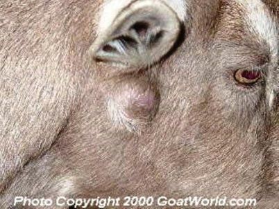
This publication is a summary of descriptions of infectious diseases goat
producers need to be aware of as possible threats to their herds.

An infectious disease is a disease which is transmitted from animal to animal,
animal to human or from equipment/clothing to animal. These diseases can be
very resilient. Many of these disease organisms (pathogens) are considered
zoonotic, which means they pose a threat to man.

It is possible that 75% of the emerging and re-emerging pathogens are zoonotic.
Therefore, the effect of these diseases on the public’s health must always be
considered. In many instances sick animals can serve as a cause of infection for
humans. At times both animals and humans may become ill from a common
source. People becoming sick can often be prevented or lessened through
effective intervention programs targeting the transfer of disease from animal to
human.

In addition to the spread of disease by natural means, the introduction of
infectious disease by mechanical means also must be considered. Therefore, it
is important to be aware of what you can do to protect yourself, your farm and
your livestock from contamination. This practice is called biosecurity or bio-risk
management. This publication covers the necessary steps recommended by the
Missouri Veterinary Medical Association and the Missouri Department of Health
and Senior Services, Division of Community and Public Health.

                                        2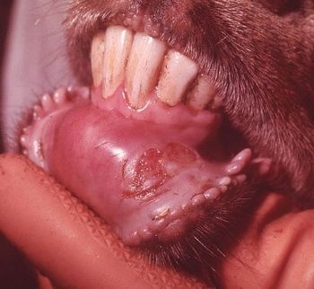
Table of Contents:

What is Biosecurity?                           Page 4

Reportable Infectious Diseases
    Scrapie                                    Page 5
    Brucellosis                                Page 6
    Tuberculosis                               Page 7
    Anthrax                                    Page 8
    Foot and Mouth Disease                     Page 9
    Vesicular Stomatitis                       Page 10

Self-Limiting Infectious Diseases
     Sore Mouth                                Page 11
     Pinkeye                                   Page 12
     Ringworm                                  Page 12
     Internal Parasites                        Page 13

Miscellaneous Diseases
    Johne's Disease                            Page 14
    Caprine Arthritis-Encephalitis Virus       Page 15
    Caseous Lymphadenitis                      Page 16
    Foot Rot, Foot Scald                       Page 17
    Q Fever                                    Page 18
    Bluetongue                                 Page 19

References                                     Page 21

                                           3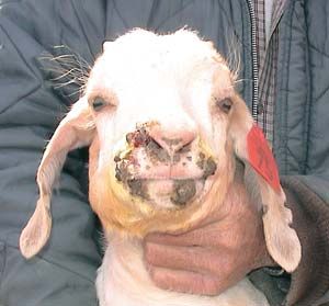
What is Biosecurity?

   Biosecurity is protecting the health of livestock by preventing the spread of
   disease on the farm and from farm-to-farm.

      ¾ Any disease that can endanger the health of your livestock is
        considered a threat to the economics of the farm and safety of the herd

      ¾ Infectious diseases are spread by numerous agents. Anything that is
        dirty is considered contaminated (i.e.clothing, shoes, needles,
        equipment, insects, wildlife, feed, water, unwashed hands) and could
        transmit diseases

How to manage and protect your herd:

      •   Keep all new animals away from other animals in the herd for at least
          21-30 days

      •   Vaccinate your farm animals against common diseases in the area

      •   Vaccinate new animals to match the program already on your farm

      •   Do not reuse dirty, contaminated needles; ALWAYS use new disposable
          needles.

      •   Clean all equipment in between uses

      •   Purchase new animals from well-known healthy herds

      •   Purchase livestock feed from trustworthy sources

      •   When anyone, including the veterinarian, visits your farm, ask that they
          have clean clothing, clean shoes and clean equipment,

      •   Supply a tub of disinfectant (freshen daily) and a brush for scrubbing
          footwear or provide plastic over-boots for visitors.

      •   Identify each animal with identification tags

      •   Keep accurate records of every animal bought and sold, medications
          and vaccinations given, breeding dates, birthing dates, death dates
          and means of disposal of dead animals

      •   Ask for help from your veterinarian or extension agent when needed

                                         4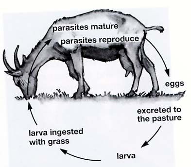
•   Keep all fences in good condition to keep your animals in and other
           farm animals and wildlife out

       •   Always tend to sick animals after working on healthy animals

       •   Report any unusual signs of animal sickness or death to your
           veterinarian

Individual Animal identification:

Good management and proper identification of animals can make managing sick
animals and/or disease outbreaks much easier. All animals on the farm should
have an individual ear tag or neck tag for identification if it becomes sick. This
will help identify and treat the animal(s) appropriately.

Accurate Record Keeping

Good record keeping is vital for successful treatment and control of disease and
the health of your animals. Records of animal identification numbers, offspring,
breeding, vaccinations, deworming schedule, documented illness and all
medications given are important when disease outbreaks occur. Information of
where animals were purchased and sold is also often helpful in investigating
disease outbreaks.

What is a Reportable Infectious Disease

Reportable Infectious Diseases are those which are highly infectious and
controlled by the USDA (United States Department of Agriculture). You are
required to report these diseases to the State Veterinarian and the USDA
Veterinarian. These diseases are controlled due to the economic threat they
pose to the agricultural industry in the United States.

Scrapie:

Scrapie is the most common reportable disease in goats and sheep in the United
States today. Scrapie is a difficult disease to diagnose and it is always fatal. It
can take up to six years or more to show clinical signs. Scrapie is in the same
category as BSE(bovine spongiform encephalopathy), or “mad cow disease”, and
chronic wasting disease (CWD) of deer and elk. There is no evidence that
Scrapie or CWD can spread to humans through consumption of meat or dairy
products nor through the handling of animals; however the industry can still be
subject to the negative public perceptions affecting the cattle industry. Scrapie is
a disease of both sheep and goats; however it is rare in goats.

                                         5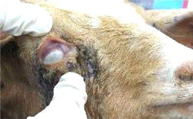
Clinical Signs: Scrapie is spread through direct contact between sheep and
goats. The infective cause is a prion, which is an organism smaller than bacteria
or a virus. It is transferred through contact with the placentas or fetal fluids of
infected sheep. The prion first invades the lymph nodes and then the nervous
system.

Clinical signs have not been seen in goats less than 2 years of age and usually
progress slowly over 1-6 months. Scrapie suspected animals will show
characteristic changes in gait, tremors of the head and neck, behavioral changes,
lip smacking, loss of coordination, increased sensitivity to noise, rubbing against
fences or feed bunks, skin/ wool biting, progressive weight loss with a normal
appetite, inability to stand and death. Genetic testing can be used in sheep to
identify a scrapie susceptibility gene; however such a gene has not yet been
identified in goats.

A video of clinical signs and information on the eradication program is available
at http://www.aphis.usda.gov/animal_health/animal_diseases/scrapie/

Brucellosis

This disease results from infection by various species of Brucella. Six species
occur in humans and animals. B.melitensis is the most important species in
sheep and goats, and B. ovis causes infertility in rams.

Brucellosis is found worldwide but it is well controlled in most developed
countries. The disease is still common in Africa, the Middle East, Central and
Southeast Asia, South America and some Mediterranean countries. B.
melitensis is rare in the United States, but B. ovis is seen in the United States,
Australia, New Zealand and many other sheep-raising regions.

Brucellosis is spread among animals by contact with the placenta, fetus, fetal
fluids, and vaginal discharges from infected animals. Animals are infectious after
either an abortion or full term birth. The organism is found in blood, urine, milk,
and semen; it can be shed in milk and semen (which can be prolonged or
lifelong). Brucella can be spread on equipment, clothing, etc. In conditions of
high humidity, low temperatures and no sunlight, these organisms can live for
several months in water, aborted fetuses, manure, wool, hay, equipment and
clothes. The Brucella organism is killed by several hours of exposure to direct
sunlight.

Clinical signs: Brucella abortus is found in cattle; and occasionally sheep, goats,
and dogs. B. melitensis is the most important cause of brucellosis in sheep and
goats. It can cause abortion, retained placenta, and swelling of the testicles.
Abortions usually occur in late pregnancy in sheep, and during the fourth month

                                          6
of pregnancy in goats. In goats, mastitis and lameness may be seen. Arthritis is
rare in sheep.

Communicability: Brucellosis is contagious to humans. Bacteria are present
in milk, placenta, fetal fluids, fetus, vaginal discharges, semen, and urine.
Ruminants and other animals can shed bacteria long-term or lifelong.

Diagnosis: By blood tests and culture of tissues listed above.

Treatment: There is no practical treatment that is successful. Long-term
antibiotic treatment can eliminate B. ovis infections in valuable rams but the
fertility may remain poor.

This is a reportable disease.

Tuberculosis

An infectious disease caused by the bacteria Mycobacterium. Before extreme
control programs and testing, this was a major disease of humans and domestic
animals. Tuberculosis can be a chronic, debilitating disease, and can also
become a very progressive disease. It is zoonotic to humans as well as other
animals. Breathing of aerosol droplets from a cough or drinking of contaminated
milk are the primary routes of infection.

Clinical signs: The clinical signs reflect the location and severity of the infection.
Weight loss, lack of energy, loss of appetite, depression, weakness, and
fluctuating fever are common symptoms. The respiratory form of the disease
causes a chronic, intermittent, moist cough that may progress to difficulty
breathing.

Diagnosis: The Intradermal Caudal-fold skin test is the most reliable diagnostic
test for tuberculosis in goats and sheep. This test must be done by a
veterinarian.

Control: Control of this disease in the United States is by test and eradication of
positive animals in a herd.

Prevention: Purchase animals from a tuberculosis-free herd.

This is a reportable disease.

                                          7
Anthrax

This disease results from infection by the bacteria Bacillus anthracis. This
bacteria forms spores and requires oxygen to survive. Anthrax is found
worldwide. In the United States areas of concern in which infection occurs are in
South Dakota, Nebraska, Mississippi, Arkansas, Texas, Louisiana, and California
with smaller areas in other states.

Anthrax is usually spread by the animals eating the Bacillus spores on plants in
pastures. Outbreaks occur in neutral or alkaline (calcium/ limestone rich) soil
and are often associated after heavy rainfall, flood, or drought. Under optimal
levels of moisture, temperature and other favorable conditions; the spores in the
soil can return to the vegetative form and grow to infectious levels. Carnivores
(such as dogs, coyotes, wolves, etc) can become infected after eating
contaminated meat. Scavengers and flies spread anthrax after feeding on
infected carcasses.

The Anthrax bacteria require oxygen in order to develop into spores. The
disease is spread when large numbers of bacteria are exposed to oxygen then
develop into spores and contaminate the soil. Therefore it is not wise to open an
infected carcass for a necropsy. Anthrax spores remain viable for decades in the
soil or on animal products such as dried or processed hides, or wool. Spores
can survive for 2 years in water, 10 years in milk and up to 71 years on silk
threads. Vegetative organisms are thought to be destroyed within a few days
during the decomposition of unopened carcasses. If you suspect an animal
dying of anthrax, contact your veterinarian immediately.

Species affected: Goats, sheep, cattle and horses are susceptible. Pigs can
become infected from eating contaminated meat. Rats and chickens are
relatively resistant.

Incubation period: Typically 1-20 days. Most infections are noticeable after 3-7
days. Incubation is only 1-2 weeks in pigs.

Clinical Signs: In ruminants (cattle, goats, and sheep) sudden death is the only
sign. Staggering, trembling, and difficulty breathing may be seen in some
animals, followed by rapid collapse, terminal convulsions, and death. Bloody
discharges from natural openings (nose, mouth, ears, penis, and rectum) in the
body are sometimes observed. Humans can become infected through the skin
leading to a dark scab. Infections obtained by inhalation or ingestion can be
highly fatal if left untreated.

Diagnosis: Typically by clinical signs. Official diagnosis is made by laboratory
identification of the organisms in samples of body fluids, skin lesions, lymph node
or spleen.

                                        8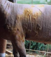
This is a highly communicable disease. However, this is NOT a highly
transmissible disease between animals. Treatment is possible with antibiotics if
started early. Vaccines are available for livestock.

This is a reportable disease.

Foot and Mouth Disease (FMD)

This is a highly contagious disease that can rapidly
spread through a region if control and eradication
practices are not put into place as soon as the disease is
identified. It primarily affects cloven-hoofed domestic and
wild animals including cattle, pigs, sheep, and goats.

The last outbreak in the US was in 1929. It does not           Figure 1 Goat, lower lip, ulcer of Foot and
                                                               Mouth Disease.
currently exist in the US. This disease is spread primarily http://www.cfsph.iastate.edu/DiseaseInfo
by respiratory spray and direct or indirect contact with
infected animals. For this disease to spread in the respiratory spray it takes the
form of an aerosol droplet (from a cough or sneeze) and requires proper
temperature and humidity. Feeding of infected animal products such as meat,
milk, bones, glands and cheese can also spread the disease. Contact with
contaminated objects such as boots, hands or clothing can be a primary source
of infection. Other sources of infection are artificial insemination, and
contaminated medications and hormone preparations.

Sheep and goats are considered maintenance hosts. They can have very mild
signs; therefore, delaying diagnosis. This can allow time for environmental
contamination by aerosol or direct contact. In pigs, FMD virus can spread rapidly
due to thousands of times higher virus particle concentration in air droplets as
compared with other species therefore pigs are considered “amplifying hosts”.
Cattle are considered “indicators” of this disease because they generally are the
first species to show signs of infection. Their lesions are more severe and
progress more rapidly.

Animals that have had contact with clinically infected animals generally show
signs of disease in 3-5 days. The virus is shed most often and the disease is
spread when the blisters rupture.

Clinical signs: Foot and mouth disease is characterized by
fever and blisters which progress to ulcers in the mouth,
nostrils, muzzle, feet, or teats. Symptoms include
depression, poor appetite, excessive salivation, clear nasal
discharge, decreased milk production, lameness, and                            Figure 2 Teat, ruptured blister. Foot and Mouth
                                                                               Disease.
reluctance to move. Abortion may occur in pregnant                             http://www.cfsph.iastate.edu/DiseaseInfo

                                                   9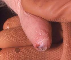
animals due to high fever. Young animals die due to severe heart damage.

Sheep and goats show very mild, if any, signs of fever, oral lesions, and
lameness. Animals generally recover in about 2 weeks with very low death rate
in adult animals.

Differential diagnosis: In sheep and goats, other diseases that may look like
FMD include Vesicular stomatitis, Bluetongue, Contagious Ecthyma (Orf or Sore
Mouth), and Lip and Leg Ulceration.

This is a reportable disease. Containing a suspected outbreak of foot and mouth
disease is vitally important. State and federal veterinarians should be
immediately informed of any suspected vesicular disease.

Vesicular Stomatitis

Another important contagious vesicular disease found in North and South
America. This disease has almost identical clinical signs to Foot and Mouth
Disease in cattle and pigs. The signs of Vesicular Stomatitis are also very
similar to Swine Vesicular Stomatitis and Vesicular Exanthema of swine. It
affects South American Camelids (Llamas, Alpacas, and Vicuñas), but sheep
and goats are resistant and rarely show clinical signs.

Clinical signs: Horses are affected most severely with oral and coronary band
blisters that progress to signs of drooling, chomping, rubbing the mouth, and
lameness. Cattle and pigs are very similar to Foot and Mouth Disease.
Vesicular Stomatitis is most likely to have lesions isolated to one part of the body
such as the mouth or feet. Recovery is usually within two weeks, and can be
longer in the presence of a secondary infection.
This is also a reportable disease.

*** Before collecting or sending any samples from a vesicular disease
suspect the proper authorities should be contacted (your veterinarian,
state veterinarian, or federal USDA veterinarian). Samples should only be
sent under secure conditions and to authorized laboratories to prevent
spread of the disease. Since vesicular diseases cannot be distinguished
clinically, and some are zoonotic, samples should be collected and handled
with all appropriate precautions and only by a veterinarian.

                                         10
Self Limiting Infectious Diseases:
The following diseases are highly infective within a herd and require intense
management for proper control; however these diseases are not reportable.
These diseases can be very costly to producers and also have a zoonotic
(contagious to man) potential.
Sore Mouth (Contagious Ecthyma, Orf)
This disease affects sheep and goats and is caused by a Parapoxvirus. Lesions
most commonly occur on the mouth and face, but can also occur on the feet,
teats, and genitalia. The poxvirus is present worldwide and can remain infective
in scabs in the environment for months to years.
The virus is spread by direct or indirect contact from environmental
contaminants. The virus enters through abrasions
(scrape) or wounds of the mouth, teat, feet, or genitalia. It
then localizes in the tissues and is shed in the scab.
Animals that are kept in the same area are at the greatest
risk.
The infection is self-limiting, with most animals developing
protective immunity, however reinfection is possible.
Figure 3 Sore Mouth (Contaious Ecthyma,
Orf) sores on lips and muzzle of young
Clinical signs: Early signs are small bumps or blisters on goat.
http://www2.luresext.edu/goats/index.htm
affected skin, usually around the mouth. Thick brown to
black crusts form and are most evident. Lesions typically resolve in 14-21 days.
Nursing lambs or kids are most likely to spread the disease to udders of
susceptible ewes or does. Oral lesions may become so severe as to cause the
animal to stop eating.
Diagnosis: Observation of clinical signs and skin biopsy.
Treatment: Treatment of individually affected animals is not provided unless
lesions are severe. Consult a veterinarian.
Prevention: Put into practice control measures immediately. Affected animals
should be separated from all the other animals. Prevent the scabs from falling off
into the environment. Vaccines are available, but not recommended in disease
free herds because the vaccine contains live virus and poses a contamination
threat.
Contagious ecthyma is highly zoonotic and may produce lesions on the hands
or fingers of the person(s) handling infected animals. Therefore it is extremely
11important to practice good hygiene. Disposable gloves should be worn when
handling these animals, and then properly disposed off of in a trash can. Hands
should be cleaned with an antimicrobial cleanser after handling.
Pinkeye
Pinkeye also known as Viral Keratoconjunctivitis
has been reported in goats. It can be a sequel
to Bluetongue Virus infection.
Pinkeye in goats is a mycoplasmal disease. A
surface infection with the mycoplasma organism
in the eye can cause pinkeye in goats and
sheep. The organism can also enter the blood Figure 4 Cloudy cornea of the eye in a goat.
Photograph provided by Terry Hutchins, University of
stream and cause septicemia, abortion, Kentucky Goat Program
These materials are the property of Goat Dairy Library,
respiratory problems and arthritis in multiple www.goatdairylibrary.org. (July 9, 2008)
joints. Flare-ups occur in times of stress,
overcrowding, kidding or lambing. Pinkeye can be spread by direct or indirect
contact with infected animals or body fluids from infected animals. Newborn
goats/lambs can spread the organisms from the mother’s mouth to her udder and
in turn become infected by ingesting contaminated milk.
Clinical signs: Cloudiness of the cornea. A mycoplasma infection should be
suspected in goats and sheep with severe pneumonia.
Diagnosis: Mycoplasma strains can be identified by bacterial culture or staining
of discharges from the eye. Examiners must be careful in interpreting results of
positive cultures as nonpathogenic mycoplasma is common (D.G. Pugh, Sheep
& Goat Medicine). Consult a veterinarian for diagnosis and treatment.
Prevention: There is not a vaccine available in the US. Provide good fly control,
preventing stress, overcrowding, and separating infected animals from healthy
ones will help prevent the spread of disease.
Ringworm
This disease is also known as dermatophytosis, lumpy wool, or club lamb fungus.
It is transmitted by direct or indirect contact with contaminated equipment or
environment. It is a fungus, caused by one of four species. The fungi invade the
skin and hair fibers. Breaks in the hair and hair loss occur due to the breakdown
12of the hair shaft. Close shearing and washing practices used
at shows leave show animals at risk.
Clinical signs: Crusty circular raised lesions on the skin’s
Figure 5 Ringworm lesions
surface. Animals may be very itchy. www.ext.vt.edu
Diagnosis: Positive culture of skin and hair samples, or a wet mount examination
by a veterinarian.
Treatment: Treatment helps limit the spread of the disease to other animals and
humans. Topical iodine compounds (2%-5%), chlorhexidine (2%), lime sulfur
(2%-5%), and topical antifungal medications are useful for local treatment of
lesions. Some severe cases require administration of medications. This
requires the help of a veterinarian.
People treating animals should always wear gloves and
protective clothing to prevent from getting this disease.
This is considered a zoonotic disease.
Prevention: Proper cleaning and disinfection of clippers,
blankets, and other equipment with antiseptic solutions, along Figure 6 Ringworm lesion on
an arm of a handler
with good sanitation practices of animal handlers will help
control the spread of this disease.
Internal Parasites:
Sheep and goats are susceptible to the same parasites and tend to share a
resistance to those that infect cattle and horses. The major gastrointestinal
worms that parasitize goats and sheep on pasture are: Haemonchus, Ostertagia,
Trichostrongylus, Cooperia species. Therefore the acronym HOTC is a
reference to the main parasites of concern. The severity of infestation of specific
parasites varies from farm to farm. Factors affecting infestation of a herd
depends on climate, age of the animal, overcrowding and pasture management.
In the United States, Haemonchus is the most common significant parasite
causing clinical disease and parasite resistance in herds resulting in economic
loss.
Life cycles of most of these parasites are
very similar, and become significant when
the larvae hatch under favorable conditions,
are eaten by the host animal while grazing
grasses in pasture, completes its life cycle
within the host animal, and produces more
eggs. These nematodes affect the
abomasums and small intestine of the
young weaned animals, and adults.
Figure 7 General Parasite Life Cycle
13If infested with significant numbers, the animals will show poor growth,
decreased weight gain, decreased milk production, weight loss, diarrhea,
anemia, lower jaw swelling (bottle jaw), midline (under-belly) swelling, and death.
Diagnosis by fecal examination and counting eggs per gram (EPG) of feces is
the best way to determine parasite load and can be done by a veterinarian.
A deworming program should be carefully designed to reduce the EPG by 90%
and use of the effective product for at least 1 year, then changing products to
avoid building resistance. A combination of strategic deworming and tactical
deworming seems to provide the best results. A veterinarian or extension agent
can help you design a program best suited for your farm.
Tapeworm (Cestode) infestation of goats in North America is most commonly
Monesia species. Tapeworms rarely cause disease in kids less than 6 months of
age but are seen in combination with other parasite infestations.
Coccidia (Protozoan) infestation of goats commonly affects young goats under
the stress of being weaned and sold. This parasite can cause severe diarrhea,
pneumonia-like symptoms, depression, weight loss, anemia, loss of appetite, and
even death. It can be easily controlled with coccidiostats added to the feed or
water. Consult a veterinarian or livestock extension agent for help.
Miscellaneous Diseases of Importance,
Not reportable in the state of Missouri.
Johne’s disease; (Paratuberculosis)
A chronic disease, that causes a wasting body condition, without diarrhea, in
goats. It is caused by the bacteria Mycobacterium avium paratuberculosis and is
spread by the fecal-oral route. Young animals are more susceptible to the
disease than adults. It is also transmitted through milk and placenta, therefore
predisposing the young of infected animals to the disease, especially those
showing symptoms. After an animal is exposed it will either clear the organism
or develop a chronic persistent infection, depending on its immune status.
The most consistent clinical sign is chronic weight loss despite a good appetite in
sheep and goats that is not caused by parasite infestation and eventually death.
Clinical signs of this disease are so subtle that it may take months or years to
realize there is a problem. Meanwhile an infected animal can be shedding the
14organism in its feces, contaminating the environment and other animals in the
herd.
Diagnosis: Culture of the organism from feces is the official test; however this
process takes up to 8 weeks or longer. Newer diagnostic methods available are
blood testing by the enzyme-linked immunosorbent assay (ELISA) and agar-gel
immunodiffusion (AGID). Both these tests are rapid and results are available in
48 hours. These tests can be useful as screening tools for whole herd
management, ELISA as a whole herd test and AGID as an individual test.
Treatment: There is no effective treatment so prevention and control are very
important. Preventing the introduction of Johne’s into a herd can be very difficult.
The best prevention is by maintaining a “closed herd” but may not be practical.
Blood testing of animals is helpful when purchasing non-infected animals, but is
not 100%. It is recommended to ask about the herd history before purchasing
and culture newly purchased animals every 6 months due to the subclinical
nature of the disease.
Control: Control is a combination of management, education, and screening
periodically. Control of the fecal-oral route of infection is a vital part of any herd
management plan. Consult a veterinarian or livestock extension agent to help
develop effective management plans, and confirm / cull any suspect animals. A
clean environment is important especially the kidding barn and pastures. Using
above ground feeders and waterers; cleaning the doe’s udder before nursing
young are a just a few ways that will help control the fecal-oral route of infection.
Caprine Arthritis-Encephalitis Virus (CAEV)
CAE is a chronic multi-systemic disease in goats. Infection is
widespread and arthritis in more than one joint is the most
common clinical signs. Infection occurs by ingestion of fluids
that contain infected cells from an infected animal to an
uninfected animal. The most common means of transmission
is the ingestion of colostrum by kids nursing infected does.
CAE can also be spread by breeding, contaminated dehorning
equipment and needles, and at parturition, has all been
documented.
Figure 8 Swollen
arthritic knees
The target tissues of CAE virus are the joints, mammary glands,
lungs, and brain. The disease results from inflammation induced by the reaction
of the immune system to the virus. Goats can develop a blood titer in 2-8 weeks
but may not show clinical signs for years.
Clinical signs: A progressive arthritis in goats over 6 months of age, usually
noted in the front pastern joints, with chronic progression over the years.
15Diagnosis: Routine diagnosis is based on specific serological (blood) testing
called agar gel immunodiffusion test (AGID) or polymerase chain reaction assay
(PCR).
There is no treatment; affected animals are a source of infection to others. It is
recommended to cull infected and positive animals in order to eradicate the
disease on the farm, otherwise rigorous management is required.
Caseous Lymphadenitis (CLA)
This can be a devastating disease caused by the bacteria Corynebacterium
pseudotuberculosis. It is more common in sheep than in goats. It causes
abscesses of the skin and subcutaneous lymph nodes that will break open to the
skin’s surface and contaminate the environment. This disease may affect the
animal internally, most commonly the respiratory system causing long-term
respiratory problems, it can also spread to the abdominal lymph nodes (weight
loss), central nervous system (neurological signs), and mammary glands
(mastitis).
This organism can survive for prolonged periods of time in dark, damp areas, soil
and manure. Most common means of infection is by injury with contaminated
shears, tail docking equipment, dip tanks, needles, and puncture wounds.
Abscesses form, then break and drain into the environment (feed bunk, or water
buckets, or on the ground), predisposing the next animal to come along and
become infected through a wound.
Clinical signs: Superficial lymph node swellings
with draining tracts; an “onion ring” appearance to
the abscess if surgically removed; a pale greenish-
cream colored pasty discharge drains from the
abscess when ruptured.
Diagnosis: Based on serological (blood) testing
and culture of organism from abscesses.
Figure 9 Abscess in a goat. Courtesy of Gary
Pfalzbot, http://www.GoatWorld.com
Treatment: The abscesses should not be opened
within the vicinity of the other animals. Isolate affected animals for treatment. If
possible, it is recommended to have the abscesses surgically removed to reduce
contamination of the environment. If the abscess(es) has/have already ruptured,
the animal should be isolated, the abscess flushed with an antiseptic solution
(3% iodine or 2% chlorhexidine) and packed with antiseptic saturated gauze (it is
best to seek the help of your veterinarian).
16Control: Affected animals should be identified and culled for best control and
prevention. Housing should be kept free of objects that can cause injury; all
equipment should be properly cleaned and disinfected after use. When handling
the contents of the abscess it is highly recommended to wear disposable exam
gloves and protective clothing to prevent spreading the organism. Abscess
contents need to be properly disposed.
Vaccination is controversial but has shown to be beneficial in reducing the
incidence of abscesses within a flock or herd already infected, but will not result
in complete disease eradication.
Foot Rot, Foot Scald:
This is a crippling infection commonly caused by bacteria that live in the soil and
can be easily carried onto a farm on the soles of shoes, or the feet of infected
animals. Two types of bacteria are commonly associated with this condition,
Bacteroides nodosus, and Fusibacterium necophorum. Both of these bacteria
thrive in moist soil conditions and are very difficult to control or eliminate once the
soil is contaminated.
Clinical Signs: Foot scald usually affects one foot and can lead to Foot Rot. The
common lesion seen is a moist raw infection of the skin between the toes which
becomes painful.
Foot Rot is a more aggressive progression of foot
scald and can occur in one or more feet causing
sever lameness. It occurs when both bacteria
together cause a dual infection of the tissues of the
foot. The foot will become very pink to almost red;
the skin between the toes will be slimy and smell foul. Figure 10 Foot Rot Infection
This infection can become severe enough to penetrate
the hoof wall and sole of the foot resulting in the hoof wall loosening and
detaching from the foot if not treated early.
Treatment: Both conditions are treatable but may take time and can be very
expensive and labor intensive. Correct hoof trimming, Koppertox®, Medicated
foot baths (10% Zinc sulfate or Copper Sulfate). Consult with a veterinarian or
extension specialist if more aggressive treatment is needed.
Control = Prevention: This is key.
• All new animals should be kept separate (quarantined) and hooves
trimmed and inspected before introduction into the farm herd after 20-30
days
• All show animals or any animals that have left the farm and possible
exposed to contaminated soil should be quarantined
17• Avoid buying animals from the sale barn; most animals that have failed
treatment are taken to the sale barn
• Provide good drainage to all areas in pastures and paddocks where water
tends to pool (around waterers, low lying areas) this is where the bacteria
tend to collect
• Keep barns dry and clean
• Practice good hoof care and management. Check the feet each time you
work the herd.
Q-Fever:
Q-fever results from infection by Coxiella burnetti. This is an unusual spore-like
organism that is highly resistant to environmental conditions. Found worldwide
except in New Zealand. It is transmitted to humans and other animals by
aerosol, direct contact with reproductive discharges, or infected milk.
Ticks can spread infection among ruminants and people.
Since the organism is so resistant in the environment it can become airborne and
travel ½ mile or more. It can survive up to 30 days in dried saliva, and 120 days
in dust.
The most common farm animal reservoirs for Q-fever are goats, sheep, and
cattle. Ticks and wild birds can also harbor this organism. It is also transmitted to
humans. Reproductive failure is sometimes the only symptom in animals.
Clinical signs: Abortion in late pregnancy, still births, retained placenta,
endometritis (inflammation of the lining of the uterus), infertility, and small or
weak offspring in ruminants.
Animals may appear asymptomatic (without signs of disease).
Goats will have decreased appetite and may be depressed 1-2
days before an abortion.
Diagnosis: With the aid of your veterinarian or extension agent submit milk,
feces, fetal tissue, placenta, vaginal discharge, blood (serology) for polymerase
chain reaction assay (PCR).
Treatment: Isolate infected animals. Antibiotics may decrease the risk of
abortion and suppress infection but not eliminate infection.
Vaccination is not available in US; it will not eliminate shedding of organism.
This disease is communicable to humans. Use extreme care when
handling these animals.
18 Wear protective clothing, disposable gloves, and a face mask to prevent
inhalation of organism when handling suspect or known infected animals and
fluids.
Dispose of placenta, birth products, fetal membranes, and aborted fetuses at
farms housing sheep and goats by burning or disposing in plastic trash bag.
Use only pasteurized milk and milk products.
Large numbers of organisms are present in placenta, fetal fluids, aborted
fetuses, milk, urine, and feces. Serologically negative animals may shed the
organisms.
Diagnosis in humans is done by serology (blood test), within the second week
of illness. Most cases occur in people exposed to farm animals or their
products.
Q-fever is a self-limiting illness, most cases resolve on their own within 2 days
-- 2 weeks.
Clinical signs in humans include fever, chills, severe headache, fatigue, non-
productive cough.
Bluetongue
A severe viral disease caused by an orbivirus transmitted mainly by gnats of the
genus Culicoides. Transmission sexually and across the placenta can also
occur. Because the vector is a gnat, the spread of this disease occurs primarily
in the late summer and fall. The virus is endemic in many areas and cattle and
wild ruminants (white-tailed deer) act as reservoirs. Goats are commonly
infected with the virus but rarely show any signs of clinical disease, it is a self-
limiting disease in goats.
Clinical signs: Affects sheep of all ages; goats rarely show clinical disease.
Clinical signs range from transient fever and swelling of the face, muzzle, and
ears; large amount of nasal discharge which may cause crusting around the
nose; oral mucus membranes become dark pink and as the disease progresses,
small hemorrhages and ulcers may form on the roof and corners of the mouth.
The tongue may become cyanotic (blue) but not as common as the name
indicates. Laminitis can develop caused by inflammation of the coronary band
and tissues of the foot to the point that some animals may slough their hooves.
Diarrhea and wool break will also occur in infected animals. Bluetongue virus will
cause abortions, stillbirths and weak lambs.
19Diagnosis: By the presence of clinical signs similar to those reported in sheep
have been documented in goats.
Treatment: Minimize animal stress and antibiotic treatment for secondary
infections.
Prevention: Control breeding areas for biting gnats. Keep animals away from
areas where biting gnats are present. Vaccines are available for sheep.
**************************************************
This publication addresses the most commonly diagnosed reportable infectious
diseases of goats in the United States and the state of Missouri. There are other
diseases that affect goats in other parts of the world not covered in this
publication. This publication was designed to provide information to help goat
producers identify certain diseases and protect their herds from infection.
20References:
1. D.G. Pugh: Sheep & Goat Medicine, Philadelphia, 2002,
W.B. Saunders Company
2. Langston University Research and Extension:
http://www2.luresext.edu/goats/index.htm
3. National Institute of Animal Agriculture: www.animalagriculture.org
4. Missouri Veterinary Emergency Preparedness Manual for Veterinarians by
the: Missouri Veterinary Medical Association,
Missouri Department of Agriculture, and
Missouri Department of Health and Senior Services
5. Center for Disease Control: www.cdc.gov/ncidod/dvrd
6. USDA/APHIS/Veterinary Services: http://www.aphis.usda.gov/animal_health
7. Goat World: http://www.goatworld.com/articles
8. Iowa State: http://www.cfsph.iastate.edu/DiseaseInfo
9. Goat Dairy Library: www.goatdairylibrary.org
10. Mike T. Collins, DVM, PhD., DACVM: http://johnes.org/authors/athors2.html
This publication was funded by the MATCH Project,
University of South Carolina, Arnold School of Public Health,
sponsored by the W.F. Kellogg Foundation 2008.
Lincoln University, U.S. Department of Agriculture and Local University Extension Councils Cooperating
Lincoln University Cooperative Extension Service offers its programs to persons
regardless of race, color, national origin, sex or handicap.
An equal opportunity employer.
21You can also read