Gorilla endoscopic sinus surgery: a life-saving collaboration between human and veterinary medicine - Kansas State University College of ...
←
→
Page content transcription
If your browser does not render page correctly, please read the page content below
ORIGINAL ARTICLE

    Gorilla endoscopic sinus surgery: a life-saving collaboration between human
                              and veterinary medicine
                         Greg E. Davis, MD, MPH1 , Fred M. Baik, MD2 , Robert M. Liddell, MD3 , Andrew G. Ayars, MD4 ,
                        Kelley R. Branch, MD, MSc5 , Paul S. Pottinger, MD6 , Allen D. Hillel, MD1 , Kelly Helmick, DVM7 and
                                                                Darin Collins, DVM8

       Background: Chronic rhinosinusitis is a common disease                      medical approach was critical to heal this ailing gorilla. This
       process in humans; however, in the primate population of                    case discusses many of the challenges and offers recom-
       gorillas, it has rarely been described. This case describes                 mendations for physicians who may be involved with similar
       lifesaving sinus surgery on a critically ill gorilla performed              care of animals in the future.
       by a human otolaryngology team in collaboration with the
       gorilla’s veterinary medicine team.                                         Conclusion: The success of the surgical and medical
                                                                                   treatment of this gorilla’s life-threatening sinus infection
       Methods: The 35-year-old western silverback gorilla was                     required many experts, careful planning, and corporate
       treated for 3 months with aggressive medical therapy for                    generosity. The interaction between human and animal
       a worsening sinus infection. When his condition became                      medicine would not have been successful without the
       severe, a computed tomography (CT) scan was performed                       close and trusting collaborations between human and vet-
       showing advanced chronic rhinosinusitis with nasal polyps                   erinary health providers. We encourage human health-
       vs other masses and some bone erosion. As his condition                     care providers to seek volunteer opportunities through
       deteriorated further, a tertiary otolaryngology team per-                   their local zoos by engaging in discussions with their local
       formed sinus surgery using the latest technology available,                 veterinarians.
                                                                                                  C 2018 ARS-AAOA, LLC.

       including image guidance, steroid-eluting sinus stents, and
       balloon sinus dilation. The postoperative course was com-                   Key Words:
       plicated by subcutaneous infection and eventual fistuliza-                   endoscopic sinus surgery; chronic rhinosinusitis; allergic
       tion. Fortunately, with culture-directed antibiotic therapy                 sinusitis; gorilla; primate; veterinary medicine; zoobiquity
       his condition gradually improved. One year later he re-
       quired revision sinus surgery. At that point allergy test-
       ing was performed followed by appropriate allergy medical                   How to Cite this Article:
       therapy. Now, 3 years out from his initial surgery, he contin-              Davis GE, Baik FM, Liddell RM, et al. Gorilla endoscopic
       ues to do well and has fathered a young female gorilla.                     sinus surgery: a life-saving collaboration between hu-
                                                                                   man and veterinary medicine. Int Forum Allergy Rhinol.
       Results: This case represents a unique collaboration be-                    2018;00:1–6.
       tween human physicians and veterinarians. The combined

                                                                                   C     hronic rhinosinusitis is a common disease in humans;
                                                                                         however, in the primate gorillas, it has rarely been
                                                                                   described. This case describes lifesaving sinus surgery on a
    1 Department  of Otolaryngology, University of Washington, Seattle,            critically ill gorilla performed by a human otolaryngology
    WA; 2 Department of Otolaryngology, Mount Sinai Health System, New
    York, NY; 3 Center for Diagnostic Imaging, Seattle, WA; 4 Department of
    Allergy and Immunology, University of Washington, Seattle, WA;
    5 Department of Cardiology, University of Washington, Seattle, WA;             Funding sources for the study: In-kind funding was provided by several
    6 Department of Infectious Diseases, University of Washington, Seattle,        companies (including Medtronic, Storz, Intersect ENT, Baxter
    WA; 7 Smithsonian Conservation Biology Institute, Washington, DC;              Pharmaceuticals, Covidien, and Cook Medical) that donated use of the sinus
    8 Woodland Park Zoo, Seattle, WA                                               surgery equipment used during the surgical procedures.
                                                                                   Potential conflict of interest: G.E.D. is a consultant for Intersect ENT and
    Correspondence to: Greg E. Davis, MD, MPH, Department of                       Medtronic.
    Otolaryngology, University of Washington, Seattle, 1959 NE Pacific Street,
                                                                                   Received: 24 December 2017; Revised: 30 January 2018; Accepted:
    Box 356515. Seattle WA 98115; e-mail: gedavis@uw.edu
                                                                                   20 February 2018
    Additional Supporting Information may be found in the online version of this   DOI: 10.1002/alr.22117
    article.                                                                       View this article online at wileyonlinelibrary.com.

1         International Forum of Allergy & Rhinology, Vol. 00, No. 0, xxxx 2018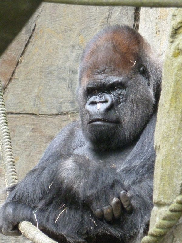
Davis et al.

                                                                   the orbit purulence suggested more extensive disease than
                                                                   isolated chronic rhinosinusitis. A nasal culture obtained by
                                                                   the gorilla keepers showed Enterobacter species. Based on
                                                                   the antibiotic sensitivity profile, both enrofloxacin (a fluo-
                                                                   roquinolone used in animals) and prednisone were admin-
                                                                   istered. Adjuvant treatments such as nasal saline irrigations
                                                                   or nasal sprays could not be feasibly performed due to a
                                                                   lack of patient cooperation. Unfortunately, his condition
                                                                   dramatically worsened after only a few days, including in-
                                                                   creased lethargy, decreased appetite, and the interpretation
                                                                   by the gorilla keepers of increased physical discomfort.
                                                                      The healthcare team of otolaryngologists and Zoo Ani-
                                                                   mal Health providers decided to obtain a computed tomog-
                                                                   raphy (CT) scan of his sinuses to obtain more information
                                                                   to help decide if surgery was a realistic option. Though the
                                                                   Zoo has ultrasound and conventional digital radiograph
                                                                   capabilities, the Zoo relies on an offsite human imaging
                                                                   center when CT scans are needed (Center for Diagnostic
                                                                   Imaging, Seattle, WA). The CT images showed complete
                                                                   opacification of his bilateral frontal, sphenoid, ethmoid,
                                                                   and maxillary sinuses, and revealed bone erosion involving
                                                                   the lateral and anterior walls of both maxillary sinuses, his
                                                                   anterior skull base, and possibly his nasal septum (Fig. 2).
                                                                      Discussion now centered on whether or not this repre-
                                                                   sented a sinus neoplasm vs severe chronic rhinosinusitis.
                                                                   Review of the veterinary medical literature yielded no ex-
  FIGURE 1. Purulent left nasal drainage seen on physical exam.
                                                                   amples of successful medical treatment for chronic rhinos-
                                                                   inusitis in gorillas. However, in the lay literature there was
  team in collaboration with the gorilla’s veterinary medicine     a case report of nasal abscesses being drained from Massa
  team.                                                            the gorilla at the Philadelphia Zoo in 1969.1 More recently,
                                                                   there was a report of sinus surgery being performed on
                                                                   Vicki the orangutan in the United Kingdom at the Chester
                       Case description                            Zoo.2 This engendered discussion of whether endoscopic
  The patient is a nearly 200-kg (435-pound) 35-year-old           sinus surgery could be performed. Unfortunately, his con-
  male silverback western lowland gorilla (Gorilla gorilla go-     dition rapidly deteriorated to the point where he ceased to
  rilla) living at the Woodland Park Zoo (Seattle, WA). In the     drink, eat, or move from a fetal position.
  spring of 2014, his keepers and Zoo veterinarians noted a           The dramatic decline in health prompted the lead veteri-
  remarkable decline in his overall health. He developed pu-       narian (D.C.) to convene an emergent discussion with the
  rulent nasal drainage from the left nostril, exhibited signs     Gorilla Species Survival Plan (SSP) (http://gorillassp.org/).
  of headache by holding his head, and decreased consensual        The Gorilla SSP is a group of dedicated professionals who
  interactions with the female gorillas.                           oversee the welfare of the gorillas in the United States. They
     The Animal Health Care team at the Zoo interpreted            provide expertise in all matters of gorilla healthcare, such
  these signs as suggestive of chronic rhinosinusitis. Despite     as developing breeding plans, directing research programs
  1 month of cephalexin followed by a second month                 to improve gorilla health, and providing veterinary medical
  of trimethoprim/sulfamethoxazole, the nasal purulence            expertise. The Zoo Animal Health Care team and the lead
  progressed to involve both nostrils and his health and           author then discussed the options, including attempting si-
  behaviors continued to decline. Thus, after 3 months of          nus surgery vs euthanasia. In the wild, western lowland go-
  symptoms without improvement, the Animal Health Care             rillas live 30 to 40 years, though in captivity they can live
  team reached out to the Department of Otolaryngology–            in to their 50s.3 Together, the decision was made to pro-
  Head and Neck Surgery at the University of Washington            ceed with surgery. The lead author reached out to industry
  for consultation.                                                representative to request emergent shipment of the neces-
     On visual examination of the gorilla by 2 otolaryngol-        sary equipment needed to perform state-of-the-art endo-
  ogists (G.E.D. and A.D.H.) in his enclosure while fully          scopic sinus surgery. This equipment included endoscopes,
  awake at the Zoo, he showed signs of chronic rhinosinusitis      a high-definition video tower, image guidance, microde-
  (Fig. 1). His nasal purulence was copious as well as pres-       brider, sinus instruments, hemostatic materials, dura repair
  ence of purulent drainage in his eyes. Given that gorillas are   materials, cautery, and mometasone-furoate steroid-eluting
  thought to have similar nasolacrimal anatomy to humans,          implants.

                                                                    International Forum of Allergy & Rhinology, Vol. 00, No. 0, xxxx 2018   2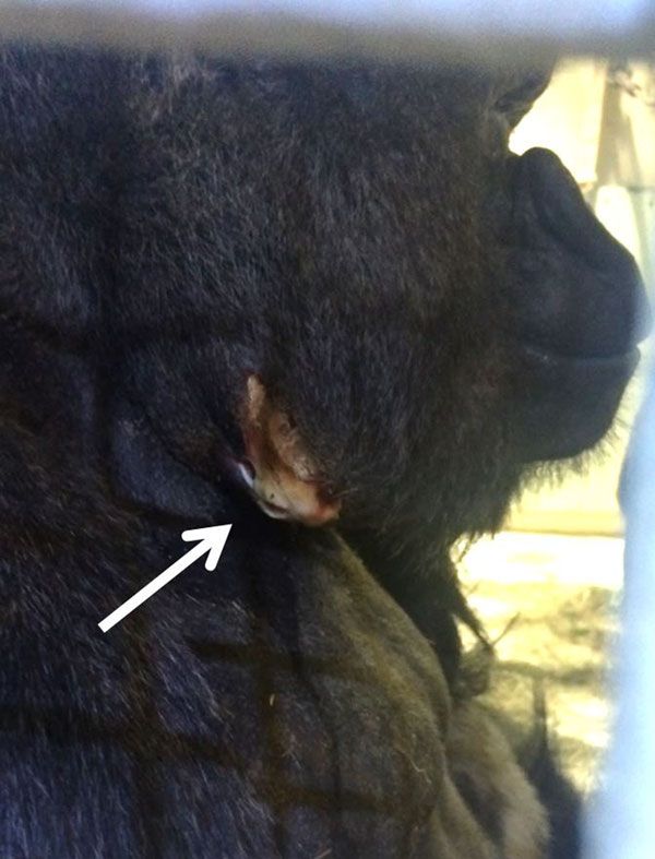
Gorilla endoscopic sinus surgery
FIGURE 2. Coronal CT scan images showing complete opacification of (A) bilateral frontal and maxillary sinuses along with erosion of the lateral maxilla
(arrow), (B) opacification of ethmoid sinuses (arrow), and (C) opacification of the sphenoid sinuses (arrow). CT = computed tomography.
The surgery was performed in the operating room at the was extracted from his sinuses (Fig. 5). Culture of this
Woodland Park Zoo’s Animal Health Care facility. The debris would later reveal Escherichia coli. Bilateral
Zoo’s Animal Health team provided the gorilla’s anesthe- maxillary antrostomies, total ethmoidectomies, and sphe-
sia while the lead author and surgical team performed the noidotomies were performed using image guidance. At this
surgery. Anesthesia was initiated by intramuscular injec- point however, his condition became unstable, with a rising
tion by a remote drug delivery system of medetomidine serum potassium and declining blood pH, necessitating the
and ketamine. Animal Health then intubated the patient need to terminate surgery fairly abruptly. Without time
and placed an intravenous (IV) line to deliver fluids and an- to perform complete frontal sinus decompressions, the
tibiotics. Anesthesia was administered using inhaled isoflu- lead author performed balloon sinus dilation (Medtronic,
rane. Animal Health monitored vital signs including heart Minneapolis, Minnesota) of the frontal sinuses and placed
rate, oxygen saturation, end tidal CO2 , body temperature, mini mometasone-furoate steroid-releasing implants (In-
noninvasive blood pressure monitoring, and frequent blood tersect ENT, Menlo Park, California) into bilateral frontal
gas monitoring. recesses. Although these implants are not approved for
Intraoperatively, the gorilla’s nasal mucosa was severely use in animals, the team concluded this was the only
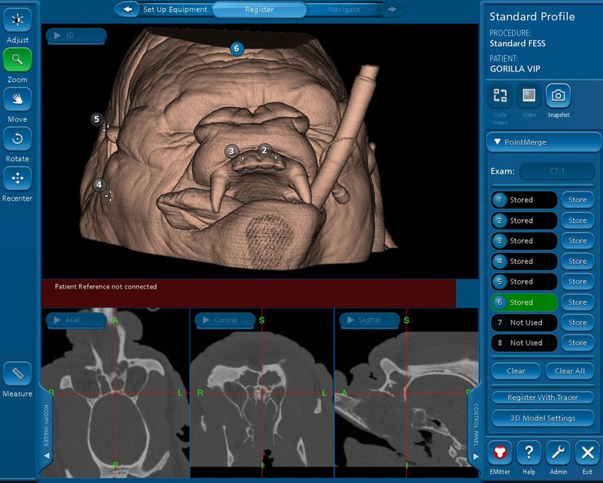
edematous and friable (Supporting Video 1). Image guid- way to deliver topical steroids to this gorilla’s sinuses. A
ance was used (Medtronic, Minneapolis, Minnesota), but gelatin-based gelfoam-thrombin hemostatic matrix (Baxter
due to the swelling of his head that occurred during the 5 Medical Deerfield, Illinois) was applied to both nasal
days between the CT scan and the time of surgery, “point cavities to assist with hemostasis. Prior to emersion from
merge” was needed to obtain the image guidance calibra- anesthesia, he received atipamezole to reverse the medeto-
tion rather than the standard point tracing with this system midine. He was transferred to his enclosure and slowly
(Fig. 3). emerged from anesthesia in the lateral decubitus position.
Gorilla sinuses have many similarities to human sinuses, His initial recovery went smoothly. On postoperative day
but also have several important distinctions from human si- 1 (POD 1), he began eating and mobilizing. However, on
nus anatomy.4 Compared to humans, gorilla frontal sinuses POD 3 he had a significant setback. His preoperative soft
are small, their ethmoid sinuses are even smaller, yet their tissue edema was, in retrospect, indicative of subcutaneous
maxillary sinuses are much taller and their sphenoid sinuses infection. He developed an open fistula in his right superior
are much deeper. In fact, preoperative planning demon- neck (Fig, 6) as well as a draining fistula from his right
strated that the distance from the nostril to the sphenoid os lateral orbit. Although gorillas do have air saccules that can
was 13 cm in this gorilla, in contrast to 7 cm in the average become infected,5 the neck fistula was thought to be more
human. Despite the increased depth of the gorilla nose, the likely related to extension of his advanced sinus infection
width of the nasal cavity is similar to that of a human. In through the maxilla erosion depicted on the CT scan.
retrospect, it would have been helpful to have a variety of The culture from surgery returned showing E. coli with
laryngeal or spine microdebrider blades as well as skull- intermediate resistance to the amoxicillin/clavulanic acid
base instruments to use during the surgery. The length of that was administered postoperatively. After discussion
those debrider blades and surgical instruments are longer with our infectious diseases colleague (P.S.P.), he was then
than standard sinus instruments and would have allowed put on enrofloxacin as well as continued on clindamycin.
easier manipulation of the deeper posterior ethmoid and It was felt that his present condition was too critical for a
sphenoid tissue. second anesthesia, so no washout of the wounds could be
Intraoperatively, his nasal cavity was packed with firm, performed. With careful attention from the gorilla keepers,
polyp-like masses (Fig. 4). Frozen specimen interpretation hand feeding and providing fluids, and continued antibiotic
was not available onsite at the Zoo. Fortunately, final support provided by intramuscular injection, his condition
histopathology revealed inflammatory polyp tissue and not gradually improved over the following week.
neoplasm. Once the polyps were removed and his sinuses Several weeks later he returned to the public viewing area
were opened, thick inspissated mucopurulent material and resumed normal male gorilla activities. True success
3 International Forum of Allergy & Rhinology, Vol. 00, No. 0, xxxx 2018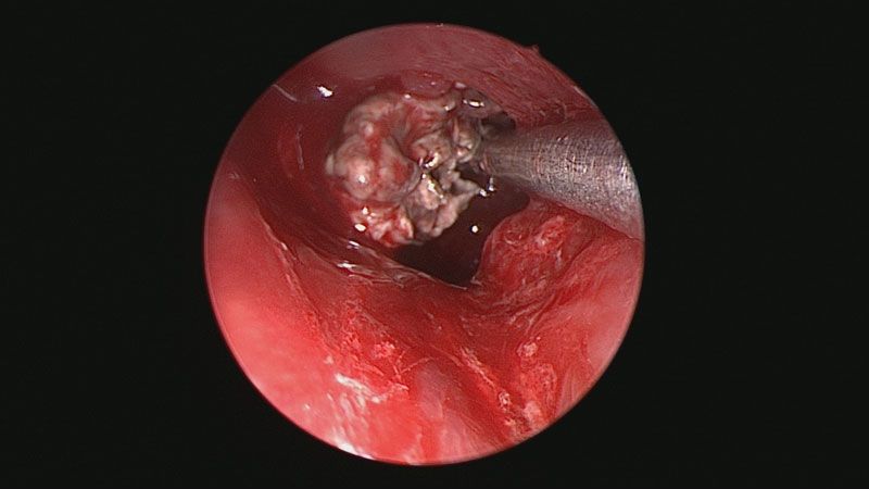
Davis et al.

  FIGURE 3. Calibration for intraoperative navigation required using point merge technique.

  FIGURE 4. Fibrous firm nasal polyps within the left nasal cavity.

  was evident a few months later when it was discovered that                     and otolaryngology team met again to discuss treatment
  1 of the females in his social group was pregnant.                             options. At this point, the primary concern was cardiac
     Approximately 1 year after the initial sinus surgery, he                    disease as this is the most common cause of death in this
  began showing symptoms of possible rhinosinusitis. He                          demographic of male gorillas.6 For a proper echocardio-
  exhibited intermittent head-holding behavior suggesting                        gram examination, a general anesthesia was required. Dis-
  headaches as well as decreased appetite, decreased interac-                    cussion centered upon whether or not to obtain a new si-
  tion with his female companions, and behavior interpreted                      nus CT scan. Critical to this decision was weighing the risk
  as possible chest discomfort. Initially, he was treated with                   of general anesthesia in these large animals. Placing them
  an empiric 3-week course of antibiotics and prednisone;                        under anesthesia can be traumatic for the animal due to the
  however, his symptoms persisted. The Animal Health Team                        intramuscular injection required and isolation from the rest

                                                                                   International Forum of Allergy & Rhinology, Vol. 00, No. 0, xxxx 2018   4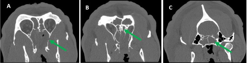
Gorilla endoscopic sinus surgery

    FIGURE 5. Endoscopic view of inspissated mucopurulent material within the left maxillary sinus.

                                                                                    before, knowing that his sinus anatomy had changed but
                                                                                    his critical skull-base and orbit anatomy would remain rel-
                                                                                    atively constant. Corporate sponsors again kindly supplied
                                                                                    the necessary equipment to perform sinus surgery.
                                                                                      In hopes of helping control his inflammatory sinus
                                                                                    disease, the lead author requested support from the
                                                                                    Immunology Division of the University of Washington
                                                                                    (A.G.A.) to attend the surgery and perform allergy skin
                                                                                    testing. Intradermal allergy testing showed a significant re-
                                                                                    action to elm tree pollen and dust mites (Dermatophagoides
                                                                                    farinae). He was subsequently started on montelukast. A
                                                                                    physician from the Cardiology Division of the University of
                                                                                    Washington (K.R.B.) performed a transthoracic echocar-
                                                                                    diogram. Fortunately, this was negative for any significant
                                                                                    abnormalities except for mild left ventricular hypertrophy.
                                                                                    Nasal endoscopy showed active chronic rhinosinusitis with
                                                                                    purulence from the frontal sinuses and some regrowth
                                                                                    of polyps. Therefore, the lead author performed revision
                                                                                    sinus surgery on all sinuses (Supporting Video 2). Again,
                                                                                    mini mometasone-furoate releasing implants were placed
                                                                                    in to bilateral frontal recesses. His recovery went without
                                                                                    incident.
                                                                                      Over the past 1.5 years since his last surgery he has
                                                                                    thrived, and he is alive and well. A recent need for emergent
                                                                                    general anesthesia to repair a life-threatening incarcerated
    FIGURE 6. Fistula tract in the right neck (white arrow).                        ventral hernia provided an opportunity to assess his si-
                                                                                    nuses. Examination showed his sinuses were healthy. He
                                                                                    has continued montelukast and was also started on fex-
    of the gorilla family. Additionally, emersion from general                      ofenadine for improved allergy control. His new daughter
    anesthesia carries a much greater risk of airway compro-                        Yola recently celebrated her 1-year birthday, highlighting
    mise compared to humans, given that jaw thrust and bag                          the successful treatment of this gorilla.
    mask ventilation are not reliable options in a partially anes-
    thetized adult gorilla. Since obtaining a new CT scan would
    require a separate general anesthesia followed likely by an-
    other anesthesia for surgery, the team decided to forgo the                                            Discussion
    CT scan and proceed directly to a general anesthesia for                        The similarities between primates and humans afford
    the cardiac exam. This also allowed an opportunity to per-                      opportunities to apply advanced human therapies when
    form additional sinus surgery if the endoscopic nasal exam-                     needed to improve the lives of our primate cousins. How-
    ination showed evidence of sinus disease. Image guidance                        ever, applications of these therapies require thought, in-
    would still be possible by using his CT scan from the year                      genuity, and preparation. Given the complexities and lack

5         International Forum of Allergy & Rhinology, Vol. 00, No. 0, xxxx 2018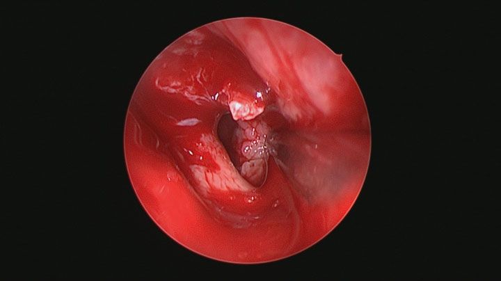
Davis et al.

  of guidance from the literature, planning for this surgery                                                              Conclusion
  required the resources of several specialists including oto-
                                                                                           The success of the surgical and medical treatment of this
  laryngology, radiology, allergy and immunology, cardi-
                                                                                           gorilla’s life threatening sinus infection required many ex-
  ology, infectious diseases, veterinary medicine, veterinary
                                                                                           perts, careful planning, and corporate generosity. The in-
  technicians, human surgical nursing and human surgical
                                                                                           teraction between human and animal medicine would not
  technicians. Further examples of similar collaborations
                                                                                           have been successful without the close and trusting collab-
  are the focus of Dr. Barbara Natterson-Horowitz’s book,
                                                                                           orations between human and veterinary health providers.
  Zoobiquity: The Astonishing Connection Between Human
                                                                                           We encourage human healthcare providers to seek volun-
  and Animal Health (New York: Vintage Publishing; April
                                                                                           teer opportunities through their local zoos by engaging in
  2013). Although this book highlights examples of human
                                                                                           discussions with their local veterinarians.
  and veterinary providers working collaboratively to care
  for animals, we report the first experience of an advanced
  endoscopic sinus surgery being performed on a gorilla.
     It is important to address the ethics of treating animals in
                                                                                                                      Acknowledgments
  advanced diseased states with modern surgical techniques.                                We thank numerous volunteers and Zoo staff who helped
  Having open communication between all parties involved                                   with the complex medical and surgical treatment of this
  is critical to help ensure mutual understanding of the risks                             gorilla: University of Washington Medical Center staff:
  and potential benefits of the procedures.                                                Jayme Richards, Corina Sisson, and Pamela Kean; Wood-
     An important note is that this surgery was made possi-                                land Park Zoo Animal Health staff: Harmony Frazier, LVT,
  ble by the generosity of human surgical device equipment                                 Linda Moneymaker, LVT, Barbara Brush, LVT, and Teri
  companies, to which we are indebted. Due to concerns for                                 Hermann, LVT, and all of the gorilla keepers who per-
  prion and other zoonotic infectious exposures, surgical in-                              formed heroic care to bring this gorilla back to health; the
  struments once used on animals are not permitted for use                                 Center for Diagnostic Imaging and staff: Kristin Kessler,
  on humans. For this reason, we elected to use equipment                                  RT, and Lisa Cook, RDMS. In addition, the authors would
  that was purposed for human cadaver dissection labs.                                     like to thank the generosity of the corporate supporters
     All medical providers and assistants donated their time                               including Medtronic, Storz, Intersect ENT, Baxter Phar-
  throughout this process.                                                                 maceuticals, Covidien, and Cook Medical.

  References
  1. Newman JL. Encountering Gorillas: A Chronicle of        3. Smithsonian’s National Zoo & Conservation Biol-         5. Hastings BE. The veterinary management of a laryngeal
     Discovery, Exploitation, Understanding, and Survival.      ogy Institute. Primates: Western Lowland Gorilla.          air sac infection in a free-ranging mountain gorilla. J
     Lanham, MD: Rowman & Littlefield Publishers, Inc.;         Washington, DC: Smithsonian’s National Zoo                 Med Primatol. 1991;20:361–364.
     2013: xv, 203 pp.                                          & Conservation Biology Institute; 2018. https://        6. Meehan TP, Lowenstine LJ. Causes of mortality in cap-
  2. Chester Zoo. And Breathe! Vicky the 29-Year-               nationalzoo.si.edu/animals/western-lowland-gorilla.        tive lowland gorillas: a survey of the SSP population. In:
     Old Orangutan Rests up After Sinus Operation.              Accessed March 7, 2018.                                    Junge RE, editor. Proceedings of the American Associa-
     Upton-by-Chester, UK: Chester Zoo; 2014. http://        4. Blaney SP. An allometric study of the frontal sinus        tion of Zoo Veterinarians and Association of Reptilian
     www.chesterzoo.org/whats-happening/zoo-news/               in Gorilla, Pan and Pongo. Folia Primatol (Basel).         and Amphibian Veterinarians: Annual Conference; Oc-
     2014/04/orangutan-sinus-operation.          Accessed       1986;47:81–96.                                             tober 22–27, 1994; Pittsburgh: PA. Yulee, FL: Ameri-
     March 7, 2018.                                                                                                        can Association of Zoo Veterinarians; 1994:216–218.

                                                                                              International Forum of Allergy & Rhinology, Vol. 00, No. 0, xxxx 2018                     6
You can also read