Hemimegalencephaly without seizures: report of a case and review of literature - Romanian ...
←
→
Page content transcription
If your browser does not render page correctly, please read the page content below
Romanian Neurosurgery | Volume XXXI | Number 3 | 2017 | July-September

Article

Hemimegalencephaly without seizures: report of
a case and review of literature

Agrawal Atul, Dutta Gautam, Singh Daljit, Sachdeva Deepashu, Gupta Robin
INDIA

DOI: 10.1515/romneu-2017-0050
306 | Agrawal et al - Hemimegalencephaly without seizures

                                                                       DOI: 10.1515/romneu-2017-0050

          Hemimegalencephaly without seizures: report of a case
          and review of literature

          Agrawal Atul1, Dutta Gautam1, Singh Daljit2, Sachdeva Deepashu1,
          Gupta Robin1
          Department of Neuro-Surgery, Govind Ballav Pant Institute of Postgraduate Medical Education
          and Research (GIPMER), New Delhi, INDIA
          1
           Senior Resident, 2Professor and Head of the Department

          Abstract: Hemimegalencephaly is a rare malformation of the brain characterized by
          enlargement of one cerebral hemisphere. The classic clinical triad consists of intractable
          epilepsy, severe psychomotor delay and hemiparesis. We report a case of a six months
          old girl, with the radiological features of hemimegalencephaly but with a comparatively
          benign clinical course. She had mild developmental delay but with no paresis or seizures.
          Literature revealed only two reported cases of hemimegalencephaly without the presence
          of seizures. We discuss the clinical and radiological findings of this third reported case.
          Key words: Brain malformations hemimegalencephaly, seizures

Introduction                                         significant mortality in infancy. [1,6] Onset of
    Hemimegalencephaly (HME) is a rare,              symptoms commonly occurs in the first week
sporadic       type    of    congenital     brain    of life. [4] Early surgical intervention
malformation, characterized by enlargement           including        hemispherectomy,          either
of one cerebral hemisphere. [1] HME is largely       anatomical or functional is warranted in these
thought to be a malformation due to abnormal         patients to achieve a better outcome in the
neuronal and glial proliferation. [2] This is        control of epilepsy and to promote
accompanied by dysplasia of the cerebral             psychomotor development. [3] However,
cortex       along      with     polymicrogyria,     there have been isolated reports of milder
agyria/pachygyria,       and     white     matter    clinical presentation [4] including only a single
heterotopia, which results in intractable            case report of a child having HME without any
epilepsy and profound disabilities. [1] The          presentation of seizures. [5] In this report, we
classical clinical triad described is of epilepsy,   describe a child who presented to our
severe     psychomotor        retardation     and    department for management of lumber
contralateral hemiparesis. [1,4] which are           meningomyelocele whose further workup
often severe in nature and associated with a         revealed HME.
Romanian Neurosurgery (2017) XXXI 3: 306 - 309 | 307
Case report generalized loss of white matter bulk with
A six months old female child was referred cortical dysplasia. There was associated
to our neurosurgical department for asymmetry of the ventricular system.
evaluation and management of a lump in Radiological features were consistent with
lower back that is present since birth along HME. MRI spine was suggestive of lumber
with macrocephaly. She was product of a full- lipomenigomyelocele. She underwent sac
term pregnancy of non-consanguineous excision and dural repair and postoperative
parents; the prenatal history was normal and stay was uneventful. The family was informed
the baby cried immediately after birth. Height that the child did not require neurosurgical
at birth was 48 cm and weight was 2500 g along intervention for HME currently, but that she
with a head circumference of 41.2 cm with a was at risk of developing epilepsy and should
flat anterior fontanelle along with lump at be closely observed for clinical seizures.
lower back. She had extra digits on both feet
Discussion
and had mildly delayed neurological
development. She had no clinical evidence of HME is a rare anomaly and there is a
paresis of any limbs neither she ever had a paucity of large series in the literature. In one
seizure or seizure-like episode as per 6 months of the comprehensive reviews, the authors
of age. On examination, a head circumference describe 14 cases, examining clinical,
over the 99th centile was noted in the child but radiological and electrophysiological features.
there was no signs or symptoms of raised [4] In this series, the authors report that one
intracranial pressure. We plotted the head patient did not have seizures. Beside this
circumference on a growth chart when she was isolated case, extensive medline search
found to have no crossing of centile lines, revealed only one report where occurrence of
consistent with normal growth of the child. HME without seizures was described, making
She was found to have 6X4cm lumber the current case only the third reported.
meningomyelocele. Non-contrast CT and Severe epilepsy is a hallmark of the
MRI of the brain and spine was obtained. condition, being present in all 10 patients
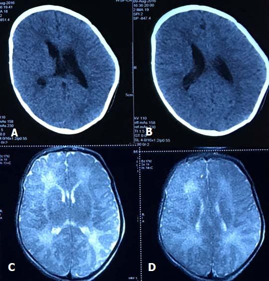
CT and MRI brain of the child described in another large series. [1] In fact,
demonstrated features typical of HME (Figure HME carries a significant mortality risk due to
1). Asymmetrical abnormalities were noted, the severity of the epilepsy [6,7] as well as
with the left hemisphere larger in volume than association with significant psychomotor
the right. This asymmetry was associated with delay and hemiparesis. The current case is
abnormalities both of grey and white matter. unusual in only having one of the clinical triad
The left hemisphere demonstrated cortical (developmental delay) and only in mild form.
malformation and polymicrogyria, as well as308 | Agrawal et al - Hemimegalencephaly without seizures
Figure 1 - Noncontrast CT (A & B) and T2W MRI brain (C & D) showing enlarged left cerebral hemisphere with
ventricular dysmorphism, white matter changes, cortical dysplasia and polymicrogyria of the left frontal and
parietal lobe
HME on brain imaging consists of cortical hemispheric hypertrophy or demyelination.
gray matter almost uniformly abnormal, areas [8] This patient presented almost all of these
of increased thickness of the cortical gray MRI findings supporting HME. The WM
matter (GM), abnormal gyral patterns, signal change may be consistent with either
increased signal intensity in the subcortical demyelination or advanced myelination. The
white matter (WM) on T2-weighted images, ipsilateral ventricle is usually enlarged and
blurring of the GM-WM transition, atrophy or dysmorphic, often with extension of theRomanian Neurosurgery (2017) XXXI 3: 306 - 309 | 309
posterior horn of the lateral ventricle across of epilepsy so that early surgical intervention
the midline. This patient did not have grossly may be undertaken.
enlarged ventricles but asymmetric.
Vigevano et al., [4] in their series of 14 Correspondence
patients, divided HME into two groups: A and Dr. Gautam Dutta, M.Ch, Senior Resident,
B. Group A was characterized by severe, Department of Neuro-Surgery, Govind Ballav Pant
uncontrollable epilepsy with marked Institute of Postgraduate Medical Education and
hemiparesis and delay (7/7), whilst group B Research (GIPMER), New Delhi- 110002
had more mild clinical features with sporadic Phone number: 9968034400
seizures and a later onset of epileptic attacks. E-mail: gautamblue@hotmail.com
Radiological features were less severe in group
B, with no patient having microgyria on their References
scan. We consider the current case represents 1.Flores-Sarnat L. Hemimegalencephaly: Part 1. Genetic,
“group B”-type child, who is as yet too young clinical, and imaging aspects. J Child Neurol.
to have had her first seizure. The other 2002;17:373–84.
2.Barkovich AJ, Kuzniecky RI, Jackson GD, Guerrini R,
possibility is that there is a population of
Dobyns WB. Classification system for malformations of
“silent” HME, with absent or mild clinical cortical development. Neurology. 2001;57:2168–2178.
signs but with radiological features of the 3.Di Rocco C, Battaglia D, Pietrini D, Piastra M, Massimi
malformation and that this was an incidental L. Hemimegalencephaly: clinical implications and
finding when the child was being investigated surgical treatment. Child’s Nerv Syst 2006;22:852–866.
4.Vigevano F, Fusco L, Granata T, Fariello G, Di Rocco C,
for unrelated reasons. As availability of MRI
Cusmai R. Hemimegalencephaly: clinical and EEG
scanning becomes ever more widespread, it characteristics. Dysplasias of cerebral cortex and epilepsy.
may be that further similar cases are detected Lippincott-Raven, Philadelphia. 1996; pp 285–294.
as children have brain imaging for various 5.Greg James, Mano Shanmuganathan, William
other reasons. Harkness. Hemimegalencephaly without epilepsy: case
report. Childs Nerv Syst. 2014;30:1617-19.
6.Vigevano F, Bertini E, Boldrini R, Bosman C, Claps D,
Conclusion
Di Capua M, Di Rocco C, Rossi GF. Hemimegalencephaly
It is important to recognize the abnormal and intractable epilepsy: benefits of hemispherectomy.
tissue organization to make the correct Epilepsia. 1989;30:833–843.
7.Bosman C, Boldrini R, Dimitri L, Di Rocco C, Corsi A.
diagnosis. In children, brain MRI is the gold
Hemimegalencephaly. Histological,
standard diagnostic tool, with brain immunohistochemical, ultrastructural and
enlargement and WM changes. Different cytofluorimetric study of six patients. Child’s Nerv Syst.
degree of changes in signal intensity revealing 1996; 12:765–775.
8.Barkovich A, Moore K, Jones B, Vezina G, Koch B,
the WM abnormalities is the most important
Raybaud C et al. Diagnostic imaging pediatric
and constant sign in HME. Regular follow up neuroradiology. Salt Lake City, Utah: Amirsys-Elsevier
and close observation is required in scarce 2007. Section 1: Cerebral Hemispheres Malformations: I-
cases like the present one who do not manifest 1-20.
seizures on presentation for later developmentYou can also read