Henipavirus Gap Analysis - Workshop Report United States Department of Agriculture
←
→
Page content transcription
If your browser does not render page correctly, please read the page content below

United States Department of Agriculture

Agricultural Research Service

November 2018

Henipavirus Gap Analysis
Workshop Report

Mention of trade names or commercial products in this report is solely for the purpose of providing
specific information and does not imply recommendation or endorsement by the U.S. Department of
Agriculture (USDA).

The USDA prohibits discrimination in all its programs and activities on the basis of race, color,
national origin, age, disability, and where applicable, sex, marital status, familial status, parental
status, religion, sexual orientation, genetic information, political beliefs, reprisal, or because all or part
of an individual’s income is derived from any public assistance program. (Not all prohibited bases
apply to all programs.) Persons with disabilities who require alternative means for communication of
program information (Braille, large print, audiotape, etc.) should contact USDA’s TARGET Center at
(202) 720-2600 (voice and TDD). To file a complaint of discrimination, write to USDA, Director,
Office of Civil Rights, 1400 Independence Avenue, SW., Washington, D.C. 20250-9410, or call (800)
795-3272 (voice) or (202) 720-6382 (TDD). USDA is an equal opportunity provider and employer.

The Agricultural Research Service (ARS) conducts research to develop and transfer solutions to
agricultural problems of high national priority and provides information access and dissemination to
ensure high-quality, safe food and other agricultural products; to assess the nutritional needs of
Americans; to sustain a competitive agricultural economy; to enhance the natural resource base and
the environment; and to provide economic opportunities for rural citizens, communities, and society as
a whole.

To cite this report:
Henipavirus Gap Analysis Workshop Report. 2018. U.S. Department of Agriculture, Agricultural
Research Service, Washington, DC. http://go.usa.gov/xnHgR.

                                                                                                             2
Contents
EXECUTIVE SUMMARY .............................................5
GROUP PICTURES ..................................................7
GLOSSARY .......................................................8
INTRODUCTION ..................................................10
BACKGROUND ...................................................11
DEFINITION OF THE THREAT ........................................13
     INFECTION IN PEOPLE...................................... 13
     INFECTION IN PIGS........................................ 13
     ECONOMIC IMPACT.......................................... 13
     BIOTERRORISM............................................. 14
GAP ANALYSIS ...................................................15
     VIROLOGY................................................. 15
     PATHOGENESIS............................................. 17
     IMMUNOLOGY............................................... 20
     EPIDEMIOLOGY............................................. 21
     BIOTERRORISM............................................. 25
SUMMARY OF OBSTACLES TO PREVENTION AND CONTROL ........................28
     DIAGNOSIS................................................ 28
     VACCINATION.............................................. 28
     SURVEILLANCE............................................. 28
     DEPOPULATION............................................. 29
COUNTERMEASURES ASSESSMENT ...................................30
    ASSUMPTIONS.............................................. 30
    DECISION MODEL........................................... 30
    VACCINES................................................. 32
    DIAGNOSTICS.............................................. 36
    DEPOPULATION............................................. 42
    SURVEILLANCE............................................. 42
    DRUGS.................................................... 43
    DISINFECTANTS............................................ 43
    PERSONAL PROTECTIVE EQUIPMENT (PPE)...................... 43
RECOMMENDATIONS ..............................................44
    RESEARCH................................................. 44
    PREPAREDNESS............................................. 45
CONCLUSION ....................................................47
FIGURES ........................................................48
TABLE I: NIPAH VIRUS INFECTION IN BATS ..................................53
TABLE II – NIPAH VIRUS CASES 2001-2018 .................................54

                                                                              3
TABLE III – VACCINE PLATFORMS .......................................55
TABLE IV – CURRENT VACCINE CANDIDATES ................................56
APPENDIX I: COUNTERMEASURES WORKING GROUP INSTRUCTIONS .................57
APPENDIX II – VACCINES ASSESSMENT ...................................58
APPENDIX III – DIAGNOSTICS ASSESSMENT .................................58
APPENDIX IV - CONTRIBUTORS .........................................60
REFERENCES ....................................................65

                                                                             4
EXECUTIVE SUMMARY
Henipavirus is the taxonomic genus for a group of viruses in the family Paramyxoviridae that includes
Hendra virus (HeV) and Nipah virus (NiV). These viruses are zoonotic agents that are highly
pathogenic in humans with case fatality rates of 40% to 70%. As such, these viruses are classified as
Biosafety Level 4 (BSL-4) agents, requiring the highest level of laboratory biocontainment.
Importantly, they have many of the physical attributes to serve as potential agents of bioterrorism, and
are also considered emerging zoonotic pathogens with increasing geographical distribution in
Australia, New Caledonia, Southeast Asia, and Madagascar.

Hendra virus first emerged in 1994 in Australia spilling over from bats to horses to humans, causing
several disease outbreaks since with significant fatality rates. Nipah virus emerged in Malaysia in
1999, resulting in nearly 300 human cases with over 100 deaths.

The Nipah virus outbreak in Malaysia was especially concerning, causing widespread panic and fear
because of the high mortality rate in people and the inability to control the disease initially. There
were also considerable social disruptions and tremendous economic loss to an important pig-rearing
industry. This highly virulent virus, believed to be introduced into pig farms by fruit bats, spread easily
and silently among pigs and was transmitted to humans who came into close contact with infected
animals. A NiV outbreak in Bangladesh in 2001 resulted from direct bat to human transmission via
contaminated date palm juice with further spread within the human population. From 2001 to 2012,
the World Health Organization (WHO) reported a total of 209 cases, with 161 deaths due to of NiV
infections. In 2014, the WHO reported a NiV outbreak in fourteen districts of Bangladesh, resulting in
24 cases and 21 deaths. In 2015, three fruit bats tested positive for NiV in New Caledonia at the
Noumea National Park, including three bats at the Noumea Zoo.

This gap analysis report focuses primarily on NiV and its potential impact on agricultural swine
production. However, information is also provided on the threat henipaviruses pose to public
health, both as emerging zoonotic agents and as potential agents of bioterrorism. Included in this
report is scientific information on Henipavirus virology, epidemiology, pathogenesis, immunology,
and an assessment of the available veterinary medical countermeasures to detect, prevent, and
control disease outbreaks. Importantly, gaps are provided to inform research needs and priorities.
Some of the major gaps and obstacles for disease control can be summarized as follows:

Diagnostics
The availability of safe laboratory diagnostic tests are limited. Virus isolation and serum
neutralization assays require live NiV; thus, BSL-4 containment laboratories are required. Nucleic
acid-based assays, such as RT-PCR are available, but genetic variation amongst henipaviruses are
reported to impact sensitivity and real time RT-PCR may not be able to detect all divergent and
novel henipavirus strains. Serological assays are limited in their ability to differentiate between
known and unknown henipaviruses, as cross-reactivity to one or more known viruses is possible.
Commercial diagnostic test kits are not available. International standards for NiV assay validation
are needed. Gaps include a lack of positive experimental and field samples for test validation (or
even evaluation) and there are restrictions on material transfer (e.g., obtaining animal samples that
could be used to validate tests) due to biosecurity concerns. Low biosafety level reference sera

                                                                                                         5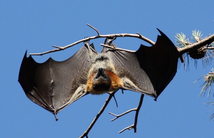
against various isolates are not yet available. There is a need for high throughput antibody assays
for disease outbreaks, recovery and surveillance purposes. There is also a need to develop operator-
safe diagnostics tests and reagents that can be produced in low biocontainment facilities.

Vaccines
There is currently a commercial vaccine available for horses, but there are no vaccines for swine or
humans. There are several experimental vaccine candidates that may be safe and effective in swine
and other domestic animals. However, all these vaccine candidates will require further research to
establish their efficacy, and they will need to be fully developed to be licensed and stockpiled for
rapid use in an emergency disease outbreak in swine.

Surveillance
Surveillance is the first line of defense against a disease outbreak. Rapid and accurate detection
affects the time when control measures can be implemented and affects the extent of the disease
outbreak. Because of limitations with laboratory diagnosis, surveillance programs are dependent on
the reporting of clinical signs in populations at risk. Diagnosis of NiV infections based on clinical
presentation has a low positive predictive value as there are numerous etiologies for encephalitis in
humans, and clinical signs in pigs are difficult to differentiate from many common endemic infectious
diseases.

Depopulation
Depopulation is the primary countermeasure to reduce virus shedding and stop the spread of NiV in
livestock. Disease outbreaks have shown that the control of NiV in pig populations through stamping
out is complex due to the zoonotic nature of the agent. In addition, depopulation may be logistically
difficult and may be impossible in a rapidly spreading outbreak in countries where there are pig dense
regions with millions of pigs, such as the states of Iowa, North Carolina, and Minnesota in the United
States, or South East China.

                                                                                                       6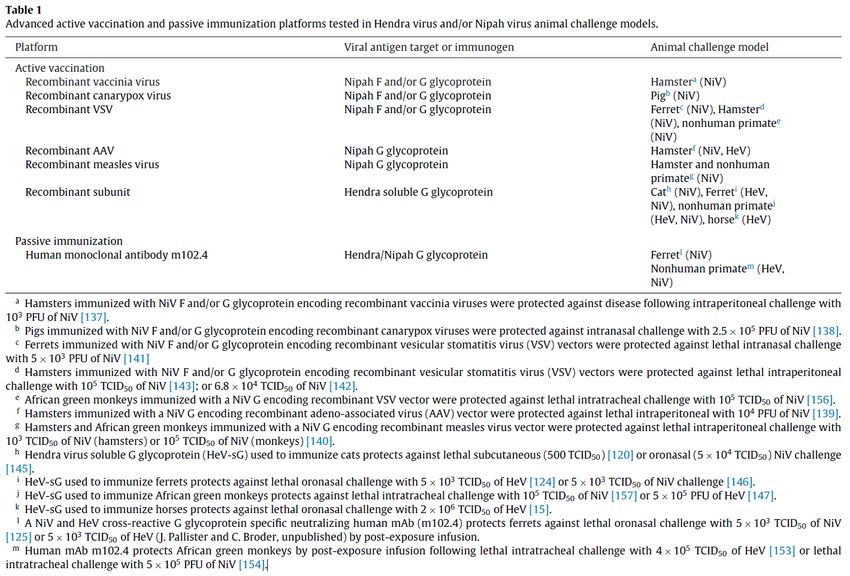
GROUP PICTURES
   Henipavirus Gap Analysis Working Group, Winnipeg, Canada
                     November 14-17, 2017

The Nipah Virus Countermeasures Working Group, Geelong, Australia
                       March 17-19, 2009

                                                                    7
GLOSSARY

APHIS: Animal and Plant Health Inspection Service, USDA, United States of America

ARS: Agricultural Research Service

AAHL: Australian Animal Health Laboratory

BSL-4: Biosafety Level 4

CDC: U.S. Centers for Disease Control and Prevention, HHS, United States of America

CFIA: Canadian Food Inspection Agency

DIVA: Differentiating between infected and vaccinated animals

ELISA: Enzyme-linked immunosorbent assay

FADDL: U.S Foreign Animal Disease Laboratory, Plum Island Animal Disease Center

FLI: Friedrich Loeffler Institute

GMP: good manufacturing practice

HeV: Hendra virus

HHS: Department of Human Health Services, United States of America

HSPD-9: Homeland Security Presidential Directive Nine

ICAR: Indian Council of Agricultural Research

Ig: Immunoglobulin

IEDCR: Institute of Epidemiology, Disease Control and Research in Bangladesh

MLV: Modified live virus vaccine

NAHLN: National Animal Health Laboratory Network, USA

NIHSAD: National Institute of High Security Animal Diseases, ICAR, India

NCFAD: National Center for Foreign Animal Disease, CFIA, Canada

                                                                                      8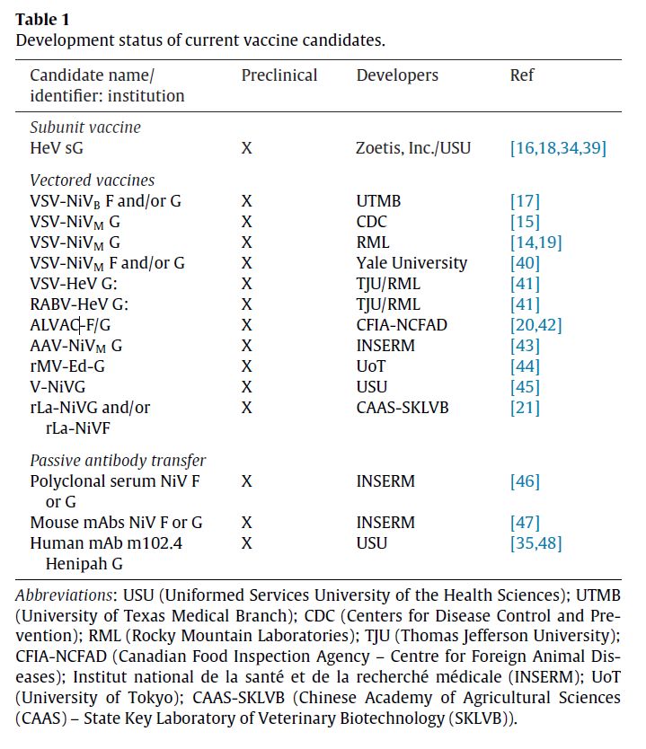
NiV: Nipah virus

NiV-B: Nipah virus Bangladesh

NiV-M: Nipah virus Malaysia

NiV N: Nipah virus nucleoprotein

NVCWG: Nipah Virus Countermeasures Working Group

NVS: National Veterinary Stockpile

OIE: World Organisation for Animal Health

PCR: Polymerase Chain Reaction.

PPE: Personal Protective Equipment

RT-PCR: Reverse transcription-polymerase chain reaction

rRT-PCR: Real-time reverse transcription-polymerase chain reaction

sHeV G: recombinant soluble Hendra virus G protein

sNiV G: recombinant soluble Nipah virus G protein

USDA: United States Department of Agriculture, United States of America

                                                                          9
INTRODUCTION
Nipah virus (NiV) is an emerging zoonotic virus. First isolated in pigs and people from an outbreak in
Malaysia in 1998 (Ang et al. 2018), this emerging virus causes severe disease in humans. The source
of transmission was determined to be from bats to pigs to humans, through close contact with infected
animals. The virus is named after the location where it was first detected in Sungai Nipah, a village in
the Malaysian Peninsula where exposed pig farmers became severely ill with encephalitis.
Nipah virus is closely related to another zoonotic virus called Hendra virus (HeV), formerly called
Equine Morbillivirus, and named after the town where it first appeared in Australia. Hendra virus
infection was first recognized in 1994, when it caused an outbreak of acute, fatal respiratory disease that
killed 14 horses. Three human cases, leading to two deaths were recorded during the outbreak. The
precise mode of virus transmission to the three Australian patients is not fully understood. All three
individuals appear to have acquired their infection as a result of close contact with horses, which were
ill and later died.
Although members of this group of viruses have only caused a few focal outbreaks, their ability to
infect a wide range of animal hosts and to produce a high mortality rate in humans has made this
emerging zoonotic viral disease a significant public health threat.
Certain species of bats of the genus Pteropus (fruit bats, also called flying foxes) are the principal
natural reservoir hosts for NiV and HeV – see Table I. Bats are susceptible to infection with these
viruses but do not develop disease. Fruit bats are distributed across an area encompassing Australia,
Southeast Asia; including Indonesia, Malaysia, the Philippines and some of the Pacific Islands, the
Indian subcontinent, and Madagascar (See Fig. 1). There is also growing evidence that viruses related
to NiV and HeV circulate in non-pteropid fruit bats across the globe (Clayton, 2017).
The exact mode of transmission of henipaviruses is uncertain, but appears to require close contact with
contaminated tissue or body fluids from infected animals. The role of domestic species other than pigs
in transmitting NiV infection to other animals has not yet been determined. In 2014, an outbreak was
reported in the Philippines involving the consumption of meat from NiV-infected horses, further
expanding the potential routes of transmission for henipaviruses.
Despite frequent contact between fruit bats and humans there is no serological evidence of human
infection among persons that are in contact with bats. Pigs were the apparent source of infection
among most human cases in the Malaysian outbreak of NiV in 1998-1999. Nipah virus has continued
to spillover over from animals with at least six outbreaks resulting in human fatalities in Bangladesh in
2013, one in India in 2014, and two in Bangladesh in 2015. The World Health Organization (WHO)
had not reported any NIV cases 2016-2017, but in 2018 twenty three new cases and 21 deaths were
reported in Kerala, India - See Table II.
The spread of henipaviruses to new geographical areas is a concern. In 2014, the Philippines reported
an outbreak with a zoonotic paramyxovirus in horses and people. There is further evidence for
broader distribution of NiV in pteropid fruit bats species. There is also growing evidence that viruses
related to NiV and HeV also circulate in non-pteropid fruit bats worldwide.
10BACKGROUND
Organization of the Gap Analysis Working Groups on Nipah Virus (2009 and 2017)
The United States Department of Agriculture (USDA) organized the first Nipah virus gap analysis
workshop in Australia in 2009 with the support of the Australian Animal Health Laboratory (AAHL).
The working group was charged by the USDA National Veterinary Stockpile Steering Committee with
making recommendations on specific materials, commercially available and in the pipeline, which will
ensure the United States has an arsenal of highly efficacious countermeasures to control and mitigate
the impact of an outbreak of Nipah virus. Nipah virus experts representing laboratories in South East
Asia, Australia, Canada, and the United States were invited to participate and contributed to this
report. The second workshop was organized in 2017 by the Special Pathogens Unit, National Centre
for Foreign Animal Disease, Canadian Food Inspection Agency (CFIA), in collaboration with
BSL4ZNet and DISCONTOOLS (http://www.discontools.eu/). The participants were charged with
assessing available veterinary medical countermeasures to control and respond to a Nipah virus
disease outbreak. In addition, the workshop participants agreed to update the gap analysis conducted
at the AAHL in Geelong, Australia, in 2009.
Report Updates
This report will be updated periodically with new scientific information, research breakthroughs,
and/or the successful development of veterinary medical countermeasures. This report was last
updated with the support of Henipavirus experts November 2018.
Reference Material
The following reports and websites are recommended:
OIE – World Organisation for Animal Health - Nipah in Animals
http://www.oie.int/en/animal-health-in-the-world/animal-diseases/Nipah-Virus/
Accessed July 22, 2018
FAO – Food and Agriculture Organization
Manual on the diagnosis of Nipah virus infection in animals
www.fao.org/DOCREP/005/AC449E/AC449E00.htm
Accessed July 22, 2018
CDC – Center for Disease Control and Prevention - Special Pathogens Branch
https://www.cdc.gov/vhf/nipah/index.html
Accessed July 22, 2018
WHO - World Health Organization
http://www.who.int/news-room/fact-sheets/detail/nipah-virus
Accessed July 22, 2018
Guidelines for Veterinarians Handling potential Hendra Virus infection in Horses (QDPI)
https://www.daf.qld.gov.au/__data/assets/pdf_file/0005/126770/2913_-Guidelines-for-veterinarians-
handling-potential-Hendra-virus-infection-in-horses-V5.1.pdf
11Accessed July 22, 2018
CFSPH – Center for Food Security and Public Health
Nipah Virus Infection
http://www.cfsph.iastate.edu/Factsheets/pdfs/nipah.pdf
Accessed July22, 2018
12DEFINITION OF THE THREAT
The threat for a natural introduction of henipaviruses in the United States is low, but there is
significant concern that henipaviruses could be used for nefarious purposes to harm agriculture and
people. Both Hendra virus and Nipah virus are on the HHS and USDA list of overlap Select Agents
and Toxins. Henipaviruses are listed as APHIS Tier 3 high-consequence foreign animal diseases and
pests. Henipaviruses are promiscuous in their ability to cause severe morbidity in several animal
species, including people, and human infections result in a very high mortality rate. The mortality rate
in pigs is actually reported as about 2.5% in adult pigs – high morbidity, but low mortality. Mortality
rates in humans range from 40% (Malaysia) to 75% (up to 100%) in Bangladesh. The animal
reservoir includes several species of bats, and henipaviruses may thus be readily available in these
wildlife reservoirs.
INFECTION IN PEOPLE
Between September 1998 and June 1999, a NiV outbreak in Malaysia resulted in severe viral
encephalitis in 105 patients (Goh et al., 2000; Epstein et al., 2006). Ninety-three percent had had
direct contact with pigs, usually within two weeks prior to the onset of illness, suggesting that there
was direct viral transmission from pigs to humans and a short incubation period. The main presenting
features were fever, headache, dizziness, and vomiting. Fifty-two patients (55%) had a reduced level
of consciousness and prominent brain-stem dysfunction. Distinctive clinical signs included segmental
myoclonus, areflexia and hypotonia, hypertension, and tachycardia. The initial cerebrospinal fluid
findings were abnormal in 75% of patients. Antibodies against Hendra virus were detected in serum
or cerebrospinal fluid in 76 percent of 83 patients tested. Thirty patients (32%) died after rapid
deterioration in their condition. An abnormal doll’s-eye reflex and tachycardia were factors associated
with a poor prognosis. Death was probably due to severe brain-stem involvement. Neurologic relapse
occurred after initially mild disease in three patients. Fifty patients (53%) recovered fully, and 14
(15%) had persistent neurologic deficits.
Unlike Malaysia, the NiV outbreaks in Bangladesh were strictly confined to human populations with
significantly higher mortality rate (Hossain et al., 2008). NiV outbreaks in Bangladesh have continued
annually since 2008 resulting in a total of 207 reported cases, 152 of which were fatal resulting in a
70% mortality rate (Clayton,. 2017). In 2018, NiV infection was confirmed in Kerala, India, where 23
confirmed cases were reported and case fatality rates were 90% (Arunkumar et al., 2018).
INFECTION IN PIGS
The NiV outbreak in Maylasia in 1999 was facilitated by the rapid spread of the virus in pig
populations. Although some pigs demonstrated a febrile respiratory illness with epistaxis, dyspnoea,
and cough, few animals exhibit neurological signs, and the majority of pigs had subclinical infections.
There are no clinical signs in pigs that are specific for NiV infection. Both, apparently healthy pigs
and pigs showing clinical signs shed significant amount of virus.
ECONOMIC IMPACT
The NiV outbreak in Malaysia in 1999 destroyed the main market for Malaysian hogs in Singapore.
The Malaysia outbreak resulted in an 80% drop in pork consumption in the domestic market. Over
half the standing pig population in the country was culled to halt the outbreak. Half the pig farms
13went out of business. The cumulative economic losses based on government figures was estimated to
be approximately $217 million USD.
BIOTERRORISM
NiV has many of the physical attributes needed for a biological weapon, including easy access to virus
resulting from its wide distribution in nature and laboratories, easy to produce, easy to disseminate,
and the potential for high morbidity and mortality in people.
14GAP ANALYSIS
The following section summarizes what we know about henipaviruses, gaps in our knowledge, and
the threat of bioterrorism.
VIROLOGY
The following summarizes our current knowledge of viral strains, taxonomy, reservoir, genome,
morphology, determinants of virulence, host range, and tissue tropism.
Virus species
Nipah virus (NiV) was first isolated in 1999 from samples collected during an outbreak of encephalitis
and respiratory illness among pig farmers. The name Nipah originated from Sungai Nipah, a village in
the Malaysian Peninsula where pig farmers became sick. There are currently two genotypes
identified: NiV-Malaysia and NiV-Bangladesh. Different strains/genotypes of NiV have emerged:
Malaysia, Bangladesh, and Cambodia. NiV Malaysia resulted in the culling of a million pigs and 250
human cases (106 fatal). NiV Bangladesh is associated with outbreaks in people (Clayton, 2017).
Hendra virus (HeV) was first isolated in 1994 from specimens obtained during an outbreak of
respiratory and neurologic disease in horses and humans in Hendra, a suburb of Brisbane, Australia.
Cedar virus (CedV) is a novel Henipavirus isolated from Australian bats, which appears to be non-
pathogenic in lab animal experiments (Marsh et al. 2012).
Ghanaian bat henipavirus (GhV) is a species of henipaviruses assembled from sequences collected
from Eidolon helvum, a bat species in the family Pteropodidae (Drexler et al. 2009; Drexler et al.
2012). No isolates have been reported, and both pathogenicity and the cross-species transmission
remain unknown. Partial sequences of 19 phylogenetically novel African henipaviruses have also been
discovered, suggestive of a further diversity of African henipaviruses.
Mòjiāng henipavirus (MojV) was discovered during retrospective surveillance for the etiologic agent
responsible for cases of fatal respiratory illness in cave-miners, China. A full-genome was assembled
from sequences detected from a cave-dwelling rodent species (Wu Z. et al. 2014). MojV is
circumstantially associated with the fatal respiratory illness, however, pathogenicity studies have not
been completed.
Taxonomy
NiV and HeV are members of the family Paramyxoviridae, order Mononegavirales. Comparison of
nucleic acid and deduced amino acid sequences with other members of the family confirms that NiV
and HeV are members of the family Paramyxoviridae, but with limited homology with members of the
Morbillivirus, Rubulavirus and Respirovirus genera (See Fig. 2). The name henipavirus was
recommended for the genus of both HeV and NiV (Wang et al., 2000). HeV appear to be less diverse
that NiV but molecular epidemiology studies are needed to identify new isolates that may bridge the
gap between HeV and NiV.
15Reservoir
The natural reservoir, or primary animal host, of the henipaviruses are fruit bats mainly from the
genus Pteropus (flying foxes). Nucleic acid and antibody signatures of exposure to NiV or NiV-like
viruses has been documented in a diversity of bat species across the globe (Table 1).
Genome
The complete genomes of both HeV and NiV have been sequenced (Wang et al., 2001).
Henipaviruses have a large non-segmented genome comprised of single-stranded negative-sense RNA.
Their genomes are 18.2 kb in size and contain six genes corresponding to six structural proteins. All
genes are of similar size to homologues in the respirovirus and morbillivirus genera, with the
exception of P which is 100-200 amino acids longer (See Fig. 3). Most of the increase in genome
length is due to longer untranslated regions between genes, mainly at the 3’ end of each gene. The
role of these long untranslated regions are not understood. Henipaviruses employ an unusual process
called RNA editing to generate multiple proteins from a single gene. The process involves the
insertion of extra guanosine residues into the P gene mRNA prior to translation. The number of
residues added determines whether the P, V or W proteins are synthesized. The C protein is made via
an alternative translational initiation mechanism. The functions of the V, W, and C proteins are
unknown, but they may be involved in disrupting host antiviral mechanisms (see Immunology below).
The function of the G protein is to attach the virus to the surface of a host cell via the major receptors
ephrin-B2 and ephrin-B3 ligands, highly conserved proteins present in many mammals. G
glycoprotein is the major neutralizing antigen and the target protein for vaccine development. X-ray
crystal structure for NiV G complex with ephrin-B3 has been determined. This interaction is highly
conserved between NiV and HeV. This interaction is a prime candidate for developing henipavirus
specific therapeutics. The F protein fuses the viral membrane with the host cell membrane, releasing
the virion contents into the cell. It also causes infected cells to fuse with neighboring cells to form
large multinucleated syncytia.
The genome size and organization of CedPV is very similar to that of HeV and NiV. The
nucleocapsid protein displays antigenic cross-reactivity with henipaviruses and CedPV uses the same
receptor molecule (ephrin-B2) for entry during infection. Clinical studies with CedPV in Henipavirus
susceptible laboratory animals confirmed virus replication and production of neutralizing antibodies
although clinical disease was not observed. In this context, it is interesting to note that the major
genetic difference between CedPV and HeV or NiV lies within the coding strategy of the P gene,
which is known to play an important role in evading the host innate immune system. Unlike NiV and
HeV, and almost all known paramyxoviruses, the CedPV P gene lacks both RNA editing and also the
coding capacity for the highly conserved, interferon pathway antagonists, V or W proteins (Marsh et
al. 2012).
Although, GhV and MojV have not yet been isolated from hosts, sequence constructed genomes are
similar in size, organization, and coding capacity to HeV, NiV, and CedV (Wu Z et al. 2014, Drexler
et al. 2012). Like HeV and NiV, both GhV and MojV are predicted to possess the RNA editing site in
the P gene and presumably coding capacity for V and W proteins. Receptor-usage studies with
recombinant GhV G glycoprotein demonstrated that like CedV, GhV G was capable of binding to
16ephrin-B2, but not ephirn-B3 (Lee B et al. 2015). A receptor remains undiscovered for MojV;
however, ephrin-B2, -B3 appear to be unlikely candidates (Rissanen I et al. 2017).
Morphology
Henipaviruses are pleomorphic ranging in size from 40 to 600 nm in diameter. They possess a lipid
membrane overlying a shell of viral matrix protein. At the core is a single helical strand of genomic
RNA tightly bound to the nucleocapsid (N) protein and associated with the large (L) and
phosphoprotein (P) proteins, which provide RNA polymerase activity during replication.
Embedded within the lipid membrane are spikes of fusion (F) protein trimers and attachment (G)
protein tetramers.
Determinants of virulence, host range, and tissue tropism
Molecular determinants of virulence, host range and cell tropism have been extensively studied and
are well understood for many paramyxoviruses. Infectivity is determined by the cell-attachment and
fusion glycoproteins and the presence of appropriate P gene products modulate virulence by
antagonizing the cellular interferon response.
Henipaviruses have a large host range, unlike other members of the Paramyxoviridae, which generally
have a very narrow host range. The cell attachment protein, unlike many other members for the
paramyxovirus subfamily, does not have haemagglutinating activity and as a consequence does not
bind sialic acid on the surface of cells.
The receptor for henipavirus is present on many different cultured cell types from many different
species. The receptors for HeV and NiV are the same and have been identified as ephrin-B2 and
ephrin-B3. Ephrin-B2 or -B3 are highly conserved across vertebrate species and are members of a
family of receptor tyrosine kinase ligands. Ephrin-B2 is highly expressed on neurons, smooth
muscle, arterial endothelial cells and capillaries, which closely parallels the known tissue tropism of
HeV and NiV in vivo. Ephrin-B3 is also widely expressed but particularly in specific regions of the
central nervous system and may facilitate pathogenesis in certain neural subsets.
Virology Research Priorities
• Molecular epidemiology and determinants of strain variation
• Need sequencing of henipaviruses from bats, especially Bangladesh
• Determine molecular basis for virulence
PATHOGENESIS
The following summarizes our current knowledge of viral pathogenesis, including routes of infection,
tissue tropism, pathogenesis, clinical signs, and clinical pathology reported in naturally acquired
infections. It should be noted that experimental infection in other animal models have been developed.
NiV and HeV (henipaviruses) are distinguished from all other paramyxoviruses particularly by their
broad species tropism and ability to cause fatal disease in multiple vertebrate hosts including humans,
monkeys, pigs, horses, cats, dogs, ferrets, hamsters and guinea pigs, spanning 6 mammalian Orders
(Broder CC et al., 2012; Geisbert TW et al., 2012).
17NiV infections in humans and pigs are linked to contact with bats. Clinical signs in human cases
indicate primarily involvement of the central nervous system with 40% of the patients in the
Malaysian outbreak having also respiratory syndromes, while in pigs the respiratory system is
considered the primary virus target, with only rare involvement of the central nervous system.
Humans
The main histopathological findings include a systemic vasculitis with extensive thrombosis and
parenchymal necrosis, particularly in the central nervous system (Wong et al., 2002). Endothelial
cell damage, necrosis, and syncytial giant cell formation are seen in affected vessels. Characteristic
viral inclusions are seen by light and electron microscopy. Immunohistochemistry (IHC) analysis
shows the widespread presence of NiV antigens in endothelial and smooth muscle cells of blood
vessels (Hooper et al., 2001). Abundant viral antigens are also seen in various parenchymal cells,
particularly in neurons. The brain appears to be invaded via the hematogenous route and virus has
been isolated from the cerebrospinal fluid of patients with NiV encephalitis (Wong et al., 2002).
Infection of endothelial cells and neurons as well as vasculitis and thrombosis seem to be critical to
the pathogenesis of this new human disease.
NiV infection can rarely cause a late-onset encephalitis up to a couple of years following a non-
encephalitic or asymptomatic infection, or a relapsed encephalitis in patients who had previously
recovered from acute encephalitis (Wong et al., 2001; Goh et al., 2000; Tan et al., 2002).
The most recent NiV outbreak, and first reported in South India, resulted in 23 human cases with a
case-fatality rate of 91% (Arunkumar et al., 2018). The clinical manifestations and high fatality rate
among people were similar to those of earlier NiV outbreaks in India and Bangladesh, and the NiV
isolate from this outbreak showed a 97% genetic similarity to the NiV-B lineage. All human cases,
following the index case, were due to nosocomial transmission in three different hospitals. Although
it was not possible to establish the exact NiV transmission event to the identified index case, the
most likely zoonotic route was from P. giganteus (Indian flying fox). It was noted that in Kerala,
date palms are not used for obtaining sap, and the narrow-mouthed vessels used to collect sap from
coconut and Asian Palmyra palm do not allow access by bats. The human-to-human transmission
rate was very high in this recent outbreak, and the index case transmitted NiV to 19 contacts
(primary cases), while three cases were reported as secondary (Arunkumar et al., 2018). These
nosocomial transmissions to the primary cases were concomitant with the index case presenting
with a persistent cough and near the terminal stage of NiV illness. Of the 23 cases, 20 (87%) had
respiratory symptoms presumably increasing the possibility of human-to-human transmission by
droplet, and it was reported that only those with direct exposure to the patient’s coughing appeared
to have acquired NiV infection.
Pigs
Experimental challenge studies in piglets conducted at the National Centre for Foreign Animal
Diseases, Winnipeg, Canada, demonstrated neurological signs in several inoculated pigs (Weingartl et
al., 2005; Berhane et al., 2008; Weingartl, H.M., personal communication of unpublished data). The
rest of the pigs remained clinically healthy. NiV was detected in the respiratory system (turbinates,
nasopharynx, trachea, bronchus, and lung), the lymphoreticular system (endothelial cells of blood and
lymphatic vessels), submandibular and bronchiolar lymph nodes, tonsil, and spleen, with observed
necrosis or lymphocyte depletion in lymphoid tissues, most importantly in lymph nodes (Hooper et al.,
182001, Weingartl et al., 2006; Berhane et al., 2008). NiV presence was confirmed in the nervous
system of both sick and apparently healthy animals (cranial nerves, trigeminal ganglion, brain, and
cerebrospinal fluid). No virus was detected urine, although NiV antigen was found in kidneys of field
swine cases (Tanimura et al., 2004). This study suggests NiV invaded the porcine host central nervous
system via cranial nerves after initial virus replication in the upper respiratory tract, and later in the
infection also by crossing the blood-brain barrier as a result of viremia. Additional information on
NiV and HeV pathogenesis in pigs are summarized in Middleton and Weingartl, 2012.
Cats
Cats were recognized as a naturally susceptible host for NiV during the 1998-99 Malaysian outbreak
(Hooper et al., 2001). Experimental infections of cats revealed they are highly susceptible to
productive infection by both HeV and NiV and disease is severe. HeV infected cats develop fever and
elevated respiratory rates, and there is rapid progression to severe illness and death within 24 hours of
the onset of clinical signs (Westbury et al., 1996). HeV disease in cats is similar to that observed in
horses, with wide-spread vasculitis and parenchymal lesions in a wide range of organ systems
particularly the lungs (Hooper et al., 2001; Hooper et al., 1997). Experimental NiV infection in the
cat is essentially identical in outcome as compared to HeV infection and closely resembles most of the
pathogenic processes seen in cases of henipavirus infection of people (Broder et al., 2012).
Dogs p
Middleton et al., 2017, conducted experimental infections with HeV in dogs and determined that the
virus is not highly pathogenic in dogs but their oral secretions pose a potential transmission risk to
people. The time window for potential oral transmission corresponded to the period of acute
infection.
Horses
The pathology caused by HeV or NiV in horses (natural or experimental infection with HeV or
natural infection with NiV) is more severe than that caused by either virus in pigs. Naturally
acquired HeV infection in horses is often associated with severe disease, and experimental
infections are essentially uniformly fatal. Animals initially become anorexic and depressed with
general uneasiness and ataxia, with a developing fever with sweating. Respiration becomes rapid,
shallow and labored with pulmonary edema and congestion with nasal discharge being a common
terminal feature 1 to 3 days following the onset of clinical signs. Neurologic disease is also present
but less frequent and noted in both terminally ill horses and in those that recovered from respiratory
infection (Rogers et al., 1996; Williamson et al., 1998). Infection is wide-spread with an endothelial
cell tropism with syncytia (Hooper et al., 2001; Hooper et al., 1997; Marsh et al., 2011; Murray et
al., 1995; Williamson et al., 1998). Experimental infection of horses with NiV has not been carried
out, but the brain and spinal cord of one naturally infected horse was confirmed and revealed non-
suppurative meningitis (Hooper et al., 2001).
Bats
Fruit bats in the Pteropus genus have been identified as the reservoir hosts for HeV, NiV, and CedV.
Henipaviruses have been isolated to date in Pteropus spp. from Australia (HeV, CedV) and
Malaysia/Bangladesh/Cambodia/Thailand (NiV). Serological evidence of NiV or NiV-like exposure
was detected in bats sampled in Madagascar and Ghana (Iehle C., et al., 2007, Hayman et al., 2008).
Subsequently, 19 novel henipavirus sequences and one full-length genome of an African henipavirus,
19GhV, were identified from related pteropodid bats, Eidolon helvum, sampled in Ghana (Drexler et al.,
2009; Drexler et al., 2012). Nucleic acid and antibody signatures of henipaviruses have been detected
serologically and by PCR in non-Pteropus, but related pteropodid bats in Central and West Africa,
China, and Southeast Asia (Table 1); however the role that these non-Pteropus spp. play in the
maintenance and transmission of henipaviruses remains unclear. The genome of MojV was
constructed from sequences collected from a rodent, Rattus flavipectus, but comprehensive surveys
have not been performed to rule out whether bats also host MojV.
There is no significant pathology in bats, and the frequency of viral shedding from wild bats is rare,
with prevalence ranging from (1-3%) with temporal variation of infection and viral shedding
observed among different bat populations (Gurley et al., 2017 and Wacharaplusadee et al. 2010,
2016). Henipavirus isolation from bat excreta is challenging, potentially due to low viral load.
Pathogenesis Research Priorities
• Identify determinants of virulence in pigs
• Develop experimental infection models in bats to study shedding
• Comparative genomic studies of contemporaneous NiV strains collected from bats and
humans during outbreaks.
• Expand knowledge of spectrum of henipaviruses in bat hosts in NiV hotspots (e.g. western
Bangladesh & West Bengal India)
• Determine whether the innate immune system in bats is responsible for limiting viral
replication
• Determine how the net reproductive value of henipaviruses are sustained in bats
• Determine how transmission effected within bats, and between bats and other species
IMMUNOLOGY
The following summarizes our current knowledge of NIV immunology, including innate and adaptive
immune responses to wild-type virus, immune evasion mechanisms, and protective immunity.
Innate and adaptive immune responses to wild-type NiV
Viral RNA can be detected by both cytoplasmic and endosomal pattern recognition receptors (PRRs),
resulting in innate immune Type I IFN induction/ and signaling pathways:
• Retinoic Acid-inducible Gene I (RIG- I)- recognizes 5’ triphosphorylated RNA
• Melanoma Differentiation Antigen 5 (Mda-5)-recognizes cytosolic dsRNA
• RNA-dependent Protein Kinase (PKR)- recognizes cytosolic dsRNA
• Toll-like Receptor (TLR) 3- recognizes endosomal dsRNA
• TLR 7-8- recognizes endosomal ssRNA
Immune evasion mechanisms
The NiV uses unusual processes called RNA editing and internal translational initiation to generate
multiple proteins from the phosphoprotein (P) gene, resulting in 4 proteins (P, C, V, and W) that
function in inhibiting Type I interferon pathways:
• NiV P, V, and W have individually been shown to bind STAT1 and STAT2, effectively
preventing STAT1 phosphorylation in type I IFN-stimulated cells.
• The V protein localizes to the cytoplasm, while the W protein localizes to the nucleus.
20• The C protein can partially rescue replication of an IFN-sensitive virus, but the mechanism is
unknown.
• Nuclear localization of W enables it to inhibit both dsRNA and TLR 3 (IRF-3 dependent) IFN-
β induction pathways.
• A single point mutation in the V protein abrogates its ability to inhibit of IFN signaling.
• The V proteins of paramyxoviruses interact with the intracellular helicase Mda-5, and inhibits
its IFN-β induction, but not with RIG-I.
• NiV V, W, and P bind polo-like kinase (PLK) via the STAT1 binding domain (Ludlow et al.,
2008).
• The P, V, and W proteins of NiV Malaysia and NiV Bangladesh inhibit IFN-stimulated
response element (ISRE), which have a role in inducing transcription of IFN-stimulated genes
(ISGs). Some of these ISGs include IRF-7, 2’5’ Oligoadenylate Synthetase (OAS), RnaseL,
p56, and double-stranded RNA-induced protein kinase (PKR). These ISGs all contribute to the
generation of an ‘antiviral state’ in the cell.
Protective immunity
The G and F protein induce neutralizing antibodies that protect against challenge. Recent evidence
from vaccination challenge studies indicates that both serum neutralizing antibody and T cell-
mediated immunity are needed for protection from NiV infection in pigs (Pickering et al., 2016).
Research needs
• Innate immunity and immunosuppression
Need studies in NiV infected cells and animal models
Need to study infection in various cell types, including cells of the immune system and bat
cells
Use infectious clone to study virulence determinants
Identify targets for antiviral agents
Cytokine response to infection in human and bat cell lines
Need to study the potential for type 1 interferon or other cytokines to provide early protection
from Nipah virus infection, transmission and/or clinical signs.
• Protective Immunity
Need to better define correlates of protection
Study T lymphocyte subset responses and cellular targets (e.g., N)
EPIDEMIOLOGY
Certain species of fruit bats of the genus Pteropus are the principal natural reservoir hosts for NiV and
HeV. Bats are susceptible to infection with these viruses but do not develop disease. Other zoonotic
viruses like Ebola, Marburg, and SARS virus, have also been identified in various bats (Leroy et al.
2005; Towner et al. 2009; Li W et al. 2005). Fruit bats are distributed across an area encompassing
Australia, Southeast Asia; including Indonesia, Malaysia, the Philippines and some of the Pacific
Islands, the Indian subcontinent, and Madagascar (See Fig. 1). There is further evidence for broader
21distribution of NiV in pteropid fruit bats species across their range (Wacharapluesadee S. and
Hemachudha T., 2007). There is also growing evidence that viruses related to NiV and HeV also
circulate in non-pteropid fruit bats worldwide.
Hendra Virus
Hendra virus infection was first recognized in 1994 in Australia, when it caused an outbreak of acute,
fatal respiratory disease that killed 14 horses. Three human cases, leading to two deaths were recorded
during the outbreak. In 1995, a horse was infected with associated human cases. The precise mode of
virus transmission to the three Australian patients is not fully understood. All three individuals appear
to have acquired their infection as a result of close contact with horses, which were ill and later died.
There have been several recognized outbreaks in Australia since 1994. Hendra virus reemerged in
1999, 2004, and 2006-2010. All known HeV cases have occurred in Queensland or northern New South
Wales. From 1994 to 2010, HeV was confirmed on 11 premises in Queensland and one premise in
northern New South Wales. In Australia, GlobalincidentMap.com reported: 21 cases in 2011; 12 cases
in 2012; 10 cases in 2013; four cases in 2014; three cases in 2015; one case in 2016; and four cases in
2017. Al cases have involved horses as an intermediate host along with some additional human
infections, resulting in several fatalities. The Australian Veterinary Association’s national president, Dr.
Ben Gardiner, was quoted as stating “no state or territory was immune from the virus.”
The natural reservoirs for HeV are flying foxes found in Australia. Bats are susceptible to infection
with these viruses but do not develop disease.
Hendra virus infection has also been detected in two dogs that were in close contact with infected horses.
Both dogs remained clinically normal with no history of related illness.
Updated statistics on HeV outbreaks, including locations, dates and confirmed human and animal cases
may be found on the Australian Veterinary Association website (Assessed July 22, 2018).
Nipah Virus
Nipah virus is a recently-recognized, zoonotic paramyxovirus that causes severe disease and high
fatality rates in people. Outbreaks have occurred in Malaysia, Singapore, India and Bangladesh, and a
putative Nipah virus was also recently associated with human disease in the Philippines (Clayton,
2017). The following summarizes our current knowledge of NiV epidemiology taking into account
disease outbreaks in Malaysia and Bangladesh.
Malaysia
Nipah virus was first described in 1999 in Malaysia. The outbreak in Malaysia resulted in over a
million pigs culled, 800 pig farms demolished, 36,000 jobs lost, $120+ million exports lost, and over
300 human cases (106 fatal, ~35% mortality) in pig farmers (Chinese) and Singapore abattoir workers
(Field et al., 2001). The NiV outbreak in pigs was described as highly infectious, frequently
asymptomatic, low mortality rate (~5%), with respiratory and neurological syndromes. The pig farm
pattern of disease included 30% morbidity and 5% mortality with sows first affected, followed by
weaners, growers and finishers. The duration of clinical disease on a farm lasted ~ 2 weeks with a
sero-prevalence approaching 100% in some cases. The outbreak in Malaysian pigs was associated
22with an increased incidence of human viral encephalitis cases, strongly associated with pig farm
workers, with temporal and spatial links to disease in pigs.
During the outbreak, evidence of NiV infection was found in domestic animals such as goats and cats,
but especially dogs (Field et al., 2001). After pig populations were destroyed, but before residents
were allowed to return to their homes, studies were undertaken in the epidemic area to determine
whether domestic animal populations maintained active infection in the absence of infected pigs (Mills
et al., 2009). Dogs were especially suspected because they live commensally with both pigs and
humans. However, serologic screening showed that in the absence of infected pigs, dogs were not a
secondary reservoir for NiV.
Although human-to-human transmission of NiV during the 1998-1999 outbreak in Malaysia was not
reported, a small number of infected people had no history of contact with pigs, suggesting human-to-
human transmission occurred in these cases (Clayton, 2017).
The reservoir and natural host of NiV was determined to be fruit bats. Fruit bats have a wide
geographic distribution, high antibody prevalence (17-30%), but no apparent clinical disease. A NiV
neutralizing antibody study (Yob et al., 2001) from 237 wild-caught bats surveyed on Peninsular
Malaysia April 1–May 7, 1999, found four different species of fruit bats, and one species of
insectivorous bats, tested positive for NiV (see Table I).
The routes of NiV excretion in bats include urine, saliva, and foetal tissues and fluids but the exact
modes of transmission have yet to be determined.
The drivers of the emergence of NiV in Malaysia were determined to be large piggery (30,000+)
adjacent to primary forest/fruit bat habitat and a network of other large farms close by. The stages of
emergence associated with the outbreak included a spillover from flying foxes to domestic pigs near
Ipoh (see Fig. 4), where farming practices and high pig densities facilitated the dissemination of the
infection. Transportation of pigs for commerce led to the southern spread of the outbreak with the
amplifying pig host facilitating the transmission of the virus to humans.
The epidemic enhancement of the outbreak included the initial introduction of infection in a naive pig
population resulting in a rapid epidemic peak, followed by burn-out and localized human infections.
Subsequent introduction(s) into partially immune pig populations resulted in a lower epidemic peak
but prolonged duration and increased total number of infectious pigs, increasing the chances of spread
to surrounding farms.
Bangladesh
Bangladesh experienced its first reported NiV outbreak in Siliguri and Naogaon in 2001 (Fig. 5).
Unlike Malaysia, outbreaks in Bangladesh appeared to be strictly confined to human populations and
significantly higher mortality rate. From 2001 to 2018, the WHO reported a total of 261 cases, with
198 deaths (76% mortality) due to NiV infection (see Table II).
The transmission of NiV to humans in Bangladesh was determined to be associated with drinking date
palm juice, considered a delicacy in this region of the world. In the Tangail outbreak of 2005, it was
23estimated that persons drinking raw date palm sap had a 7.0 odds ratio of developing a NiV infection
when compared to controls (95% confidence level, 1.6).
NiV cases in Bangladesh have been seasonal, with human cases reported between the months of
January and April. This coincides with the season for collecting date palm sap, late November
through April. However, there is significant heterogeneity in the number of spillovers detected by
district and year that remains unexplained. Cortes et al., in 2018 analyzed data from all 57 spillovers
occurring during 2007–2013 and found that temperature differences explained 36% of the year-to-year
variation in the total number of spillovers each winter, and that distance to surveillance hospitals
explained 45% of spatial heterogeneity. January, when 40% of the spillover events occurred, was the
month with the lowest mean temperature during every year of the study.
Bats are recognized as a nuisance and frequently drink the juice, defecate into juice, and occasionally
drown in the palm sap collecting pot. Measures have been put in place to prevent bats access to the
sap collecting pot, which has been very effective in reducing the spread of NiV from bats to humans in
Bangladesh.
India
In 2001, an outbreak occurred within a hospital in Siliguri, West Bengal. Nosocomial transmission
likely occurred, though it is unknown how primary cases were infected. Another outbreak in 2007
was reported in Nadia, West Bengal. Consumption of date palm sap was identified as the likely route
of infection of primary cases there. In May of 2018, another outbreak was reported in Kerala. A total
of 85 cases were reported in these three outbreaks in 2001, 2007, and 2018, with 62 deaths (73%
mortality) due to NiV infection (see Table II).
In 2012, Yadav et al. surveyed the Indian states of Maharashtra and West Bengal to evaluate the
presence of viral RNA and IgG against NiV in different bat populations belonging to the species
Pteropus giganteus, Cynopterus sphinx and Megaderma lyra. The authors found NiV RNA in
Pteropus bat thus suggesting it may be a reservoir for NiV in India.
In 2018, an outbreak of 23 cases of NiV disease was reported in Kerala, India. This was the first
spillover in NiV in South India. 18 cases were lab-confirmed and the case fatality rate during this
outbreak was 91% (Arunkumar G et al. 2018).
Philippines
In 2014, the Philippines reported an outbreak with a zoonotic paramyxovirus in horses and people that
is very closely related to NiV based on sequence analysis. Virus isolation was unsuccessful so it was
impossible to confirm that there was transmission from presumably bats to horses, from horses to
people, and also human to human (Ching P.K., et al., 2015; Clayton, 2017).
New Caledonia
In 2015, three fruit bats tested positive for NiV in New Caledonia at the Noumea National Park,
including three bats at the Noumea Zoo.
24Research needs
• Improved understanding of infection dynamics in flying foxes: modes of transmission, immune
response, evidence of disease, and the implications of co-infection with NiV and other
henipaviruses
• Better understanding of co-circulation of different strains / species of henipaviruses within
Pteropus populations and the effect of waning herd immunity on outbreaks.
• Other animals such as infected dogs and cats need to be further studied to determine their
potential role in the transmission of NiV.
• Improved understanding of infection dynamics in humans: modes of transmission, implications
of genetic diversity of the virus for infection, transmission & pathogenicity
• Research into bat populations: additional research regarding bat distributions & ecological
impacts
• Research aimed at improving the capacity to diagnose henipavirus infections and improve
human health outcomes
• Research into infection and clinical signs in pigs in Bangladesh and potential for pig to human
and human to pig transmission.
BIOTERRORISM
The following summarizes the rationale for considering NiV as a potential agent of bioterrorism.
NiV is classified by CDC as a Category C pathogen – emerging pathogens that could be engineered
for mass dissemination in the future. Category C include pathogens are readily available, easy to
produce, easy to disseminate, and have the potential for high morbidity and mortality with major
health impact.
NiV has many of the physical attributes to serve as a potential agent of bioterrorism. The outbreak in
Malaysia caused widespread panic and fear because of its high mortality and the inability to control
the disease initially. There were considerable social disruptions and tremendous economic loss to an
important pig-rearing industry. This highly virulent virus, believed to be introduced into pig farms by
fruit bats, spread easily among pigs and was transmitted to humans who came into close contact with
infected animals. From pigs, the virus was also transmitted to other animals such as dogs, cats, and
horses.
Nipah Virus Bioterrorism Threat Assessment
Virology
• Reverse genetic methods are available for negative strand RNA viruses, including Nipah, and all
genomic sequence data is available.
• Many laboratories are actively engaged in research programs on the cell biological properties of
the henipaviruses.
25• Virus can be amplified to reasonably high unconcentrated titers (>107). Several cell culture lines
can be used, Vero cell use most often reported, and wild-type virus can be grown and harvested
from cell cultures.
• A major constraint in handling Nipah is the requirement for BSL4 facilities; , however, potential
terrorists may not respect this need.
• Inactivation of virus can be achieved with a variety of agents typically used for envelope viruses;
but extensive environmental stability testing not reported.
• Vaccines and passively-delivered countermeasures are under development both for human and
veterinary use. A commercial Hendra virus vaccine is available for horses, and the soluble G
protein based vaccine has shown experimental efficacy against Nipah virus in nonhuman primates.
• Bats are sold (often live) in markets throughout their range, providing a potential source of virus;
and serological tests are available for identifying henipaviruses
Economic Impact
• Destroyed the main market for Malaysian hogs in Singapore
• ~80% drop in pork consumption in the domestic market.
• Over half the standing pig population in the country was culled to halt the outbreak.
• Half the pig farms went out of business.
• During the outbreak cumulative economic losses based on government figures >$217 million
USD.
• Cumulative government costs in operations and lost revenues >$298 million USD.
• Other countries in South East Asia often have a higher pig density than Malaysia. China, with
approximately half of the pigs in the world, is especially vulnerable to an economic and public
health disaster if NiV were to emerge and be rapidly transmitted between pigs and from pigs to
people.
Epidemiology and Clinical Disease
• In outbreaks to date henipaviruses do not appear to be highly infectious. Infection requires close
contact with secretions of diseased animals. Many infections can be mild to asymptomatic.
• In the initial 1998-99 outbreak the virus was initially misdiagnosed as Japanese Encephalitis;
amplification occurred from veterinary reuse of needles in immunization programs to control JE,
increasing outbreak severity and extent.
• Time from exposure to signs of infection averages ~2 weeks for humans and seroconversion
occurs within a month of onset (dose / route dependent).
• Transmission directly to the vascular system could occur through bites from infected animals or
broken skin exposed to secretions of infected animals.
• It is quite likely that an outbreak in animals would result in transmissions to humans.
• An outbreak of Nipah pneumonia or ARDs-like disease with human-to-human transmission as
demonstrated in the Bangladesh outbreak could cause significant mortality. Nipah could cause
more severe or different disease presentations in older or sick populations.
Viral Transmission
• Deliberate release of virus in some manner is possible.
26You can also read