High-intensity ultrasound catheter ablation achieves deep mid-myocardial lesions in vivo
←
→
Page content transcription
If your browser does not render page correctly, please read the page content below

High-intensity ultrasound catheter ablation achieves
deep mid-myocardial lesions in vivo
Babak Nazer, MD,* David Giraud, MEng,* Yan Zhao, MD,* James Hodovan, MS,*
Miriam R. Elman, MPH, MS,*† Ahmad Masri, MD, MS,*
Edward P. Gerstenfeld, MD, MS, FHRS,‡ Jonathan R. Lindner, MD*
From the *Knight Cardiovascular Institute, Oregon Health and Science University, Portland, Oregon,
†
  School of Public Health, OHSU/Portland State University, Portland, Oregon, and ‡Division of
Cardiology, University of California San Francisco, San Francisco, California.

BACKGROUND Radiofrequency ablation of epicardial and mid-                        mm; 95% CI 10.6–12.4 mm) (P 5 .56). Lesion volume decreased
myocardial ventricular arrhythmias is limited by lesion depth.                   postablation to 30 days (from 255 [95% CI 198–440] to 162
                                                                                 [95% CI 133–234] mm3; P 5 .05), yet transmurality increased
OBJECTIVE The purpose of this study was to generate deep mid-                    from 58% (95% CI 50%–76%) to 81% (95% CI 74%–93%), attribut-
interventricular septal (IVS) lesions using high-intensity ultrasound            able to a reduction in IVS thickness (from 16.0 6 1.7 to 10.6 6 2.4
(US) from an endocardial catheter-based approach.                                mm; P 5 .007). Magnetic resonance imaging confirmed dense
METHODS Irrigated US catheters (12 F) were fabricated with 3 ! 5                 septal ablation by delayed enhancement, with increased T1 time
mm transducers of 5.0, 6.5, and 8.0 MHz frequencies and compared                 post-ablation and at 30 days and increased T2 time only post-abla-
in an ex vivo perfused myocardial ablation model. In vivo septal                 tion. Voltage mapping of both sides of IVS demonstrated reduced
ablation in swine (n 5 12) was performed via femoral venous access               unipolar (but not bipolar) voltage along the IVS.
to the right ventricle. Lesions were characterized by echocardiogra-             CONCLUSION High-intensity US catheter ablation may be an effec-
phy, cardiac magnetic resonance imaging, and electroanatomic                     tive treatment of mid-myocardial or epicardial ventricular arrhyth-
voltage mapping pre- and post-ablation, and at 30 days. Four ani-                mias from an endocardial approach.
mals were euthanized immediately post-ablation to compare acute
and chronic lesion histology and gross pathology.
                                                                                 KEYWORDS Catheter ablation; Mid-myocardial; Ultrasound; Ven-
RESULTS In ex vivo models, maximal lesion depth and volume was                   tricular arrhythmia; Ventricular tachycardia
achieved by 6.5 MHz catheters, which were used in vivo. Lesion
depth by gross pathology was similar post-ablation (10.8 mm;                     (Heart Rhythm 2021;-:1–9)        ©   2020 Heart Rhythm Society.
95% confidence interval [CI] 9.9–12.4 mm) and at 30 days (11.2                    All rights reserved.

Introduction                                                                     of the basal interventricular septum (IVS) from a right ventric-
Ventricular arrhythmias (VAs) often arise from deep mid-                         ular (RV) approach may increase the risk of damage to the His-
myocardial or epicardial sites, particularly in patients with                    Purkinje system, which is often subendocardial in location.7
nonischemic cardiomyopathy1,2 or those with idiopathic                               Therapeutic high-intensity ultrasound (HIU) is achieved
VAs from the left ventricular (LV) summit.3 Radiofrequency                       by applying ultrasound (US) energy, often in the diagnostic
(RF) ablation is limited by shallow depth (1.9–6.7 mm),4–6                       frequency range (1–10 MHz) but at sufficiently high ampli-
often precluding mid-myocardial ablation or epicardial abla-                     tude (w10 MPa) to generate localized tissue heating and
tion from an endocardial approach. Furthermore, RF ablation                      thermal necrosis.8 By virtue of the natural focusing and
                                                                                 deep tissue penetration of US energy, HIU ablation has
                                                                                 been shown to demonstrate deep, nearly transmural lesions
     Dr. Nazer is supported by a grant (K08-HL138156) from the National In-      from an epicardial approach, while sparing the immediately
stitutes of Health (NIH; United States). Dr. Nazer and Mr Giraud are sup-
ported by an E21 Physician-Engineer Partnership Award from the
                                                                                 adjacent tissue.9 The application of HIU for arrhythmias
American Society of Echocardiography (Durham, NC). Dr. Lindner is sup-           has focused primarily on epicardial US device placement,
ported by grants (R01-HL078610, R01-HL130046, and P51-OD011092)                  which allows ablation without near-field injury to epicardial
from the NIH. The rest of the authors report no conflicts of interest. This       vessels.9,10 HIU ablation from an endocardial approach has
study was partially supported by an investigator-initated grant from Bio-        not been fully explored, but has the potential to make deep
sense-Webster International (United States). Address reprint requests
and correspondence: Dr. Babak Nazer, Knight Cardiovascular Institute, Or-
                                                                                 mid-myocardial lesions from an endocardial catheter
egon Health and Science University, 3181 SW Sam Jackson Park Rd, Port-           location. The sparing of the near-field myocardium during
land, OR 97239. E-mail address: nazer@ohsu.edu.                                  HIU ablation may also be advantageous for avoiding damage

1547-5271/$-see front matter © 2020 Heart Rhythm Society. All rights reserved.                               https://doi.org/10.1016/j.hrthm.2020.12.027

                                      FLA 5.6.0 DTD  HRTHM8634_proof  5 February 2021  9:47 pm  ce
2                                                                                        Heart Rhythm, Vol -, No -, - 2021

to the His-Purkinje system when performing ablation of the         parallel to the LAD and a second anterior line of 4 just 1 cm
IVS from an RV approach.                                           lateral to the first line. US frequency (n 5 32 sonications for
    In this study, we designed, fabricated, acoustically charac-   each frequency) was randomized on a per-heart basis.
terized, optimized, and tested HIU ablation catheters suitable
for ablation of the IVS from a femoral venous approach to the
                                                                   In vivo myocardial ablation protocol
RV. We hypothesized that this approach could produce deep
                                                                   This study was approved and monitored by the Oregon Health
mid-myocardial lesions without His-Purkinje injury and iat-
                                                                   & Science University Institutional Animal Care and Use Com-
rogenic atrioventricular (AV) block.
                                                                   mittee under guidelines set forth by the Association for
                                                                   Assessment and Accreditation of Laboratory Animal Care In-
Methods                                                            ternational and consistent with the Guide for the Care and Use
Catheter assembly and acoustic testing                             of Laboratory Animals (National Research Council). Female
Side-facing HIU ablation catheters were fabricated from piezo-     farm swine (Sus domesticus) (40–45 kg) were anesthetized
electric crystals, electrical cable, modified stainless steel       with tiletamine and zolazepam and intubated. Anesthesia
tubing, nylon tubing, and electrical connectors (Figure 1A).       was maintained with inhaled isoflurane (1.5%–3.5% via 2 L/
Each catheter contained a planar 5.0, 6.5, and 8.0 MHz             min O2). Isothermia was maintained with water-heated blan-
single-element PZT-4 piezoceramic crystal 3 ! 5 mm in              kets. The HIU catheter was advanced via femoral venous ac-
dimension (EBL Products Inc., East Hartford, CT), which            cess through a 14-F steerable sheath to the RV and guided
was soldered to 34 AWG micro coaxial electrical cable (Alpha-      to the RV septum using fluoroscopic and transthoracic echo-
wire, Elizabeth, NJ) and a BNC connector (Amphenol, Wall-          cardiography (TTE) guidance. Eight sonications at a power
ingford, CT). Edges of the US crystal were bonded to a             of 30 W were performed for 60 seconds, approximately 1
partially cut stainless steel hypodermic tubing that served as     cm apart in a 4 ! 2 pattern with 4 lesions extending from
a rigid transducer platform with air backing. The transducer       the basal to mid-anteroseptum and a second row from the basal
platform assembly was then bonded to 12-F flexible nylon            to mid-inferoseptum. Continuous electrocardiographic moni-
catheter tubing (Freelin-Wade Inc., McMinnville, OR). The          toring was performed during the procedure, with attention to
transducer assemblies were encapsulated within a thin-             AV conduction, QRS width, and morphology. Four swine
walled polyester balloon through which closed-loop internal        were euthanized immediately post-ablation per protocol for
irrigation was performed with deionized and degassed water         the assessment of acute lesions. The remainder were survived
at 10 C with a flow rate of 50 mL/min supplied by a peristaltic    for 30 days.
pump. Finally, impedance matching networks were built to
achieve .99% electrical power transmission.                        Imaging and electroanatomic mapping
    Catheters were operated in the continuous wave mode using      TTE (Epiq CV, Philips Ultrasound, Andover, MA) was per-
signals produced by a waveform generator (Agilent 33250A,          formed with a phased array probe and tissue harmonic
Santa Clara, CA), amplified by a power amplifier (Amplifier           filtering pre-ablation, immediately post-ablation, and, when
Research 600A225, Souderton, PA), and monitored using a            applicable, at 30 days. Parasternal short-axis views at the
high-voltage passive detection US probe (LeCroy PPE 1.2            basal and mid-ventricular planes and apical 4-chamber views
kV, Chestnut Ridge, NY) connected to an oscilloscope (LeCroy       were obtained at a transmit frequency of 1.6 MHz.
Waverunner 44 MXi-A, Chestnut Ridge, NY). After fabrica-              A subset of swine underwent cardiac magnetic resonance
tion, the US pressure field for each catheter was characterized     imaging (MRI) preablation (n 5 4), immediately post-ablation
in filtered, deionized, and degassed water at room temperature      (n 5 6), and at 30 days (n 5 6). MRI was performed on a 3.0 T
using a needle hydrophone (75 mm, Precision Acoustics, Dor-        Siemens Prisma system (Erlangen, Germany) using the body
chester, UK) to map peak negative acoustic pressures (Figure       coil for RF transmitting as well as spine and body matrix coils
1B-C).                                                             for signal receiving and with electrocardiographic gating. After
                                                                   standard T1- (spin-lattice relaxation time) and T2- (spin-spin
Ex vivo myocardial ablation protocol                               relaxation time) weighted scans, 0.1 mmol/kg of ProHance
Ovine hearts were obtained from animals receiving intrave-         gadolinium contrast agent (Bracco, Milan, Italy) was adminis-
nous heparin immediately before killing. Hearts were studied       tered intravenously. First-pass perfusion as well as T1-
within 30 minutes of explantation. A 5-F perfusion catheter        weighted delayed enhanced images were acquired w20 mi-
was sutured in the proximal left anterior descending coronary      nutes postinjection with phase-sensitive inversion recovery
artery (LAD) to perfuse the anterior myocardium. The heart         reconstruction. MRI analysis was performed using dedicated
was suspended in a degassed water bath at room temperature.        MRI software (Circle Cardiovascular Imaging, Calgary, Can-
A solution of 1% bovine serum albumin in normal saline was         ada). The number of lesions and their locations were first as-
perfused through the LAD at 30 mL/min using a non-peristaltic      sessed on short-axis slices of delayed enhancement images.
syringe pump. Epicardial HIU lesions were applied at a power       The corresponding regions on the native (precontrast) T1 and
of 30 W for 60 seconds. Eight separate lesions were made in the    T2 maps were sampled for T1 and T2 times at lesion locations.
LAD perfusion territory: one anteroseptal line of 4 extending      We used MOdified Look Locker Inversion recovery (MOLLI
from the basal to the apical anteroseptum 1 cm lateral and         sequences) to measure T1. These measurements were also

                                FLA 5.6.0 DTD  HRTHM8634_proof  5 February 2021  9:47 pm  ce
Nazer et al      Mid-Myocardial Catheter Ablation by High-Intensity Ultrasound                                                                               3
print & web 4C=FPO

                     Figure 1 High-intensity ultrasound (HIU) catheter and acoustic fields. A: Twelve-French internally irrigated HIU catheters were built and directed to the right
                     ventricular septum under transthoracic echocardiogram and fluoroscopic guidance (left anterior oblique 15o view demonstrates the transducer in the profile and
                     aiming toward the septum). B: Acoustic field maps measured using a needle hydrophone in degassed water, where the catheter surface is at Z 5 0 mm and HIU
                     pressure propagates in the positive Z direction, indicating the 6.5 MHz (middle) catheter to generate the deepest lesions and largest acoustic field. C: Further
                     characterization of the 6.5 MHz HIU catheter’s acoustic field in 2 orthogonal planes. PNP 5 peak negative pressure.

                     normalized to their respective T1 and T2 times directly oppo-                      end of the lesion (Figure 2B). RV subendocardial sparing mea-
                     site in the non-ablated, inferolateral LV myocardium to ac-                        surement also started at the RV endocardium, but extended to-
                     count for variations between swine. T1 and T2 times were                           ward the proximal (toward the RV) end of the lesion
                     made in the middle third of the ventricular myocardium to                          (Figure 2B). Lesion volume was calculated using the visual
                     avoid partial volume effects.                                                      observation of lesion boundaries as input to the revolved ellip-
                         Electroanatomic voltage mapping of the RV and LV                               soid volume formula (4/3 ! p ! L/2 ! W/2 ! T/2), where L
                     septum was also performed in a subset of 4 swine using the                         is the lesion length (measured in the axis going away from the
                     PentaRay catheter (Biosense Webster International, Dia-                            transducer from the RV to the LV endocardium), W is the width
                     mond Bar, CA) pre- and post-ablation and at 30 days. Bipolar                       (from the antero- to the inferoseptum), and T is the thickness
                     voltage mapping was assessed at the standard threshold of 0.5                      (along the basal-to-apical axis). This equation has been vali-
                     and 1.5 mV and unipolar voltage mapping at 1.0 and 5.5 mV                          dated for the assessment of ablation lesions.10 SLesion trans-
                     for the RV septum and 1.0 and 8.3 mV for the LV septum.                            murality (defined as percentage of the IVS thickness bounded
                                                                                                        by the lesion at its cross-section) and near-field (RV side of
                                                                                                        the mid-septum) vs far-field (LV side of the mid-septum) depo-
                     Gross pathology and histology                                                      sition of necrosis was quantified by determining the IVS
                     For both ex vivo and in vivo studies, the heart was perfused                       midline with a custom image processing algorithm using spline
                     with 10 g of 2,3,5-triphenyl-2H-tetrazolium chloride in 50                         analysis (Online Supplement).
                     mL of saline (through the perfusion catheter for ex vivo                               For histological analysis of necrosis, inflammatory infil-
                     studies and via central venous access for in vivo studies) to                      trate, hemorrhage, and fibrosis, lesion blocks were further
                     aid visual inspection of myocardial necrosis. Hearts were                          sectioned at a thickness of 10 mm and stained with hematox-
                     fixed in 10% formalin at 4oC for 1 week and then sectioned                          ylin and eosin and Masson’s trichrome.
                     in 3-mm-thick short-axis segments. Segments containing le-
                     sions were further sectioned to measure individual lesion
                     sizes in 3 dimensions and then further fixed and stained                            Statistical analysis
                     with Masson’s trichrome and hematoxylin and eosin.                                 Data were analyzed using GraphPad Prism software (version
                        Lesion depth was measured starting from and orthogonal to                       8.4.1; San Diego, CA) and SAS (version 9.4; Cary, NC).
                     the RV endocardium and ending at the distal (toward the LV)                        Groupwise differences were assessed using the

                                                           FLA 5.6.0 DTD  HRTHM8634_proof  5 February 2021  9:47 pm  ce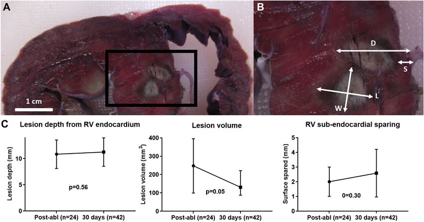
4                                                                                                             Heart Rhythm, Vol -, No -, - 2021
print & web 4C=FPO

                     Figure 2 In vivo myocardial lesion characteristics. A: Short-axis gross pathology of 2 adjacent lesions in the interventricular septum (IVS). B: Magnification of
                     IVS demonstrating lesion measurements (D, depth; S, subendocardial surface sparing; L, length; W, width; T, thickness is not shown, but was measured by
                     sectioning serial short-axis cross-sections). C: Quantification of gross pathological lesion characteristics. Graph bars represent means, and error bars represent
                     SD. RV 5 right ventricular.

                     Mann-Whitney U test for data that were determined to be                             137 6 95 mm3 in volume; P 5 .028) than at 8 MHz (Online
                     nonnormally distributed. For data with normal distribution,                         Supplemental Figure 1). Sparing of the RV subendocardial
                     groupwise differences were assessed using the Student t                             surface tissue immediately adjacent to the transducer was
                     test (paired for comparisons at different time points) or anal-                     similar between both frequencies (1.6 6 1.2 mm vs 1.5 6
                     ysis of variance (ANOVA). These data are presented as mean                          0.6 mm distance from the RV endocardial surface to ablation
                     6 SD. A 1-way analysis of variance test was used when                               lesion; P 5 .797). To increase the study efficiency, in vivo
                     comparing multiple groups per time points. One survival                             studies were performed only with 6.5 MHz HIU catheters.
                     swine died at 14 days. Gross pathological data from this
                     swine were included with the 30-day survival swine. When
                                                                                                         In vivo ablation: Pathological analysis
                     comparing repeated measures (multiple ablation lesions per
                                                                                                         In the 4 animals euthanized after immediate post-ablation im-
                     animal) among multiple swine, fixed effects regression
                                                                                                         aging, histology detected 24 necrotic lesions out of 32 HIU
                     models were fit and SEs adjusted to address the correlation
                                                                                                         ablation attempts (75% efficiency); while 42 necrotic lesions
                     among measurements from the same animal. Means with
                                                                                                         were found out of 64 HIU ablation attempts (66% efficiency)
                     bootstrapped 95% confidence intervals (CIs) based on 500
                                                                                                         in survival animals. Sonication locations that were more
                     replications are reported from these models. All statistical
                                                                                                         apically located were least likely to result in visible lesions
                     tests were 2-sided, with P  .05 considered significant.
                                                                                                         on gross pathology, largely because of the inability of the
                                                                                                         custom steerable sheath to reach the apical septum.
                     Results                                                                                 HIU lesions were typically ellipsoid in shape (Figure 2),
                     Comparison of US frequencies                                                        similar to prior descriptions of HIU epicardial LV lesions,
                     The acoustic fields of HIU catheters with fundamental fre-                           and in contrast to the typical half-ellipsoid shape of RF le-
                     quencies of 5.0, 6.5, and 8.0 MHz were mapped (Figure 1)                            sions.9 The mean lesion depth was similar immediately posta-
                     and predicted that 6.5 MHz would generate the largest and                           blation (10.8 mm; 95% CI 9.9–12.4 mm) and at 30 days (11.2
                     deepest acoustic field. Ablation at 5 MHz did not reproducibly                       mm; 95% CI 10.6–12.4 mm) (P 5 .56), although lesion vol-
                     result in necrotic lesions (32 sonications yielded only 4 iden-                     ume decreased over time (from 255 [95% CI 198–440] to
                     tifiable small lesions on gross pathology), likely because of                        162 [95% CI 133–234] mm3; P 5 .05) (Figure 2). The RV sub-
                     the reduced thermal effects of US at lower frequencies having                       endocardial surface was similarly spared at both time points
                     fewer harmonics formed during nonlinear acoustic propaga-                           (2.2 mm; 95% CI 1.7–3.3 mm vs 2.7 mm; 95% CI 2.3–3.4
                     tion.11,12 Ablation at 6.5 MHz generated lesions that were                          mm; P 5 .3). Similar to lesion depth, lesion length (L, going
                     deeper (10.7 6 3.2 mm vs 8.3 6 2.6 mm from the RV endo-                             away from the transducer from the RV to the LV endocardium
                     cardium; P 5 .0015) and larger in volume (223 6 171 mm3 vs                          and excluding RV subendocardial sparing) was similar at both

                                                           FLA 5.6.0 DTD  HRTHM8634_proof  5 February 2021  9:47 pm  ce
Nazer et al Mid-Myocardial Catheter Ablation by High-Intensity Ultrasound 5
print & web 4C=FPO
Figure 3 Gross pathology images of in vivo lesions immediately postablation (left) and at 30 days (right). Bottom row images demonstrate image processing
with spline analysis defining the portion of the lesion surface area to the right (RV; red) and left (LV; green) ventricular side of the septum, with the mid-septum
defined as the white dotted line.
time points. However, lesion width (W) decreased from 6.4 days, dense midseptal fibrosis was noted without a surround-
(95% CI 5.9–7.0) to 5.5 (95% CI 5.0–6.1) mm (P 5 .006) ing inflammatory infiltrate (Online Supplemental Figure 2).
and lesion thickness (T) decreased from 7.8 (95% CI 7.2–
8.4) to 6.2 (95% CI 5.6–6.8) mm at 30 days (P , .0001). TTE, MRI, and electroanatomic mapping
Based on spline analysis image processing, a similar TTE confirmed gross pathological findings: diastolic IVS
portion of the lesion surface area was deposited on the LV thickness by TTE increased from 11.8 6 1.3 mm at baseline
side of the IVS (30%; 95% CI 22%–46% postablation vs to 13.4 6 1.6 mm (P 5 0.002) immediately post-ablation (pre-
39%; 95% CI 33%–51% at 30 days; P 5 .29) (Figure 3). sumably because of edema) but decreased to 10.0 6 1.3 mm at
Spline analysis showed that lesion transmurality increased 30 days (P 5 .0003 by ANOVA for between-group differ-
from 58% (95% CI 50%–76%) to 81% (95% CI 74%– ences; P 5 0.004 for comparison between pre-ablation and
93%) over 30 days (P 5 .002). Increased transmurality 30 days post-ablation) despite swine growth. The LV ejection
despite decreased lesion volume at 30 days compared with fraction at baseline was 58% 6 6%, decreased to 49% 6 4%
immediately post-ablation was attributable to IVS remodel- immediately postablation (due to hypokinesis of the basal
ing resulting in reduced thickness: IVS thickness at the level IVS), and recovered to 58% 6 5% at 30 days (P 5 .0002 by
of the lesions decreased from 16.0 6 1.7 mm post-ablation to ANOVA for between-group differences; P 5 .92 for compar-
10.6 6 2.4 mm at 30 days (P 5 .0074) (Figure 3). ison between pre-ablation and 30 days post-ablation).
Histology demonstrated myocyte disruption without MRI also confirmed IVS remodeling: IVS thickness
fibrosis or inflammation immediately post-ablation. At 30 increased from 11.0 6 0.8 mm at baseline to 13.8 6 1.7
FLA 5.6.0 DTD HRTHM8634_proof 5 February 2021 9:47 pm ce6 Heart Rhythm, Vol -, No -, - 2021
Figure 4 Short-axis magnetic resonance imaging images of high-intensity ultrasound (HIU) lesions immediately postablation (left) and at 30 days (right). Late
gadolinium enhancement in the septum (top row) corresponded to increased precontrast T1 time (bottom row). Ablation had been performed from the right
ventricle in the direction of white arrows.
mm immediately postablation (presumably because of edema) (10.6 6 6.1 cm2) and at 30 days (7.0 6 3.5 cm2) (P 5 .35).
but decreased to 10.6 6 1.1 mm at 30 days (P 5 .003 by AN- Similar unipolar voltage findings were present in the LV
OVA) despite swine growth. First-pass MRI contrast perfu- septum (7.6 6 0.5 cm2 vs 6.0 6 1.8 cm2; P 5 .31) confirming
sion imaging demonstrated dense perfusion defects in the deep lesions (Figure 5). No fractionated or late electrograms
basal to mid-IVS immediately post-ablation (Online were noted, suggesting fairly homogeneous, deep ablation.
Supplemental Videos 1 and 2). Late gadolinium enhancement
(LGE) detected areas of HIU ablation in the IVS, but resolu-
tion was insufficient to quantify individual lesion size on a Procedural complications
per-lesion basis: 3.6 6 1.1 regions of LGE were present post- No AV block, QRS widening, pericardial effusions, or ven-
ablation and 3.4 6 1.1 regions at 30 days (Figure 4; Online tricular septal defects were noted intra- or post-procedure.
Supplemental Figure 3), likely because of the coalescence of All survival swine recovered uneventfully from anesthesia
multiple HIU lesions into single LGE regions. T1 time at the after the procedure. One survival swine died suddenly at 14
basal IVS HIU ablation sites increased from 1176 6 18 ms days postprocedure, with no structural abnormalities deter-
preablation to 1408 6 87 ms postablation and remained mined at autopsy.
elevated at 30 days (1319 6 128 ms; P 5 .008 by ANOVA).
T2 time increased from 40.4 6 5.1 to 64.2 6 6.0 ms postabla-
tion but then returned to near baseline 47.1 6 5.8 ms at 30 days Discussion
(P 5 .1169 for comparison between pre-ablation and 30 days; There are spatial challenges related to catheter ablation of
P , .0001 by ANOVA for groupwise comparison). Data were VAs. Mid-myocardial sites of VA origin and epicardial sites
similar when normalizing septal ablation site T1 and T2 values of origin in patients unsuitable for percutaneous subxiphoid
with basal inferolateral (non-ablated) T1 and T2 values. pericardial access pose significant challenges for RF ablation.
Electroanatomic voltage mapping did not reveal any bipolar Several novel strategies have arisen to improve upon shallow
voltage abnormalities at standard voltage thresholds, but lesion depth of standard RF ablation, including an RF needle
demonstrated new areas of reduced unipolar voltage along catheter,13 stereotactic body radiation therapy,14 use of hypo-
the basal and mid-RV septum immediately post-ablation tonic irrigated RF ablation,15 bipolar RF ablation,16,17 and
FLA 5.6.0 DTD HRTHM8634_proof 5 February 2021 9:47 pm ceNazer et al Mid-Myocardial Catheter Ablation by High-Intensity Ultrasound 7
print & web 4C=FPO
Figure 5 Electroanatomic unipolar voltage mapping of the right (RV) and left (LV) ventricular septum. New unipolar voltage abnormalities noted postablation
and at 30 days, confirming the presence of deep mid-myocardial lesions.
alcohol venous ablation,18 although access to these therapies heterogeneous necrosis in the IVS with lesions spaced 1 cm
is limited, and recurrence and complication rates remain high. apart, although late AV block cannot be excluded.19
There are several potential advantages of using HIU over HIU lesions were successfully identified by cardiac MRI,
RF for targeting mid-myocardial and epicardial sites from the which may serve as a means of assessing lesion extent clini-
endocardium. Lower US absorption in tissue allows deeper cally using either LGE or noncontrast T1 imaging, as has pre-
penetration. Moreover, US transducers can be geometrically viously been shown for RF.20 In our study, T1 and T2 times at
focused, or the natural focus of the flat US element may the IVS were both elevated immediately post-ablation whereas
permit an “offset” that, with catheter cooling by irrigation, only T1 time remained elevated at 30 days, suggesting an
can spare tissue immediately adjacent to the transducer, acute edematous and/or inflammatory reaction (although sig-
thereby avoiding the subendocardial His-Purkinje system nificant inflammation not noted histologically post-ablation),
with septal ablation. Finally, US-based ablation may permit resolving to dense, mid-septal fibrosis without edema or
the real-time monitoring of the region of thermal injury that inflammation at 30 days (which was confirmed by histology).
could help guide lesion placement or size. It should be noted that our MRI lacked the resolution to confi-
In our study, HIU ablation from the RV side of the IVS re- dently measure individual lesion dimensions for correlation
sulted in lesions of w11 mm depth, initially spanning 58% of with gross pathology.
the IVS acutely increasing to 81% transmural at 30 days. This Limitations of our study include lack of an RF control
increase in transmurality was, in part, explained by remodeling group, although the shallow depth of RF ablation has been
of the IVS, which was noted to markedly decrease in thickness well-documented,4–6 including in our prior study9 where it
in response to septal ablation. Indeed, 38% of the lesion sur- was 4.7 6 4.0 mm and significantly less deep than epicardial
face area was deposited on the far-field, LV side of the IVS, HIU. Swine were not monitored with telemetry or implant-
with the first 2–3 mm of the RV septal subendocardium able loop recorder monitoring over the 30-day survival
spared. No QRS widening, bundle branch block, or AV block period; we cannot exclude delayed transient AV block. We
was noted, suggesting protection of the subendocardial also did not test a large range of frequencies, transducer sizes
His-Purkinje system. However, it should be noted that while (restricted to 3 ! 5 mm transducers that would accommodate
extensive ablation of the basal-mid RV anteroseptum was per- a 12-F catheter), or differences in acoustic power or lesion
formed, sonications were not performed in a dedicated manner duration time. Instead, we used HIU frequencies within a
over the His and right bundle potentials, but simply in their range known to promote thermal necrosis21 and our previous
general region. Furthermore, we did not histopathologically experience to narrow our range of HIU variables, compared
define the His-Purkinje system after euthanasia and thus 5, 6.5, and 8 MHz in ex vivo models, which identified 6.5
were unable to define the proximity of our ablation lesions MHz as the optimal frequency for the transducer size in the
to the conduction system. No acute complications were noted, in vivo study.
but there was 1 autopsy-negative sudden death event at 14 Another limitation is the large variability in lesion sizes.
days that we presume was due to VA secondary to While TTE was used to confirm placement against the RV
FLA 5.6.0 DTD HRTHM8634_proof 5 February 2021 9:47 pm ce8 Heart Rhythm, Vol -, No -, - 2021
endocardial surface, we cannot exclude the periodic noncon- Appendix
tact, which could explain the variability as well as why lesion Supplementary data
efficiency was not 100% (ie, fewer histological lesions than Supplementary data associated with this article can be found
ablation procedures performed; most evident for sonications in the online version at https://doi.org/10.1016/j.hrthm.202
made in the most apical portions of the RV) and also why 0.12.027.
lesion size varied. Furthermore, our side-facing transducer
may not have always been exactly parallel to the IVS. Future
iterations of the catheter should use a contact- and
orientation-sensing mechanism. Integration of the catheter
References
into electroanatomic mapping may also aid in assessing con- 1. Oloriz T, Silberbauer J, MacCabelli G, et al. Catheter ablation of ventricular
tact as well as allow us to guide HIU ablation directly over the arrhythmia in nonischemic cardiomyopathy: anteroseptal versus inferolateral
His-Purkinje system to formally demonstrate sparing of the scar sub-types. Circ Arrhythm Electrophysiol 2014;7:414–423.
2. Haqqani HM, Tschabrunn CM, Tzou WS, et al. Isolated septal substrate for ven-
conduction system with surface-sparing lesions. Finally, tricular tachycardia in nonischemic dilated cardiomyopathy: incidence, character-
our studies were not performed in a model of arrhythmia or ization, and implications. Heart Rhythm 2011;8:1169–1176.
cardiomyopathy. It has been shown that RF applications 3. Romero J, Shivkumar K, Valderrabano M, et al. Modern mapping and ablation
techniques to treat ventricular arrhythmias from the left ventricular summit and
within the myocardial scar are less likely to generate reliable interventricular septum. Heart Rhythm 2020;17:1609–1620.
and large necrotic lesions compared with applications per- 4. Jauregui-Abularach ME, Campos B, Betensky BP, Michele J,
formed in healthy myocardium because of heterogeneous Gerstenfeld EP. Comparison of epicardial cryoablation and irrigated radiofre-
quency ablation in a swine infarct model. J Cardiovasc Electrophysiol 2012;
electrical impedance of scar tissue.22 HIU thermal necrosis 23:1016–1023.
relies on tissue acoustic impedance, which is more homoge- 5. John RM, Connell J, Termin P, et al. Characterization of warm saline-enhanced
neous across tissue types than electrical impedance.23 Thus, radiofrequency ablation lesions in the infarcted porcine ventricular myocardium.
J Cardiovasc Electrophysiol 2014;25:309–316.
HIU lesions may perform more efficiently in scar, although 6. D’Avila A, Houghtaling C, Gutierrez P, et al. Catheter ablation of ventricular
this has yet to be tested. epicardial tissue: a comparison of standard and cooled-tip radiofrequency energy.
Future investigations of HIU should assess lesions in large Circulation 2004;109:2363–2369.
7. Nagarajan VD, Ho SY, Ernst S. Anatomical considerations for His bundle pacing.
animal cardiomyopathy models, use long-term ambulatory Circ Arrhythm Electrophysiol 2019;12:e006897.
rhythm monitoring for proarrhythmia, and assess endocardial 8. Nazer B, Gerstenfeld EP, Hata A, Crum LA, Matula TJ. Cardiovascular applica-
to epicardial lesions with ablation over the non-septal LV tions of therapeutic ultrasound. J Interv Card Electrophysiol 2014;39:287–294.
9. Nazer B, Salgaonkar V, Diederich CJ, et al. Epicardial catheter ablation using
endocardium. While we previously demonstrated a dose- high-intensity ultrasound: validation in a swine model. Circ Arrhythm Electro-
response relationship between HIU powers of 15–30 W physiol 2015;8:1491–1497.
and lesion depth and volume9 (and thus chose 30 W for 10. Koruth JS, Dukkipati S, Carrillo RG, et al. Safety and efficacy of high-intensity
focused ultrasound atop coronary arteries during epicardial catheter ablation. J
this study as it is the maximal acoustic power that the trans- Cardiovasc Electrophysiol 2011;22:1274–1280.
ducer and circuitry can tolerate in the continuous wave 11. Swindell W. A theoretical study of nonlinear effects with focused ultrasound in
mode), further control of lesion depth and size may also be tissues: an “acoustic bragg peak.” Ultrasound Med Biol 1985;11:121–130.
12. Hynynen K, Roemer R, Anhalt D, et al. A scanned, focused, multiple transducer
achieved with adjusting transducer size or ablation time ultrasonic system for localized hyperthermia treatments. Int J Hyperthermia 2010;
and should be investigated. Our finding of reduction in IVS 26:1–11.
thickness is intriguing and warrants further investigation 13. Stevenson WG, Tedrow UB, Reddy V, et al. Infusion needle radiofrequency abla-
tion for treatment of refractory ventricular arrhythmias. J Am Coll Cardiol 2019;
with a consolidated lesion set over the basal septum to deter- 73:1413–1425.
mine whether HIU may be a suitable approach for septal 14. Cuculich PS, Schill MR, Kashani R, et al. Noninvasive cardiac radiation for abla-
reduction therapy in hypertrophic cardiomyopathy. tion of ventricular tachycardia. N Engl J Med 2017;377:2325–2336.
15. Nguyen DT, Tzou WS, Sandhu A, et al. Prospective multicenter experience with
cooled radiofrequency ablation using high impedance irrigant to target deep
myocardial substrate refractory to standard ablation. JACC Clin Electrophysiol
2018;4:1176–1185.
Conclusion 16. Koruth JS, Dukkipati S, Miller MA, Neuzil P, D’Avila A, Reddy VY. Bipolar irri-
This first study of HIU endocardial ventricular ablation demon- gated radiofrequency ablation: a therapeutic option for refractory intramural atrial
and ventricular tachycardia circuits. Heart Rhythm 2012;9:1932–1941.
strates deep, nearly transmural mid-myocardial lesion forma- 17. Nguyen DT, Tzou WS, Brunnquell M, et al. Clinical and biophysical evaluation
tion with sparing of the subendocardium and His-Purkinje of variable bipolar configurations during radiofrequency ablation for treatment of
ventricular arrhythmias. Heart Rhythm 2016;13:2161–2171.
system.
18. Kreidieh B, Rodríguez-Ma~nero M, Schurmann P, Ibarra-Cortez SH, Dave AS,
Valderrabano M. Retrograde coronary venous ethanol infusion for ablation of re-
fractory ventricular tachycardia. Circ Arrhythm Electrophysiol 2016;9.
19. Bleszynski PA, Goldenberg I, Fernandez G, et al. Risk of arrhythmic events after
Acknowledgments alcohol septal ablation for hypertrophic cardiomyopathy using continuous
implantable cardiac monitoring. Heart Rhythm 2020;18:50–56.
We thank Xin Li, PhD, William Woodward, MS, Chris Die- 20. Kholmovski EG, Silvernagel J, Angel N, et al. Acute noncontrast T1-weighted
derich, PhD, and Peter Douglas Jones, BS, for their technical magnetic resonance imaging predicts chronic radiofrequency ablation lesions. J
assistance. We also thank Traci L. Schaller BS, LVT, LAT, Cardiovasc Electrophysiol 2018;29:1556–1562.
21. Salgaonkar VA, Diederich CJ. Catheter-based ultrasound technology for image-
for veterinary and anesthesia care and Thomas Murphy, guided thermal therapy: current technology and applications. Int J Hyperthermia
BA for assistance with electroanatomic mapping. 2015;31:203–215.
FLA 5.6.0 DTD HRTHM8634_proof 5 February 2021 9:47 pm ceNazer et al Mid-Myocardial Catheter Ablation by High-Intensity Ultrasound 9
22. Barkagan M, Leshem E, Shapira-Daniels A, et al. Histopathological characteriza- 23. Sarvazyan AP, Rudenko OV, Swanson SD, Fowlkes JB, Emelianov SY. Shear
tion of radiofrequency ablation in ventricular scar tissue. JACC Clin Electrophy- wave elasticity imaging: a new ultrasonic technology of medical diagnostics. Ul-
siol 2019;5:920–931. trasound Med Biol 1998;24:1419–1435.
FLA 5.6.0 DTD HRTHM8634_proof 5 February 2021 9:47 pm ceYou can also read