Histological and histomorphometric study of the cranial digestive tract of ostriches (Struthio camelus) with advancing age
←
→
Page content transcription
If your browser does not render page correctly, please read the page content below
Original Paper                                                   Veterinarni Medicina, 66, 2021 (04): 127–139

                                                                     https://doi.org/10.17221/120/2020-VETMED

Histological and histomorphometric study
of the cranial digestive tract of ostriches
(Struthio camelus) with advancing age
Zaima Umar1*, Anas Sarwar Qureshi1, Rehmatullah Shahid1,
Farah Deeba2
1
    Department of Anatomy, University of Agriculture, Faisalabad, Pakistan
2
    Department of Clinical Medicine and Surgery, University of Agriculture,
    Faisalabad, Pakistan
*Corresponding author: zaimaumar@gmail.com

Citation: Umar Z, Qureshi AS, Shahid R, Deeba F (2021): Histological and histomorphometric study of the cranial diges-
tive tract of ostriches (Struthio camelus) with advancing age. Vet Med-Czech 66, 127–139.

Abstract: The present study was conducted to determine the histological and histomorphometric variations
in the tongue, oesophagus, proventriculus, and gizzard of ostriches (Struthio camelus) with regards to the sex
and advancing age. A total of 40 healthy ostriches of both sexes and five age groups; young (up to 1 year,
1 to 2 years and 2 to 3 years) and adult (3 to 4 years and above 4 years) in equal numbers (n = 8) were used in this
study. The organs under study were collected immediately after slaughtering the birds. Overall, the colour, shape,
weight and various dimensions (length, width, and diameter) of the collected organs were recorded. The mean
values of the gross anatomical variables of the studied organs increased (P < 0.05) among all the young groups
(i.e., from 1 to 2 years, 2 to 3 years). Similarly, the organs under study in the adult groups (birds aged 3 to 4 years
and above 4 years) grew (P < 0.05) as well. However, the differences between the adults were not significant.
The histological analysis and histometric measurements were conducted on paraffin embedded tissue sec-
tions with Image J ® analysis software. The statistical analysis revealed a significant increase in the thickness
of the different tunics of the digestive organs in all the groups except those the adult groups. These findings
may be of importance for the strategic manipulation of feed and nutrition to enhance the growth rate and also
to diagnose pathological processes.

Keywords: biometry; oesophagus; gizzard; histometry; proventriculus; tongue

  Globally, ostrich farming is gaining importance              The crop, a storage place for feed, is absent in os-
on the market with food animal commodities, mak-             triches when compared to other avian species. The
ing it more diversified. However, there is still a gap       proventriculus of ostriches is large enough to store
in our knowledge of precise nutrient requirements            the food and, hence, compensates for the function
of ostriches. Their meat production depends on the           of the crop.
development of the entire digestive tract, including           The bird’s stomach consists of two parts: the
the stomach. A detailed study of the gastrointesti-          glandular (pars glandularis) and muscular (pars
nal tract (GIT) limited to the early postincubation          muscularis) part. The true stomach of the ostrich
period, up to day 72, in ostrich chickens showed             is sac-shaped and located in the cranial part of the
a rapid growth and development of its individu-              abdomen in the left hypochondrium (Bezuidenhout
al parts (Iji et al. 2003).                                  2001). In this part, protein digestion begins through

127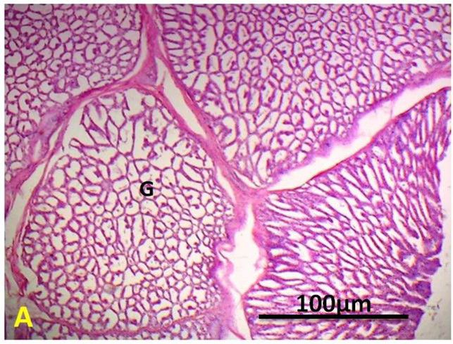
Original Paper                                              Veterinarni Medicina, 66, 2021 (04): 127–139

                                                                 https://doi.org/10.17221/120/2020-VETMED

the secretion of pepsinogen and hydrochloric acid       Table 1. Grouping of the young and adult birds
(Camiruaga et al. 2003). These two parts of the
                                                        Groups              Age        Male   Female     Total
stomach are separated by constriction, called isth-
                                                                        up to 1 year    4       4         8
mus gastris (Rossi et al. 2005). The deep glandular
                                                        Young groups     1–2 years      4       4         8
region (regio glandularis) is situated on the greater
curvature of the proventriculus. The junction be-                        2–3 years      4       4         8
tween the oesophagus and the proventriculus has                          3–4 years      4       4         8
                                                        Adult groups
a narrowed cranial end and a wide rounded cau-                         above 4 years    4       4         8
dal end.                                                Total                           20      20        40
  The thick-walled gizzard is situated to the left
of the midline, and it contains grit (small piec-       Experimental groups
es of stone) important for grinding and mixing
of food (Speer 2006; Oliveira et al. 2008). The pars      A total of 40 clinically healthy ostriches of either
muscularis of the gizzard is bilaterally curved with    sex (20 males, 20 females) comprising five age groups
a complex structure. In most birds, the gizzard         of equal size, namely young (up to 1 year, 1 to 2 years
develops with two-layers of smooth musculature.         and 2 to 3 years) and adult (3 to 4 years and above
Moreover, the innermost layer is covered by koilin,     4 years) in equal numbers (n = 8) were used in this
a substance formed by protein secreted from the         study (Table 1).
glands combined with entrapped sloughed cells
and cellular debris (Shanawany and Dingle 1999).
A simpler gizzard is observed in piscivorous and        Collection of samples
pray birds. The caudodorsal and caudoventral areas
of the thick muscles of the ostrich gizzard are par-      All the cranial digestive tract organs studied
ticularly well developed, and its thickness is 5.2–     (tongue, oesophagus, proventriculus, gizzard) were
6.5 cm, in hens, its thickness is only 1.5–2.1 cm       collected from the Signature Meat Shop Lahore and
(Sales 2006).                                           Riphah College of Veterinary Sciences, Lahore,
  To the best of our knowledge, the biometrical and     Pakistan. Immediately after slaughtering, the or-
histometric variations in the tongue, oesophagus,       gans were washed with normal saline for use in the
proventriculus, and gizzard of ostriches (Struthio      anatomical and histological studies.
camelus) with regards to the advancing age and
sex have not been studied yet in detail beyond the
early postincubation period.                            Biometric characteristics
  Therefore, this study was designed to perform
the biometry and histometry of the digestive or-          Following the collection, the gross features (i.e.,
gans in ostriches over a life span longer than four     shape, colour), weight, biometric characteristics
years, concerned with the sex and progressing age,      (length, width, and diameter) of the collected or-
to illustrate the changes also at the tissue and cel-   gans were measured with the help of a measuring
lular levels.                                           tape. The length of the tongue (cm) was measured
                                                        from the apex to the root, and the width (cm) was
                                                        measured from the basis of the tongue, as shown
MATERIAL AND METHODS                                    in Figure 1. The oesophageal length (cm) was taken
                                                        from the pharyngeal opening to the proventricular
Ethical concern                                         opening, and outer diameter was also measured.
                                                        The width (cm) of the proventriculus and gizzard
  This research was conducted according to the          were recorded dorsoventrally, whereas the length
standards of the research ethics committee of the       (cm) of proventriculus was determined from the
University of Agriculture, Faisalabad-Pakistan          oesophageal distal termination to the proventricu-
(Letter No. 962).                                       lar-gizzard (PG) junction. Similarly, the gizzard
  No bird was otherwise harmed and no health            length (cm) was measured from the PG junction
hazard was caused to the handlers during the            to the duodenal opening (Figure 1). An electronic
slaughtering process.                                   weighing balance was used to weigh (g) the organs.

128
Original Paper Veterinarni Medicina, 66, 2021 (04): 127–139
https://doi.org/10.17221/120/2020-VETMED
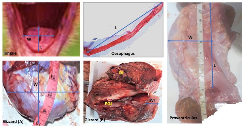
Figure 1
Figure 1. Photomicrograph of the different digestive organs of ostriches
The length of the tongue (L) was measured from the apex to the root, the width (W) was measured from the base
of the tongue. The oesophageal length (L) was taken from the oesophageal distal termination to the proventricular opening,
and the outer diameter was also measured. The width (W) of the proventriculus and gizzard was measured dorsoventrally
while the length (L) of the proventriculus was determined from oesophageal opening to the proventricular-gizzard junc-
tion (PGJ). Similarly, the gizzard length (L) was measured from the proventricular-gizzard junction (PGJ) to the duodenal
opening (DJ). The fibrous muscular wall thickness (WT) of gizzard was also measured
Histological study lamina propria, lamina muscularis), tela submu-
cosa, tunica muscularis, and tunica adventitia/se-
The collected organs (tongue, oesophagus, rosa of each organ with the help of the automated
proventriculus and gizzard/ventriculus of each image analysis system Image J ® v1.80 (Research
bird) were cut into small cubes of 1 cm 3 using Service Branch, National Institute of Mental Health,
a sharp knife/blade. These pieces were washed Bethesda, Maryland, USA).
in normal saline. All the organs were fixed in neu-
tral buffered 10% formalin, followed by the forma-
tion of paraffin blocks by an embedding technique Statistical analysis
(Bancroft and Gamble 2008). Thin slices of 5–7 µm
thickness were obtained using a rotary microtome. A factorial one-way analysis of variance (ANOVA)
These sections were mounted on glass slides and was used to compare the means of the parameters.
stained with haematoxylin and eosin. The stained Tukey’s honest significance test was used to com-
sections were observed under × 100 (10 × 10) mag- pare the group means at a 5% level of significance
nification. (Rushing et al. 2013).
Histometric analysis RESULTS
Photomicrographs of each sample of each studied Biometric characteristics
organ were captured using a Nikon optiphot mi-
croscope at × 100 magnification These photos were The mean ± SEM values of weight (g), length
used to determine the thickness of four histologi- (cm), width (cm), diameter, and wall thickness (cm)
cal layers, namely the tunica mucosa (epithelium, of the selected digestive organs (i.e., the tongue,
129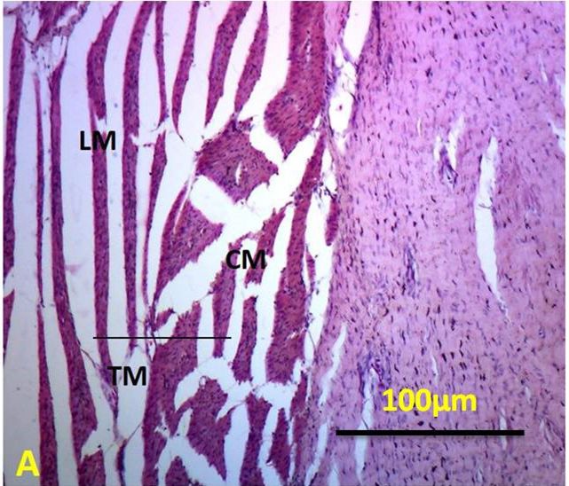
Table 2. Mean (± SEM) of the weight, length, width, diameter and the wall thickness of the digestive organs of the ostriches (Struthio camelus) of the different age-

130
        groups and sexes

                                                                          Sex groups                                                   Age groups
                                         Mean ± SEM
        Organs           Parameters                              male                   female     up to 1 year       1–2 years         2–3 years          3–4 years        above 4 years
                                                                                                                                                                                                                                         Original Paper

                                          (n = 40)
                                                               (n = 20)                (n = 20)      (n = 8)           (n = 8)           (n = 8)            (n = 8)            (n = 8)
                           weight
                                           3.95 ± 0.21        3.94 ± 0.20a         3.94 ± 0.19a     2.89 ± 0.02a      3.38 ± 0.14b      3.75 ± 0.15c       4.80 ± 0.06d       4.93 ± 0.09d
                            (g)
                           length
                                           5.56 ± 0.33        3.55 ± 0.23a         3.54 ± 0.22a     1.77 ± 0.11a      2.82 ± 0.21b      3.97 ± 0.12c       4.58 ± 0.08d       4.63 ± 0.07d
                            (cm)

      Tongue
                           width
                                           2.33 ± 0.16        2.34 ± 0.17a         2.32 ± 0.15a     1.50 ± 0.04a      1.73 ± 0.05b      2.54 ± 0.12c       2.92 ± 0.01d       2.94 ± 0.01d
                           (cm)

                           weight
                                        198.89 ± 11.44     192.66 ± 10.87a      193.44 ± 10.67a    118.0 ± 4.16a      142 ± 8.19        195 ± 2.89c     218.33 ± 6.01d     223.33 ± 6.17d
                            (g)
                           length
                                        118.23 ± 12.15     122.74 ± 8.89a       121.72 ± 8.85a     77.57 ± 2.29a    101.00 ± 3.52    115.11 ± 4.67c     131.13 ± 7.12d     132.96 ± 7.55d
                            (cm)

      Oesophagus
                          diameter
                                           3.13 ± 0.14        3.12 ± 0.13a         3.11 ± 0.11a     2.52 ± 0.05a      2.73 ± 0.06       3.00 ± 0.05c       3.70 ± 0.06d       3.72 ± 0.12d
                            (cm)

                           weight
                                         687.2 ± 21.9         750 ± 28.12a         752 ± 29.11a   586.00 ± 6.66a    620.51 ± 9.27b   681.62 ± 5.49c      777.3 ± 28.3d     776.45 ± 30.7d
                            (g)
                           length
                                         26.07 ± 0.51       27.16 ± 0.61a         27.06 ± 0.61a    23.46 ± 0.23a     24.77 ± 0.04b    25.97 ± 0.35c      28.01 ± 0.57d      28.16 ± 0.64d
                            (cm)
                           width
                                         12.89 ± 0.18       11.99 ± 0.15a         12.71 ± 0.17a    11.93 ± 0.20a     12.47 ± 0.02b    12.97 ± 0.04c      13.57 ± 0.06d      13.67 ± 0.07d
                           (cm)

      Proventriculus
                       wall thickness
                                           3.30 ± 0.11        3.27 ± 0.12a         3.26 ± 0.12a    2.762 ± 0.02a      3.06 ± 0.09b      3.51 ± 0.01c       3.66 ± 0.005d      3.67 ± 0.008d
                            (cm)

                           weight
                                        1 086.2 ± 39.3     1 250.1 ± 6.81a        1 251 ± 6.80a    888.3 ± 1.74a    984.56 ± 6.84b   1 050.4 ± 21.10c   1 253.8 ± 6.92d    1 254.1 ± 6.71d
                            (g)
                           length
                                         26.45 ± 0.61         27.1 ± 0.71a        27.12 ± 0.70a     23.3 ± 0.06a      24.6 ± 0.25b    26.83 ± 0.56c        28.5 ± 0.31d     29.03 ± 0.56d
                            (cm)

      Gizzard
                           width
                                         16.72 ± 0.31       16.77 ± 0.42a         16.75 ± 0.39a    14.80 ± 0.05a     16.13 ± 0.15b    16.97 ± 0.07c      17.73 ± 0.17d      17.96 ± 0.11d
                           (cm)
                       wall thickness
                                           3.92 ± 0.19        3.99 ± 0.21a         3.97 ± 0.20a     2.87 ± 0.01a      3.34 ± 0.12b      3.96 ± 0.02c       4.68 ± 0.06d       4.75 ± 0.04d
                            (cm)
        a–d
                                                                                                                                                                                              https://doi.org/10.17221/120/2020-VETMED
                                                                                                                                                                                                                                         Veterinarni Medicina, 66, 2021 (04): 127–139

                  Mean values having a different letter of the alphabet differ significantly from one another (P ≤ 0.05)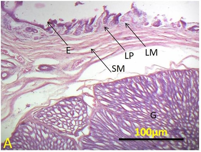
Original Paper                                                       Veterinarni Medicina, 66, 2021 (04): 127–139

                                                                       https://doi.org/10.17221/120/2020-VETMED

oesophagus, proventriculus, and gizzard) of either         did not affect any of the variables within the same
sex and five age progressive groups of ostriches           age group (Table 2). A rapid increase (P < 0.05)
(Table 2). The statistical analysis showed that the        in the morphometrical parameters of the cranial
age is directly related to the macroscopic values          digestive organs was observed during the young age
of the organs. The mean values of the gross ana-           period, but a non-significant (P > 0.05) increase was
tomical variables of the studied organs increased          recorded in the adults (3 to 4 years and > 4 years).
(P < 0.05) among all young groups (i.e., up to 1 year,
1 to 2 years, 2 to 3 years). The values for young
groups were significantly (P < 0.05) lower than            Histometric characteristics
those for the adult groups of birds (3 to 4 years
and above 4 years). The mean values of gross ana-            The histometric analysis of the organs under
tomical parameters of studied organs followed              study, including the tongue, oesophagus, proven-
an increasing trend (P < 0.05) within young group,         triculus, and gizzard of the different groups of
however, mean values were significantly (P < 0.05)         ostriches, are presented in Figures 2, 3, 4-A, B, C
lower than that of adults group (Table 2). The sex         and 5-A, B.

         Figure 2

(A)                                                            (B)
Figure 2. Tongue photomicrograph (A) of an adult ostrich. Tongue photomicrograph (B) of a young ostrich; Haema-
toxylin and eosin (H&E) × 100
E = epithelium; G = salivary glands; P-S = propria-submucosa

        Figure 3

 (A)                                                           (B)
Figure 3. Oesophagus photomicrograph (A) of an adult ostrich. Oesophagus photomicrograph (B) of a young ostrich;
Haematoxylin and eosin (H&E) × 100
E = epithelium; LM = lamina muscularis; LP = lamina propria; OG = oesophageal glands; SM = submucosa; TM = tunica
muscularis

131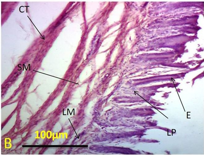
Original Paper                                                    Veterinarni Medicina, 66, 2021 (04): 127–139

                                                                      https://doi.org/10.17221/120/2020-VETMED

        Figure 4-A

 (A)                                                           (B)
Figure 4-A. Proventriculus (glandular part) photomicrograph (A) of a young ostrich. Proventriculus (glandular part)
photomicrograph (B) of an adult ostrich; Haematoxylin and eosin (H&E) × 100
CT = connective tissue; E = epithelium; G = gastric glands; LM = lamina muscularis; LP = lamina propria; SM = sub
mucosa; TM = tunica muscularis

         Figure 4-B

 (A)                                                           (B)
Figure 4-B. Proventriculus (glandular part) photomicrograph (A) of an adult ostrich. Proventriculus (non-glandular
part) photomicrograph (B) of an adult ostrich; Haematoxylin and eosin (H&E) × 100
E = epithelium; G = gastric glands; LM = lamina muscularis; LP = lamina propria; SM = sub mucosa; TM = tunica muscularis

        Figure 4-C

  (A)                                                           (B)
Figure 4-C. Proventriculus photomicrograph (A) of tunica muscularis of a young ostrich. Proventriculus photomi-
crograph (B) of tunica muscularis of an adult ostrich; Haematoxylin and eosin (H&E) × 100
CM = circular muscularis; LM = longitudinal muscularis; TM = tunica muscularis

132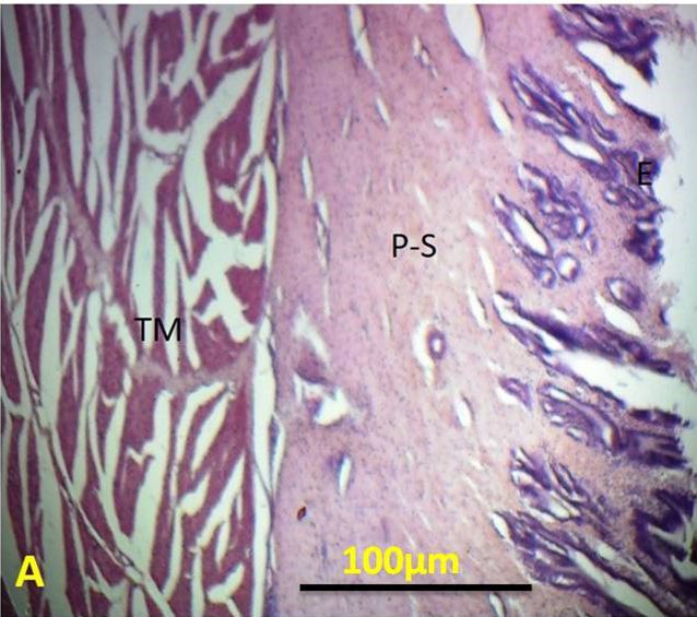
Original Paper                                                  Veterinarni Medicina, 66, 2021 (04): 127–139

                                                                  https://doi.org/10.17221/120/2020-VETMED

      Figure 5-A

(A)                                                       (B)
Figure 5-A. Gizzard photomicrograph (A) of a young ostrich. Gizzard photomicrograph (B) of an adult ostrich; Hae-
matoxylin and eosin (H&E) × 100
E = epithelium; LM = lamina muscularis; P-S = propria-submucosa; TM = tunica muscularis

      Figure 5-B

(A)                                                       (B)
Figure 5-B. Gizzard photomicrograph (A) of a young ostrich. Gizzard photomicrograph (B) of an adult ostrich; Hae-
matoxylin and eosin (H&E) × 100
CM = circular muscles; LM = longitudinal muscles; TM = tunica muscularis

  The different histometric parameters, includ-           DISCUSSION
ing the thickness of the epithelium, cartilage,
lamina muscularis propria-submucosa, inner cir-              The ostrich (Struthio camelus), the largest non-
cular, outer longitudinal, and tunica adventitia/         flying bird, is a preferred meat source due to its
serosa in the young and adult ostriches, are pre-         very low cholesterol and fat contents.
sented in Table 3.                                           Ostrich by-products, feathers and skin, are
  The statistical analysis revealed that the mean val-    in great demand in the leather industry because
ues of the thickness of all layers of all groups grew     it requires less acreage in comparison to livestock.
(P < 0.05) except between the adult (3 to 4 years and     The meat production of animals is directly related
above 4 years) age groups, but these values were          to different factors, like the health status, meta-
non-significantly (P > 0.05) different between the        bolic processes and digestive tract development.
sexes (Table 3).                                          Therefore, the digestive tract of ostrich was sub-

133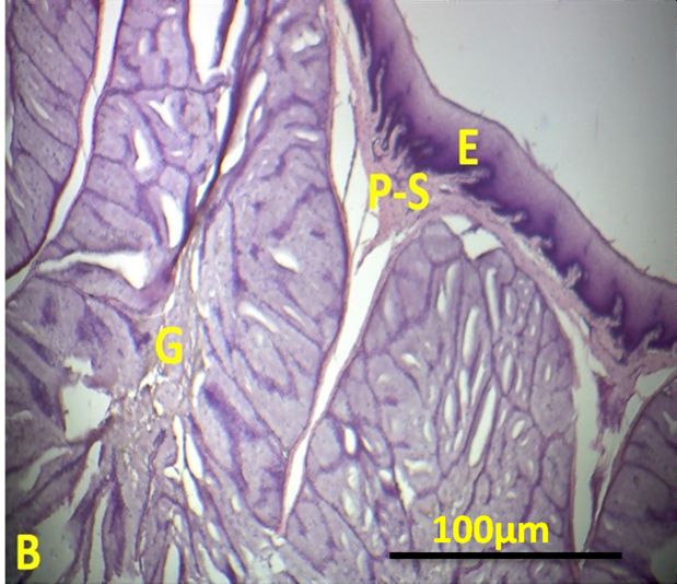
Table 3. Mean (± SEM) thickness (µm) of various cellular layers of the digestive organs of the ostriches (Struthio camelus) at different ages and both sexes
134
Sex groups Progressive age groups
Organs Parameters mean ± SEM male female up to 1 year 1–2 years 2–3 years 3–4 years above 4 years
(n = 40) (n = 20) (n = 20) (n = 8) (n = 8) (n = 8) (n = 8) (n = 8)
Original Paper
epithelium 96.56 ± 9.33 121.55 ± 8.23a 120.54 ± 8.22a 57.77 ± 4.11a 63.82 ± 5.21b 98.97 ± 6.36c 131.58 ± 7.08d 130.63 ± 7.06d
cartilage 458.89 ± 39.21 449 ± 35.8a 448.9 ± 34.23a 168.4 ± 13.9a 230.2 ± 57.4b 372.2 ± 25.1c 472.1 ± 40.5d 472.6 ± 41.5d
a a a b c d
propria submucosa 498.45 ± 31.16 485.34 ± 29.17 484.32 ± 29.15 211.4 ± 23.5 306.4 ± 25.2 496.55 ± 7.85 522.3 ± 24.5 524.4 ± 25.2d
Tongue
tunica muscularis (circular) 373.4 ± 45.4 381.4 ± 37.4a 383.4 ± 38.4a 127.26 ± 7.38a 167.26 ± 10.38b 291.3 ± 30.3c 383.4 ± 35.4d 393.4 ± 36.4d
a a a b c d
tunica muscularis (longitudinal) 333.4 ± 24.6 349.4 ± 29.6 348.4 ± 29.5 130.99 ± 9.33 269.17 ± 11.96 339.5 ± 14.7 359.5 ± 19.7 359.4 ± 19.6d
tunica adventitia 168.26 ± 11.38 156.36 ± 10.38a 157.26 ± 10.37a 115.68 ± 9.70a 130.02 ± 9.16b 159.4 ± 23.0c 199.3 ± 19.6d 198.3 ± 19.9d
epithelium 45.92 ± 15.625 47.92 ± 16.62a 48.92 ± 15.25a 9.92 ± 0.625a 24.11 ± 1.63b 36.66 ± 4.89c 49.71 ± 5.92d 49.92 ± 5.63d
lamina propria 73.36 ± 8.02 77.36 ± 7.02a 76.36 ± 6.02a 11.36 ± 1.02a 47.07 ± 1.20b 70.09 ± 2.20c 79.95 ± 4.09d 78.36 ± 4.02d
laminae muscularis 40.68 ± 15.92 42.68 ± 8.89a 41.68 ± 15.97a 44.68 ± 5.92a 43.7 ± 5.11b 101.46 ± 8.32c 99.74 ± 8.08d 44.68 ± 5.92d
a a a b c d
submucosa 140.02 ± 24.20 144.02 ± 14.20 143.02 ± 14.10 46.02 ± 4.20 89.58 ± 8.39 122.6 ± 15.9 151.2 ± 14.4 146.02 ± 14.20d
tunica muscularis (circular) 221.1 ± 19.21 224.1 ± 8.81a 222.1 ± 8.41a 67.1 ± 10.2a 136.00 ± 7.28b 204.95 ± 7.24c 227.84 ± 9.42d 226.1 ± 9.21d
Oesophagus
a a a b c d
tunica muscularis (longitudinal) 286.1 ± 31.0 291.1 ± 29.80 290.1 ± 29.70 76.1 ± 12.0 178.9 ± 14.0 250.1 ± 26.3 292.8 ± 20.1 296.1 ± 21.0d
tunica serosa 110.47 ± 20.47 112.47 ± 14.41a 111.77 ± 13.68a 40.47 ± 2.48a 70.12 ± 7.17b 91.6 ± 10.7c 114.6 ± 13.1d 114.47 ± 12.48d
epithelium 89.07 ± 23.80 92.27 ± 13.31a 92.11 ± 13.31a 39.07 ± 3.80a 79.95 ± 5.35b 84.49 ± 6.94c 95.8 ± 11.3d 99.07 ± 13.80d
lamina propria 80.50 ± 39.51 85.50 ± 18.51a 84.50 ± 19.24a 57.50 ± 2.51a 69.22 ± 3.69b 78.47 ± 4.49c 88.11 ± 9.93d 87.50 ± 9.51d
laminae muscularis 75.93 ± 23.54 78.83 ± 13.64a 77.30 ± 13.54a 48.93 ± 3.54a 63.78 ± 4.30 b 71.98 ± 6.69c 82.8 ± 13.1d 83.93 ± 13.54d
a a a b c d
submucosa 500.7 ± 47.2 505.23 ± 25.2 506.7 ± 25.2 360.7 ± 19.2 417.0 ± 20.2 456.9 ± 21.9 514.6 ± 26.7 510.7 ± 27.2d
tunica muscularis (circular) 512.8 ± 59.6 518.8 ± 28.6a 517.67 ± 27.6a 144.2 ± 24.1a 327.8 ± 29.6 b 465.6 ± 13.3c 513.6 ± 26.4d 522.8 ± 29.6d
Proventriculus
a a a b c d
tunica muscularis (longitudinal) 517.8 ± 49.1 520.8 ± 29.91 521.8 ± 30.21 365.8 ± 21.1 450.1 ± 28.9 495.1 ± 33.9 528.7 ± 38.0 527.8 ± 39.1d
tunica serosa 145.21 ± 41.78 150.21 ± 13.78a 151.21 ± 14.64a 75.21 ± 4.58a 105.21 ± 5.78b 142.6 ± 9.27c 157.7 ± 10.7d 158.21 ± 11.78d
epithelium 290.6 ± 49.0 294.6 ± 54.0a 2933.6 ± 57.0a 114.6 ± 11.70a 210.5 ± 14.81b 255.3 ± 27.9c 293.3 ± 46.1d 294.6 ± 47.0d
laminae muscularis 201.2 ± 45.6 210.2 ± 25.6a 208.2 ± 25.56a 110.2 ± 12.6a 142.0 ± 13.4b 64.0 ± 14.6c 199.9 ± 15.9d 216.2 ± 15.6d
a a a b c d
propria submucosa 214.9 ± 56.2 220.9 ± 16.2 219.7 ± 16.241 124.9 ± 10.2 156.07 ± 17.06 198.5 ± 22.8 222.6 ± 25.7 224.9 ± 26.2d
Gizzard
tunica muscularis (circular) 979.2 ± 71.6 982.2 ± 51.4a 981.2 ± 51.89a 406.2 ± 25.6a 654.0 ± 35.4b 842 ± 42.80c 1 012.7 ± 52.5d 999.2 ± 51.6d
tunica muscularis (longitudinal) 877.1 ± 69.6 878.1 ± 36.6a 880.1 ± 39.6a 656.1 ± 29.6a 794.5 ± 33.1b 826.49 ± 47.7c 886.7 ± 59.4d 887.1 ± 59.6d
a a a b c d
tunica serosa 80.83 ± 30.56 93.13 ± 14.68 92.53 ± 14.56 58.83 ± 4.86 63.25 ± 8.81 79.37 ± 9.06 90.17 ± 10.89 98.83 ± 10.86d
a–d
https://doi.org/10.17221/120/2020-VETMED
Veterinarni Medicina, 66, 2021 (04): 127–139
Mean values having a different letter of the alphabet differ significantly from one another (P ≤ 0.05)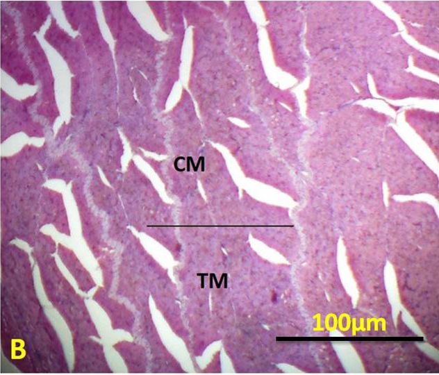
Original Paper                                               Veterinarni Medicina, 66, 2021 (04): 127–139

                                                                https://doi.org/10.17221/120/2020-VETMED

jected to morphometric and histological evalua-          prised of skeletal muscles that were divided into
tion in order to illustrate the changes at tissue and    two layers, inner circular and outer longitudinal
cellular level.                                          ones, which can be a link to its strong prehension
                                                         and mixing of the food with the saliva. The tunica
                                                         adventitia was composed of loose connective tissue.
Tongue                                                   Jackowiak and Ludwig (2008) and Guimaraes et al.
                                                         (2009) also described similar findings in three-
  The tongue of the ostriches was quite thick and        to five-year old and one and a half year old os-
short. The gross biometric results revealed that the     triches, respectively.
tongue was significantly (P < 0.05) longer among all
the young groups and also the mean values of the
adult group were significantly (P < 0.05) higher         Oesophagus
than the young group’s readings and remained
non-significant (P > 0.05) between the sexes in the        The gross biometrical results revealed that the
same age group (Table 2). These findings were            mean oesophagus length differed significantly
similar to studies by Rossi et al. (2005), who meas-     (P < 0.05) among all the young groups and also the
ured the length of the tongue in adult partridges        mean values of the adult group were significantly
(1.70 ± 0.08 cm in males and 1.65 ± 0.072 cm in fe-      (P < 0.05) higher than the young group’s readings
males) and Tadjalli et al. (2008), who measured the      and remained non-significant (P > 0.05) between
length of two-year-old ostriches (1.92 ± 0.086 cm).      the sexes in the same age group (Table 2). No lit-
In this study, these findings were determined in         erature is available on the oesophagi of ostriches
immature (> 2 years) as well as in adult. There was      to compare these findings to. Swallowing is accom-
no significant difference in tongue length between       plished by oesophageal peristalsis, and, in most
different sexes all ages group which is in line with     birds, appears to be aided by the extension of the
the findings of Rossi et al. (2005). The current study   neck. This study conflicted with the study of Bailey
indicated that the width of the tongue was signifi-      et al. (1997), who described the oesophagus as being
cantly (P < 0.05) wider in the adult ostriches than      significantly longer in male bustards when com-
in the young ostriches. The tongue width did not         pared to females. The present study indicated that
differ between males and females. Tadjalli et al.        the oesophagus was significantly (P < 0.05) longer
(2008) measured the width of the tongue (1.92 cm)        in the young ostriches than in the adult ostriches
in young ostriches. In this study, the weight of the     (225.60 ± 8.13 g) without any ingesta.
tongue was determined in the young and adult os-           The oesophagus of ostriches consisted of four
triches. The mean values remained non-significant        layers, i.e., mucosa, sub-mucosa, muscularis and
(P > 0.05) between the sexes in a group, but were        adventitia. The oesophagus epithelium was com-
highly significantly (P < 0.05) different between        posed of squamous shape cells. Simple straight tu-
the young and adult groups. A larger length and          bular glands and connective tissue (loose to dense)
width of the tongue may be linked with the high-         were present in the lamina propria. The lamina
er production of saliva which helps the digestion        muscularis layer was formed of a well-developed
of food. The strong intrinsic muscles control the        layer of longitudinally arranged smooth muscles.
movement of the tongue (Beason 2003; Olsen et al.        The tela submucosa was a thin layer and consist-
2011). Tactile and temperature receptors are pre-        ed of a DICT, the sub-mucosal nerve and vascular
sent on the surface of their tongue, which helps         plexus. Similar trends were found in other species
to position and identify (sensitive to cold and hot)     of birds like the duck, chicken, geese, and pheasant
the food before swallowing it (Beason 2003; Olsen        (Srisai et al. 2002; Nagy et al. 2005; Shiina et al.
et al. 2011).                                            2005; Qureshi et al. 2017). Guimarraes et al. (2009)
  The present study revealed that the epithelium         also supported analogous findings in ostriches. The
of the tongue had squamous shaped cells with             muscularis tunic was composed of smooth mus-
stratifications and are not keratinised. The pro-        cles and divided into two layers according to the
pria-submucosa contained salivary glands, dense          orientation, either inner circular and outer longi-
irregular connective tissue (DICT), arteries, veins      tudinal, the same as reported in chickens (Rossi
and lymphatics. The tunica muscularis was com-           et al. 2005). Shiina et al. (2005) reported that the

135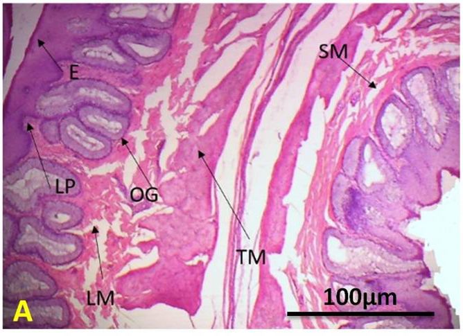
Original Paper                                              Veterinarni Medicina, 66, 2021 (04): 127–139

                                                               https://doi.org/10.17221/120/2020-VETMED

oesophagus consisted of two regions (cervical and       the feed and water for up to twenty hours. In older
thoracic) in all birds and was composed of smooth       birds and larger young birds with a slower metabo-
muscles. In this study, the thickness of all the lay-   lism, these water reserves may be stored for an even
ers was measured in the ostriches. All the layers       longer time. The present study indicated that the
were (P < 0.05) thicker in the adult group as related   proventriculus length and weight were (P < 0.05)
to the young group, but these values did not change     increased among all the young groups and also
within the sexes of the ostriches. Qureshi et al.       the mean values of the adult group were signifi-
(2017) also reported congruent findings in ducks.       cantly (P < 0.05) higher than the young group’s
                                                        readings and remained non-significant (P > 0.05)
                                                        between the sexes in the same age group (Table 2).
Proventriculus and gizzard                              Wu et al. (2010) reported the proventriculus length
                                                        and weight was 60 mm and 35.9 g, respectively,
  Ostriches do not have a crop. The proventriculus      in pheasants.
and gizzard can fulfil the crop’s function (stor-          The shape of the gizzard was like a biconvex
age) (Bezuidenhout 2001). The shape of the proven-      lens and was located left to the midline of in the
triculus was elongated and oval, so that it was not     body cavity. The internal membrane of the gizzard,
constricted in the middle like other avian species.     the koilin layer was very rigid, and detachable. Its
Its wall thickness was variable, the cardiac region’s   colour was yellowish-green, which was due to the
thickness had a minimum and membranous thick-           bile reflux and its feed content. Elnagy and Osman
ness, contrarily, but the half pyloric region was       (2010) revealed that the gizzard was an elongated
thin-walled, yet half of it was thick-walled. The       dilation between the oesophagus and the intestines
proventriculus gland was dumbbell-shaped.               of the digestive tract in rabbits. Wu et al. (2010)
The same organ shape was reported in doves and          reported a dark purple colour of the gizzard in the
owls by Mol (2010) and in falcons by Abumandour         pheasant, which was due to its feed. These length
(2013). Similar results were reported in ostriches      and weight values in the gizzard and proventriculus
by Mahdy (2009). Hassan and Moussa (2012) re-           are different in different species because it depends
vealed that the proventriculus was cone-shaped          upon the feed intake and body size. Szczepanczyk
in the pigeon. The actual stomach of the ostrich        (2005) reported that the shape and length of the
was sac-shaped and located at the cranial part of the   gizzard and proventriculus of the long-tailed duck
abdomen in the left hypochondrium (Bezuidenhout         depended upon its feed intake. Nasrin et al. (2012)
2001). In this part, the protein digestion begins       revealed that the weight of the gizzard was 5.32 g
by the secretion of pepsinogen and hydrochloric         in broiler chickens.
acid (Camiruaga et al. 2003), but the main func-           In comparison, the ostrich gizzard shape looked
tion of the gizzard is mechanical grinding of the       like a hen’s gizzard, but the ostrich gizzard weight
hard food particles. The present study indicated        (1 001–1 150 g) was 12 times heavier than the
that the proventriculus was significantly (P < 0.05)    weight in the hen’s gizzard (52.0–81.0 g). The com-
longer from one to the next age group and the mean      parative analysis with other running birds revealed
values of adult group were also (P < 0.05) higher       that the area of the glandular part of the ostrich
than the young group’s readings and remained non-       was proportionally more extensive than the area
significant (P > 0.05) between the sexes in the same    of the muscular part and the area of the deep
age group (Table 2). Hassan and Moussa (2012) re-       glandular region of the ostrich proventriculus was
ported the length of the proventriculus in pigeons      smaller than that of other running birds as well
to be 26 ± 2.16 mm and was 60 ± 5.16 mm in ducks.       as the stomach mucosal surface area was only
The length of the proventriculus in the Muscovy         25% as compared to other running birds (Cooper
duck was 56 ± 6.43 mm, as reported by Madkour           and Mahroze 2004; Sales 2006). Ostriches have two
(2015). For ostriches, the efficiency of the dry feed   parts of the stomach; the proventriculus secretes
digestibility is enhanced due to the presence of the    gastric juice and the gizzard acts like a grinder.
large glandular parts with a thin wall stomach.         Variations have occurred in many stomach char-
This is related, in large birds like the ostrich and    acteristics in different species of animals during
emu, to the fact that crops are not present in these    the development of the digestive tract (Fukuda and
birds and the stomach plays its part and stored         Yasugi 2005; Mason et al. 2013; Garcia et al. 2014).

136
Original Paper Veterinarni Medicina, 66, 2021 (04): 127–139
https://doi.org/10.17221/120/2020-VETMED
The proventriculus epithelium (in ostrich) was of the cranial GIT segments. Close scrutiny of the
composed of columnar shape cells. The present data indicates that the development and growth
study results revealed that the epithelium was sig- of the cranial digestive organs is rapid during the
nificantly (P < 0.05) higher the adult groups when young age; however, it maintains a plateau with
compared to the young groups, but these values minor increments in the adult age. The sex of the
were non-significant (P > 0.05) within both ostrich birds had no significant effect on the development
sexes. The advancing age was positively correlated and growth of the different cranial digestive organs
with the thickness of all the layers of the proven- in ostriches (Struthio camelus).
triculus and the gizzard. Qureshi et al. (2017) re- These findings can serve e.g. for the manipula-
ported comparable findings in ducks. The lamina tion in ostrich feeding and nutrition, and diagnosis
muscularis was composed of well-developed layers of pathological processes.
of longitudinally arranged smooth muscles. The
data in Table 3 may serve as orientation values
e.g. in diagnosis of gastric impaction of ostriches Acknowledgement
which is a life-threatening condition (Irfan et al.
2020). The lamina muscularis in the gizzard was We are grateful to the Signature meat shop
significantly (P < 0.05) thicker in the adult groups Lahore and Riphah Veterinary College, Lahore
when compared to the young age groups. The re- in Pakistan for allowing us to carry out the study
sults of this study were similar to those of Wang and their practical support.
et al. (2017). They measured the thickness of the We also thank Dr. Muhammad Usman and Dr.
proventriculus glands and muscularis mucosae Adeel Sarfraz for help in the conduct of laborato-
in ostriches. The tunica muscularis was composed ry work.
of thick layers of smooth muscles and divided into
two layers according to the orientation, outer lon-
gitudinal and inner circular. The mean thickness Conflict of interest
values of circular and longitudinal muscles were
significantly (P < 0.05) thicker within young group, The authors declare no conflict of interest.
however, mean values were significantly (P < 0.05)
lower than that of adults group (Table 3). The layers
of the tunica muscularis were found to be posi- REFERENCES
tively related to the progressing age. Rahman et al.
(2003) measured the muscularis tunic’s thickness Abumandour MM. Morphological studies of the stomach
of gizzards in two-week-old quail. Qureshi et al. of falcon. Sci J Vet Adv. 2013;2(3):30-40.
(2017) and Starck and Rahman (2003) reported Bailey TA, Diamond JM, Fonkalsrud EW. Comparative mor-
similar results for the tunica muscularis in imma- phology of the alimentary tract and its glandular deriva-
ture, adult and old age quail groups. The tunica tives of captive bustards. J Anat. 1997 Oct;191:387-98.
serosa was composed of connective tissues and Bancroft JD, Gamble M. Theory and practice of histolog-
a layer of mesothelial cells. The outermost layer ical techniques. 6 th ed. London: Churchill Livingstone;
tunica serosa was also significantly (P < 0.05) de- 2008. p. 303-20.
veloped in the adults as compared to the young Beason RC. Through a bird’s eye – Exploring avian sensory
groups. Wang et al. (2017) reported data similar perception. Bird Strike Commit tee USA/Canada.
to these results on tunica serosa in ostriches. All 5th Joint Annual Meeting. Toronto: Internet Center for
the layers of the proventriculus and gizzard were Wildlife Damage Management; 2003.
thicker (P < 0.05) in the adult groups when com- Bezuidenhout AJ. Anatomia [Anatomy]. In: Deeming DC,
pared with immature groups, but these values editor. El avestruz: Biologia, produccion y sanidad [The
were not significantly different between males and ostrich: Biology, production and health]. Zaragoza: Ac-
females. ribia; 2001. p. 13-50. Spanish.
The current study was aimed at a comprehensive Camiruaga M, Garcia F, Elera R, Simonetti C. Respuesta pro-
histomorphometric evaluation of the organs of the ductiva de pollos Broilers a la adicion de enzimas exogenas
cranial digestive system in ostriches of both sexes a dietas basadas en maiz o triticale [Productive response
and various ages along with the basic measures of broiler chickens to the addition of exogenous enzymes
137Original Paper Veterinarni Medicina, 66, 2021 (04): 127–139
https://doi.org/10.17221/120/2020-VETMED
to corn or triticale-based diets]. Cien Inv Agr. 2003; Nasrin M, Siddiqi MNH, Masum MA, Wares MA. Gross
28(1):23-6. Spanish. and histological studies of digestive tract of broilers dur-
Cooper RG, Mahroze KM. Anatomy and physiology of the ing postnatal growth and development. J Bangladesh
gastrointestinal tract and growth curves of the ostrich Agril Univ. 2012;10(1):69-77.
(Struthio camelus). Anim Sci J. 2004;75(6):491-8. Oliveira D, Colaco Filho MAC, Santos JF, Oliveira D, Barbosa
Elnagy TMMA, Osman DI. Anatomical study on the post- AMS. Anatomic description of the proventriculus and
natal development of the gastrointestinal tract in rab- gizzard of an ostrich (Struthio camelus). Braz J Morphol
bits. J Vet Med Anim Prod. 2010;1(2):174-83. Sci. 2008;25(1-4):1-34.
Fukuda K, Yasugi S. The molecular mechanisms of stom- Olsen P, Joseph L, CSIRO (Australia), Australian Biologi-
ach development in vertebrates. Dev Growth Differ. 2005 cal Resources Study. Stray feathers: Reflections on the
Aug;47(6):375-82. structure, behavior and evolution of birds. Collingwood,
Garcia A, Rodriguez P, Masot J, Franco A, Redondo E. Vic.: CSIRO Publishing; 2011. 286 p.
Histomorphometric study of the goat stomach during Qureshi AS, Faisal T, Saleemi MK, Ali MZ. Histological
prenatal development. Anim Sci J. 2014 Nov;85(11): and histometric alterations in the digestive tract and ac-
951-62. cessory glands of duck (Anas platyrhynchos) with sex and
Guimaraes JP, Mari Rde B, Carvalho HS, Watanabe IS. Fine progressive age. J Anim Pla Sci. 2017;27(5):1528-33.
structure of the dorsal surface of ostrich’s (Struthio Rahman ML, Islam MR, Masuduzzaman M, Khan MZI.
camelus) tongue. Zoolog Sci. 2009 Feb;26(2):153-6. Lymphoid tissues in the digestive tract of deshi chicken
Hassan SA, Moussa EA. Gross and microscopic studies (Gallus domesticus) in Bangladesh. Pak J Biol Sci. 2003;
on the stomach of domestic duck (Anas platyrhynchos) 6(13):1145-50.
and domestic pigeon (Columba livia domestica). J Vet Rossi JR, Baraldi-Artonii SM, Oliveira D, Cruz D, Franzo
Anat. 2012;5(2):105-27. VS, Sagula A. Morphology of glandular stomach (Ven-
Iji PA, van der Walt JG, Brand TS, Boomker EA, Booyse D. triculus glandularis) and muscular stomach (Ventriculus
Development of the digestive tract in the ostrich (Struthio muscularis) of the partrigde Rhynchotus rufescens.
camelus). Arch Tierernahr. 2003 Jun;57(3):217-28. Cienc Rural. 2005 Nov-Dec;35(6):1319-24.
Irfan M, Mukhtar N, Ahmad T, Munir MT. Gastric impac- Sales J. Digestive physiology and nutrition of ratites. Avian
tion: An important health and welfare issue of growing Poult Biol Rev. 2006;17(3):41-55.
ostriches. Agricultura Trop Subtrop. 2020;53(4):149-55. Shanawany MM, Dingle JH. Ostrich production systems.
Jackowiak H, Ludwig M. Light and scanning electron mi- FAO Animal Production and Health Paper 144 Part 1.
croscopic study of the structure of the ostrich (Strutio Rome: Food and Agriculture Organization of the United
camelus) tongue. Zoolog Sci. 2008 Feb;25(2):188-94. Nations; 1999. 256 p.
Madkour FA. Morphological studies on the stomach of the Shiina T, Shimizu Y, Izumi N, Suzuki Y, Asano M, Atoji Y,
post hatching ducks [PhD thesis]. Qena: Faculty of Vet- Nikami H, Takewaki T. A comparative histological study
erinary Medicine, South Valley University; 2015. on the distribution of striated and smooth muscles and
Mahdy EAA. Some anatomical studies on the stomach glands in the esophagus of wild birds and mammals.
of ostrich (Struthio camelus) [thesis]. Egypt: Faculty of J Vet Med Sci. 2005 Jan;67(1):115-7.
Veterinary Medicine, Zagazig University; 2009. Speer B. Ratite medicine and surgery. Proceedings of The
Mason F, Pascotto E, Zanfi C, Spanghero M. Effect of di- North American Veterinary Conference, 2006. Orlando,
etary inclusion of whole ear corn silage on stomach de- Florida: NAVC, Gaylord Palms Resort & Convention
velopment and gastric mucosa integrity of heavy pigs Center; 2006. p. 1593-7.
at slaughter. Vet J. 2013 Dec;198(3):717-9. Srisai D, Juntaravimol S, Pongkete P, Koonjaenak S, Supra-
Mol M. Morphological aspects of digestive apparatus sert A. Histological and histochemical studies on es-
in owl (Asio flammeus) and dove (Columba livia). Luc ophagus of the germain’s swiftlet (Collocalia germani
Stin Med Vet. 2010;8(2):364-7. Oustalet, 1878). Kasetsart Vet. 2002;12:16-21.
Rushing H, Wisnowski J, Karl A. Design and analysis of ex- Starck JM, Rahmaan GH. Phenotypic flexibility of struc-
periments by Douglas Montgomery: A supplement for ture and function of the digestive system of Japanese
using JMP. 6 th ed. Cary, North Carolina, USA: SAS In- quail. J Exp Biol. 2003 Jun;206(Pt 11):1887-97.
stitute Inc.; 2013. p. 26. Szczepanczyk DE. Morphological and morphometric char-
Nagy N, Igyarto B, Magyar A, Gazdag E, Palya V, Olah I. acteristics of the glandular stomach and gizzard in the
Oesophageal tonsil of the chicken. Acta Vet Hung. 2005; long-tailed duck (Clangulahyemalis). Zool Poloniae.
53(2):173-88. 2005;50(4):49-61.
138Original Paper Veterinarni Medicina, 66, 2021 (04): 127–139
https://doi.org/10.17221/120/2020-VETMED
Tadjalli M, Mansouri SH, Poostpasand A. Gross anatomy Wu B, Li T, Yu X. Winter diet and digestive tract of the
of the oropharyngeal cavity in the ostrich (Struthio Golden pheasant (Chrysolophus pictus) in the Qinling
camelus). Iran J Vet Res. 2008;9(4):316-23. mountains, China. Chin Birds. 2010;1(1):45-50.
Wang JX, Li P, Zhang XT, Ye LX. Developmental morphol-
ogy study on the stomach of African ostrich chicks. Poult Received: May 29, 2020
Sci. 2017 Jul 1;96(7):2006-12. Accepted: December 16, 2020
139You can also read