IC AN Notes Angle Resolved XPS - ARXPS - U. Hagemann, ICAN Notes 3, 1 - 2 (2021) Uni-DUE
←
→
Page content transcription
If your browser does not render page correctly, please read the page content below

IC AN
Notes

    Angle Resolved XPS - ARXPS

          U. Hagemann, ICAN Notes 3, 1 - 2 (2021)
           https://doi.org/10.17185/ican.notes/3

                              Open - Minded
                                                    4
IC AN
       Notes

Angle Resolved XPS - ARXPS
by Ulrich Hagemann* - Interdisciplinary Center for Analytics on the Nanoscale (ICAN)
The three-step model of photoemission breaks down the quantum-
mechanical process of the absorption of a photon and the emission
of an electron into subsequent processes of excitation in the solid,
transport to the surface, and transmission through the surface
barrier into the vacuum [1]. Hereby, the number of excited photo-
electrons that reach the sample decreases exponentially with the
depth of the emitting atom relative to the surface. The corresponding
depth-dependent yield can be described as

                                     -a
                            Ya = Y0 e / lIMFP

where Ya is the electron yield after the electron beam had to travel
the distance a through the sample, and lIMFP is the inelastic mean
free path (IMFP) of the excited electrons [1]. The IMFP depends
mainly on the kinetic energy of the electron and follows the famous
universal curve [2] that is roughly element-independent. In the
kinetic energy range of photoelectrons excited by X-rays (~200 –
1200 eV) the IMFP is on the order of a few nm. The equation for
Ya predicts that when photoelectrons have to travel three times the
IMFP to reach the sample surface, only about 5% of those originally
excited will do so.

In XPS it is usually assumed that the signal from electrons that
travelled a distance of l ≈ 3 ∙ lIMFP ≈ 10 nm through the solid is still               Figure 1: Correlation of the emission angle a
detectable. This traveling distance equals the maximal attainable                         and the resulting information depth d.
information depth l = dmax. However, the traveling distance does
not equal the information depth d in all cases. Figure 1 illustrates        changed, and the detected electrons must have left the sample under
how a tilt of the sample relative to the analyser affects the correlation   a much smaller angle. However, as lIMFP in the sample is usually
between the electron emission angle a, the information depth d and          unchanged, the resulting information depth d is decreased as
the traveling distance l. The analyser is positioned in the direction
of the red arrow above the sample surface. For the case shown in                                   d = dmax cos(90°− a).
Fig. 1a, the analyser dominantly detects electrons that are being
emitted along the sample surface’s normal, and the information              Hence, recording XPS spectra at different sample tilt angles yields
depth is d = dmax = 3 ∙ lIMFP . If the sample is tilted, like in Fig. 1b    spectra with different information depths. This allows for a thick-
the relative angle between the analyser and the sample surface is           ness determination of ultrathin films or layered structures of a
                                                                            few nm thickness while still obtaining chemical state information
* Corresponding author; eMail: ulrich.hagemann@uni-due.de                   (and without destroying the surface).

1                             U. Hagemann, ICAN Notes 3, 1 - 2 (2021); https://doi.org/10.17185/ican.notes/3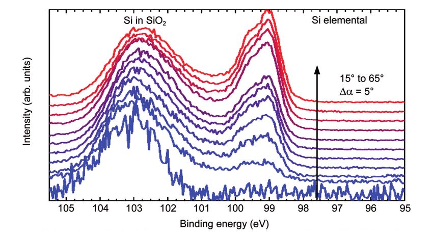
Figure 2: Si 2p spectra of a thin SiO2 film on a Si substrate.
   The emission angle is changed in steps of 5° between 15° and 65° from bottom to top, illustrated by the color change from blue to the red.

Figure 2 shows XPS scans of the Si 2p region of a thin SiO2 layer on         (i)   The sample must have a rather flat surface.
a silicon substrate. The emission angle a is gradually changed in 5°         (ii)  Layers on the sample must be continuous. If the covering
steps from 15° (blue curve) to 65° (red curve). The dominant photo-                layers consist of islands smaller than the beam diameter,
electron signal at 15° emission angle and ~103 eV binding energy                   the data interpretation is likely flawed.
corresponds to oxidized Si. The elemental Si signal at around 99 eV          (iii) The electron attenuation lengths are considered to be
binding energy is barely visible at a detection angle of 15°. Upon                 constant within each layer of the sample and are assumed
increasing the emission angle towards normal emission, the elemental               to be independent of the detection angle.
Si peaks appears and becomes stronger, while the corresponding               (iv) The density of atoms is assumed to be constant within
signal from oxidized Si becomes weaker in comparison. From this                    each layer.
data, the SiO2 layer thickness can be calculated to be about 1 nm.           (v) The X-ray intensity is presumed to be constant in the
                                                                                   analysed volume.
This example demonstrates that ARXPS is a powerful technique to
determine a layer thickness, but it is important to note that this          In conclusion, ARXPS provides a non-destructive depth-profile of
technique cannot be applied to all types of samples. Rather, some           the topmost few layers (about 10 nm) of a sample surface. It allows
requirements of the sample structure have to be fulfilled and some          the determination of the thickness of oxide films, the order of
assumptions have to be made for the analysis of the results:                multilayer structures, and elemental gradients at the surface.

References                                                                  Acknowledgements
[1] S. Hüfner. Photoelectron Spectroscopy. Springer Series: Advanced        ICAN is a registered open core facility (DFG RIsources reference:
    Texts in Physics. Springer-Verlag, Berlin, Heidelberg, 2003.            RI_00313). Funding by the German Research Foundation (DFG,
[2] M. P. Seah and W. A. Dench. Quantitative electron spectroscopy of       grant HA 2769/7-1) is gratefully acknowledged.
    surfaces: a standard data base for electron inelastic mean free paths
    in solids. Surf. Interface Anal. 1, 2 - 11, 1979.

          Contact
                                       ICAN | CENIDE                                         Tel.: +49 (0) 201 379 8080
                                       Universität Duisburg-Essen                            Fax: +49 (0) 201 379 8046
                                       Carl-Benz-Str. 199                                    eMail: ican@uni-due.de
                                       47057 Duisburg                                        www.uni-due.de/ican

                              U. Hagemann, ICAN Notes 3, 1 - 2 (2021); https://doi.org/10.17185/ican.notes/3                                    2
Imprint
          ICAN | CENIDE                                    Published by:
          Universität Duisburg-Essen                       ICAN Scientific Director
          Carl-Benz-Str. 199                               Frank Meyer zu Heringdorf
          47057 Duisburg                                   © 2021, all rights reserved.

          Cover image: Detail of the XPS instrument in ICAN. © ICAN 2016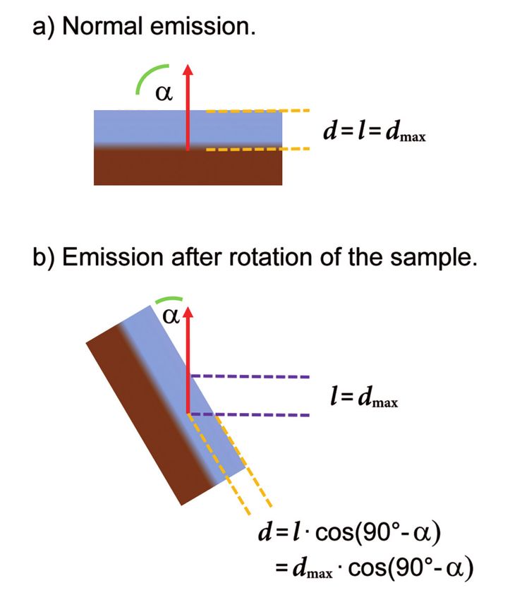
Dieser Text wird über DuEPublico, dem Dokumenten- und Publikationsserver der
Universität Duisburg-Essen, zur Verfügung gestellt. Die hier veröffentlichte Version der E-
Publikation kann von einer eventuell ebenfalls veröffentlichten Verlagsversion abweichen.

DOI:      10.17185/ican.notes/3
URN:      urn:nbn:de:hbz:464-20210303-143710-5

Alle Rechte vorbehalten.
You can also read