Infectious coryza (IC) - Haemophilus paragallinarum infection Fowl coryza
←
→
Page content transcription
If your browser does not render page correctly, please read the page content below
Infectious coryza (IC)
Haemophilus paragallinarum
        infection
       Fowl coryza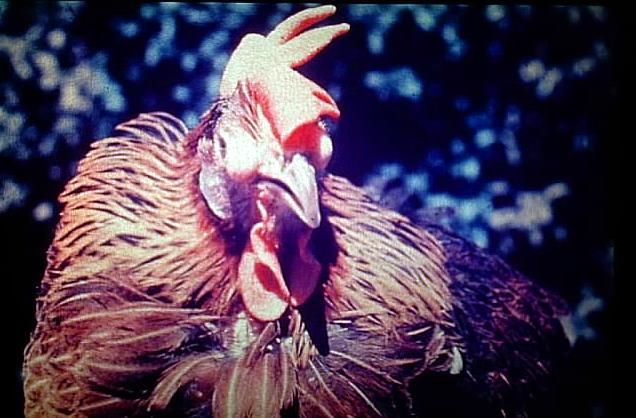
It is a highly contagious acute or sub acute
rapidly spreading disease of upper
respiratory tract of chickens,
 characterized by conjunctivitis, oculo-
nasal discharge, sneezing, swelling of the
infraorbital sinuses, swelling of the face,
swollen wattle, growth retardation, reduced
egg production, and some times by
infection of the lower respiratory tract.
Economic importance:
1-I.coryza can turn into a chronic respiratory disease.
2-Inferior food conversion in broilers with an increased
  number of culls (Stunted birds- air sacculitis).
3-Poor growth performance in growing birds.
4-A marked reduction from 10- to more than 40% in
egg production in layers and persists for at least 1
month.
Etiology:
Haemophilus paragallinarum (HP).
-It is a small gram-negative, aerobic, non-motile, non
spore forming, bipolar staining and capsulated
bacterium. It appears as short rods or coccobacilli.
-The organism requires V-factor (Nicotinamide
adenine dinucleotide-NAD) for growth in enriched
artificial media. A number of bacterial species
excrete V-factor (Staph epidermidis, Staph aureus).
Cultures of HP appear adjacent or next to the feeder
culture (Satellite phenomenon).
-Blood agar to which supplements are added is used for the
isolation of HP. The organism is grown at 37ºC-38ºC in an
atmosphere of high humidity, 5% CO2 or under reduced O2
tension (microaerophilic environment). Use candle jar if CO2
incubators are not available.
-On Blood agar colonies of HP are
tiny small up to 0.3-1 mm in diameter, dew drop-like, non-
hemolytic.
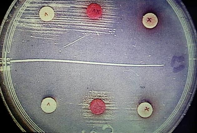
Satellite phenomenon. Tiny dewdrop colonies of HP growing
  adjacent to staph.culture (broad streak) on blood agar plate
Antigenic structure and strain classification:
-3 antigenic types (A, B, and C) have been defined by
plate agglutination and heamagglutination typing using
known
antisera.
-All types share certain antigens (Common antigenic
components exist) and are utilized in the examination of
sera for antibodies by agglutination tests.
Virulence: variation from very low to high virulence
occurs.
Resistance to chemical and physical agents: HP is
a delicate bacterium that does not survive for more
than 5hrs outside of birds. It is easily destroyed by
many disinfectants.
Susceptibility:             chickens

 Chicken 4 weeks to       IC is seldom seen        IC occurs in
3 years are susceptible   during brooding       growing chickens
                          period. Older birds       and layers
                          tend to react more
                             severely.
-Chickens 14 weeks of age and older are most susceptible.
-I.Coryza has been reported in Pheasants, Guinea fowl and
 Quails
Complicating factors and stresses:
Intercurrent infection with other pathogens such as viruses of
Pox, I.B, ILT, ND and bacteria such as Mycoplasma,
P.multiocida, E.coli plus cold, wet conditions, over crowding,
parasitism, inadequate nutrition, and Vit.A def, and others are
factors that predispose to more severe and prolonged disease.
Mode of infection and transmission: -
-The main sources of disease spread are: -
1) Clinically affected birds.
2) Carrier birds.(recovered birds remain carriers and
shedders for life)
-Transmission occurs by: -
1) Inhalation (Air-borne disease) 2) Through ingestion.The disease spreads from bird to bird and flock to flock by:
-
1) direct bird to bird Contact.
2) inhalation of infectious aerosols coughed into the air.
3) ingestion of Contaminated feed and water.
4) mechanically throw contaminated Equipment and
personnel.
Incubation period: -
1 to 3 or 4 days.
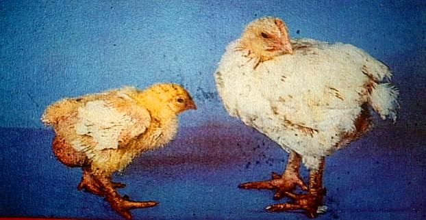
the course of the disease is 4 to 12 weeksClinical signs: - -the severity of infection varies depending on: the age, breed, environmental stress and presence of concurrent infections. 1- in the mild form seen in young chickens, birds become depressed and have nasal discharge and mild swelling of the face. 2- in the sever form seen in young adults, there is an acute infection of the upper respiratory tract.
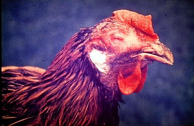
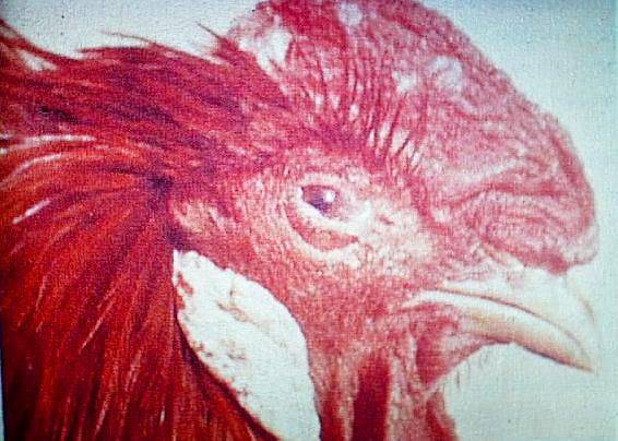
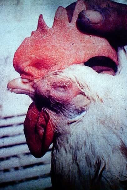
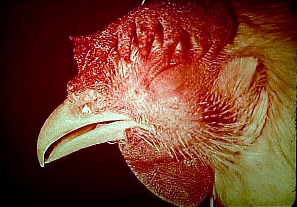
Clinical signs: -
-Usually there is a rapid onset, depression, serous to mucoid nasal
 discharge that become thick and purulent of very offensive odor,
 conjunctivitis “red eye”, lacrimation, in some cases the eyes are
 partially or completely closed, sneezing, swollen face, swollen
 wattles, and swollen infra orbital sinuses.
-Rales (Resp. noises), difficult breathing and coughing are
indicative of lower resp. tract involvement (trachea, airsacs).
-Birds may have diarrhoea, and feed and water intake is usually
reduced leading to loss of weight and in growing birds, this means
an increased number of culls (stunted birds), and in laying flocks
this means a drop in egg production (10-40%).
-A very offensive odor may be detected in flocks especially when
the disease has bean become chronic and complicated with other
bacteria.
-All the susceptible birds will show signs within 7-10 days.
-IC is characterized by high morbidity and low mortality if not
complicated by other infections. Mortality is very low (1%) and the
course of the disease is not more than about 10 days in the mild
form and approximately 3 weeks in the more severe form.
-IC is usually more severe and prolonged 1-2 ms or more with
resulting increased mortality rate (10-50%) with picture of chronic
resp. disease when complicated with other agents or when the birds
are exposed to stress factors.
-Recovered birds are frequently carriers.
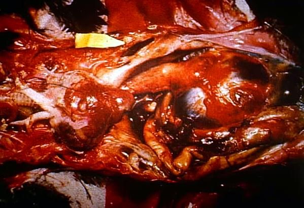
Lesions: -
In uncomplicated cases: H. paragallinarum produces:
1) Acute catarrhal inflammation of the mucous ms of the
nasal passages and infraorbital sinuses (distended with
serous or mucous exudates).
2) Catarrhal conjunctivitis. Eyelids may stick together.
3) S/C oedema of the face and wattles (swollen face and
wattles).
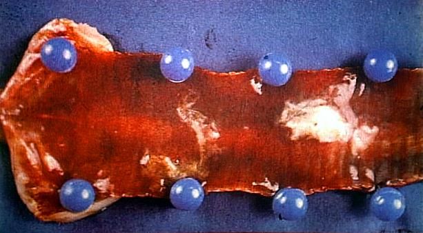
4) Typically pneumonia or air-sacculitis is rarely present.In complicated cases:(chronic inflammatory process) A mucopurulent or caseous exudates is present in the sinuses, nasal passages and conjunctival sac, catarrhal tracheitis, pneumonia or air sacculitis.
Diagnosis: -
I-History of a rapidly spreading disease.
II-Signs and lesions are suggestive.
III-Demonstration of the organism.
-A smear of nasal exudates or sinus exudates should be
made and gram stained, it should reveal Gram –ve bipolar
staining rods.
IV- Isolation and identification of the organism, this
is usually done by:
Swabbing from the infraorbital sinus, trachea or airsac in
the acute stage of the of the disease (1-7 days post
infection) onto blood agar plates containing NAD or cross
streaked with a feeder organism
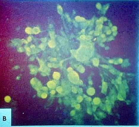
-Culture plates are incubated at 37C in a candle jar
(large screw cap jar in which a candle is allowed to burn
out) or in an atmosphere of 5% CO2 (CO2 incubator) for
24-48hr
Identification: -
 1) Colonial morphology: colonies appear as tiny very
small 0.3-1 mm, translucent, dewdrop like. When a
feeder organism is used, colonies of HP appear adjacent
or next to the feeder colonies → (satellitic growth).
2) Cellular morphology: Gram –ve, non-motile, polar
staining. …etc
.

    4) Serological identification: To identify the serotypes
    of the organism by the use of known antisera with the
    aid of the tube aggl. Test, plate aggl. Test, agar gel
    precipitin test. The organism can be identified by
    immunofluorescent technique by staining colony imprints
    with fluorescein-labeled antisera.
    5) A polymerase chain reaction (PCR) test can be applied
    either to suspect colonies or directly to samples from
    chickens.
V- Bird inoculation: Reproducing of the disease: Inoculate sinus exudates or culture of the organism into the infraorbital sinus or in the nostrils of chickens 4 wks. If HP is present in the inoculum, the clinical signs are observed in 1-2 days up to 6 days. VI- Response to sulfathiazol treatment: -If disease responded well to this drug, it was felt it was sure to be IC. VII- Serological testing: -PAT, TAT, AGPT, HI, FAT, and ELISA can be used to detect HP antibodies in serum → 7-14 days post-infection to check flocks for evidence of past infection.
Prevention:
I-Good management and sanitation: should be considered to
prevent the introduction of infection including:
 a-Allow good ventilation.
 b-Avoidance of stress factor.
 c-Control of complicating factors (other pathogens).
 d-An all in, all out prevention program is the best way for
   avoiding this disease.
 e-Avoiding carrier stock, the introduction of new stock
   should be limited to 1 day old chick, or older birds which
   are known to be free from infection, such birds should be
   reared so that there is no direct or indirect contact with
   other chickens.
f-Cleaning and disinfection of building after one flock of
birds
 have left and before others are housed and good hygienic
 prevention should be applied
II- Vaccination: This is only applied in endemic areas. IC
 bacterins (Coryza vaccine, IC inactivated vaccine) can be
 used to immunize chickens in areas where the diseases are
 endemic.
IC bacterins may be autogenous (prepared specifically from
the flock with the organism isolated from it) or may contain
 one serotype (Monovalent) or 2 types (bivalent), 3 types
(Poly-valent).
Field vaccination protocol and regimes:
1- inactivated vaccine:
A-oil emulsion vaccine
B- aluminum hydroxid vaccine
Bacterins are injected S/C (under the skin) on back of neck or
I/M (leg muscle) in chickens between 10-20 wks of age. All
birds should receive two injections of the bacterin at 3-4 wks
intervals before 20 wks of age, Example (vaccinate each
chicken 2 times starting at 10 wks of age, then at 13 or 14 wks
of age).
Dose:follow the instructions of the producer.
Immunity period: 9 months.
Live vaccines: Vaccines involving live HP organisms are said to
stimulate a more protective response but will give rise to
carriers and may produce disease.
Control and treatment: (If you have an outbreak)
I-Apply strict sanitation and sound management to
 prevent the spread of infection:
 - Properly dispose of dead birds by incineration,
cleaning and disinfection of houses after the occurrence
of an outbreak.
 -vaccinate all replacement flocks at least 3 weeks
before housing.
-During an outbreak add potassium permanganate or
 iodophor 1:10000 in D.W.
-control requires attention to flock sanitation and
biosecurity
-remove manure and clean house, control rodent, flies
II- Treatment:
Various sulfonamide and antibiotics are used in alleviating
the severity and course of IC.
 A-Sulfa Drugs
 - Sulfathiazol: is the drug of choice, dose for birds under
14 wk is 0.5% in feed for days), it can be used also in water
at a rate of 1-1.5 gm/ liter for 5 –7 days.
 - Sulfadimethoxine: 2-3 gm initial dose per liter water for
the first day and subsequent dose up to 4-6 gm/L.W. for 3-
5 days.
-Sulfadimidine: 1-2 gm/L.W for 3-5 days.
-Sulfadimidine Sol. 33.5%: 3ml/L.W (3 days treatment,
2 days rest, treat again 2 days)
-Sulfaquinoxaline 20%: 1.5-2.5gm/L.W for 3 days,
 withdrawal for 2 days and administered again for 3
 days.
-Sulfadiazine+trimethoprim: 1gm/5lieter for 5 days.
-Trisulfa(Sulfamethoxanie,sulfathiazol,sulfamerazine):
 3 Kg/ ton feed for 4-5 days.
-Drugs in combination: Found effective include
(Sulfachloropyrazine+S.dimidine,S.dimethoxaine+Chlorte
trcy-cline, and S.dimethoxaine+trimethoprim).
N.B. → Very important
- The sulfa drugs are not approved for chickens
older than 14 weeks or for commercial layer hens.
B) Antibiotics:
-Erythromycin: 1 gm/Liter for 3-5 days, 40-50 mg/bird
 by injection.
- Terramycin: 1gm/liter for 3-5 days up to 7 days.
- Lincospectin inj. 20-30 mg/Kg B.W for 3 days.
- Streptomycin inj 100-200 mg/ bird for 3-5 days.
- Terramycin long acting inj: 40-50-mg/ birds S/C.
-Ampicillin or Amoxycillin: 0.5-1.5-gm/Liter water for
 3-5 days.
- Aureomycin: 10-20 mg / bird for 5-10 days in water.
- Spectam: 20 mg/Kg B.W. for 3-5 days in water
- Flumiquin: 12 mg/Kg B.W. in water for 3-5 days
-Recently the new second generation quinolone
 derivatives that inhibits bacterial replication are
 used for IC treatment including:
i- Enrofloxacin (Baytril, Cidotril, Spectramavit): at a rate
   of 10 mg/ Kg B.W. for 3-5 days.
ii- Danofloxacin (Advocin): 5 mg/Kg B.W for 3 days in
   water.
iii-Norfloxacin (Norotril): 12 mg/Kg B.W for 3-5 days
   During treatment give vit. A. After treatment give
    diuretics (nephryle, hexavit, nephrotonic, …. Etc).
N.B. -No drugs have been found to prevent production of carriers and signs reappear after treatment (Drug resistant does develop). If condition reoccurs, retreat. The performance of antimicrobial sensitivity test is recommended. - Control can not be accomplished with drug alone. Mangement is equally important.
III- Eradication:
Eradication method is applied in non-endemic area to
eliminate the agent from the farm by the removal of
infected or recovered birds and the thorough cleaning,
disinfection and resting of building for at least 2-3
weeks before restocking with clean birds.
You can also read