Infestation of Mangifera indica by the mango gall fly, Procontarinia matteiana, (Kieffer & Cecconi) (Diptera: Cecidomyiidae)
←
→
Page content transcription
If your browser does not render page correctly, please read the page content below
Infestation of Mangifera indica by the mango gall fly, Procontarinia matteiana,
(Kieffer & Cecconi) (Diptera: Cecidomyiidae)

W.A. Augustyn *, W. du Plooy , B.M. Botha & E. van Wilpe
             1              2            1               3

1
  Department of Chemistry, Tshwane University of Technology, P.O. Box 56208, Arcadia, 0007 South Africa
2
  John Bean Technologies, South Africa (Pty) Ltd, P.O. Box 891, Brackenfell, 7561 South Africa
3
  Electron Microscope Unit, Faculty of Veterinary Science, University of Pretoria, Private Bag X04,
 Onderstepoort, 0110 South Africa.

                            Mango gall fly (Procontarinia matteiana Kieffer & Cecconi, 1906) is an orchard pest that infests
                            flush leaves of mango, forming wart-like structures on the leaves. Serious outbreaks may
                            result in reduced fruit yield. A natural parasite (Chrysonotomyia pulcherimma Kerrich, 1970)
                            of the gall fly lays its eggs inside the gall and the larvae feed on the gall fly. Mango cultivars
                            present varying susceptibilities to gall fly infestation, with cultivars ranging from completely
                            resistant, highly susceptible to intermediate stages where pseudo-galls are formed. The
                            latter cultivars are ovipositioned by the gall fly, but secondary metabolites within the leaves
                            possibly halt the development, thereby preventing the development of true galls. Micros-
                            copy was used to identify characteristic features of the gall fly and its parasite inside the gall,
                            to study the development of the insects and to distinguish them. Evidence was obtained that
                            the use of insecticides curbs the development of the larvae. Tissue development within true
                            and pseudo-galls was studied to provide insights into the role of secondary plant metabo-
                            lites in arresting true gall formation. This study will contribute to a more holistic approach to
                            pest management of mango.
                            Key words: mango gall fly, scanning electron microscopy, gall structures, systemic insecti-
                            cide, parasite.

INTRODUCTION

  Mangifera indica (mango) is often referred to as                distribution of populations of M. indica (Raman
the king of fruit, because of the succulent, exotic               et al. 2009).
flavour and delicious taste. Over 25 million tonnes                  The female gall fly oviposits on flush leaves,
of mangoes are produced annually by 87 countries                  maggots hatch from the eggs and tunnel into the
with Africa contributing 9 % of world production                  leaf tissue where the insects develop into mature
(Saãco 2004). Mango is a profitable source of revenue             gall flies. Tumour-like growths develop on the
for South Africa; however, there is a growing                     host plants as a result of chemical stimuli from the
threat that fruit yields may be reduced as a result               galling insects. These stimuli can be maternal secre-
of mango gall fly infestations. Numerous species                  tions injected during ovipositioning or stimuli
of mango gall fly have been identified worldwide                  produced by larvae developing within the plant
(Raman et al. 2009). The predominant species                      tissue (Pascual-Alvarado et al. 2008; Stone &
occurring in South Africa is Procontarinia matteiana              Schönrogge 2003). Colonized plants provide the
(Schoeman et al. 1996). Almost all of the Procontarinia           insect with food and shelter to the detriment of the
midges induce galls on leaves, but did not evolve                 host (Tooker & De Moraes 2008). In this study
to parasitize other plant organs or taxa. Galls were              it was confirmed that a group of volatile com-
discovered on fossil leaves of an ancestor of                     pounds, produced by the plant is associated with
M. indica from Upper Palaeocene sediments of                      gall fly infestation (Augustyn et al. 2010a).
northeastern India. This indicates that the feeding
                                                                     Differential susceptibility of mango cultivars to
behaviour of species of Procontarinia has not signif-
                                                                  gall fly infestation is a worldwide occurrence.
icantly changed over time. An explanation for
                                                                  Githure et al. (1998) classified 11 South African
this apparent feeding specialization may be low
                                                                  mango cultivars into categories of susceptibility to
selection pressure due to the abundance and
                                                                  P. matteiana. Cultivars that are highly susceptible to
*Author for correspondence. E-mail: augustynw@tut.ac.za           gall fly infestation exhibit large numbers of galls
                                                                                        African Entomology 21(1): 79–88 (2013)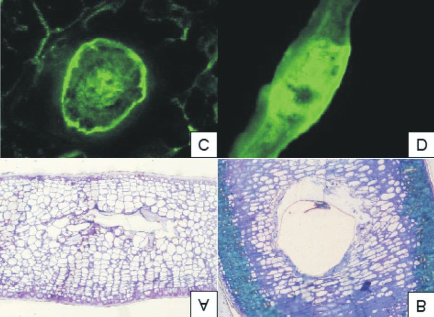
80                                   African Entomology Vol. 21, No. 1, 2013

per leaf, while other cultivars display signs of           pseudo-galls were fixed in 10 % phosphate-buffered
apparently unsuccessful gall development in the            formalin and the relevant areas dissected for
form of so-called pseudo-galls (Githure et al. 1998;       further processing. The fixative was replaced by
Schoeman et al. 1996)                                      0.13 M Millonig’s phosphate buffer (pH 7.2) for
  Resistant cultivars were found to be generally           30 min, whereafter the samples were rinsed in
free of galls. This observed resistance has been           distilled water for 30 min. This was followed by
attributed to antixenosis properties of the cultivar,      dehydration in a graded absolute ethanol series
rendering it unsuitable for feeding, shelter or            up to 100 % absolute ethanol, critical-point drying
ovipositioning by insects.                                 (Polaron E3100, West Sussex, U.K.), mounting
  The mango gall fly is not a serious economic             onto aluminium stubs and sputter-coating
problem in India to which they are indigenous,             (Polaron E5100, Watford, U.K.) with palladium.
because parasitoids are able to control their numbers      Samples were examined with a JEOL 840 scanning
(Sankaran 1988). Although the insect was not of            electron microscope (JEOL, Tokyo, Japan) operated
concern in the past, areas such as Oman, Mauritius,        at 8 kV.
Kenya, Réunion, Italy and recently South Africa,
have experienced serious outbreaks. This is largely        Light microscopy
due to a lack of natural enemies, combined with              Leaf sections displaying galls or pseudo-galls
the favourable ecological conditions prevailing in         were fixed in 4 % formaldehyde in 0.075 M phos-
these regions. In the past, gall fly has been of little    phate buffer (pH 7.4) for 1 h. Samples were first
consequence to producers as only flush leaves              rinsed three times for 10 min, in 0.075 M phos-
were attacked, leaving the fruit unharmed (San-            phate buffer and were followed by an ascending
karan 1988). However, in 2004 a newly identified           series of ethanol solutions from 50 % to 100 %
species, Procontarinia frugivora, that attacks only        (15 min, in each solution) and twice more in addi-
fruit, was reported (Gagné & Medina 2004). Though          tional fresh 100 % ethanol. The plant material was
this species is currently thought to be restricted to      first infiltrated with 50 % LR White resin (SPI
the Luzon Island of the Philippines, its emergence         Supplies, West Chester, PA, U.S.A.) in ethanol for
has placed mango gall fly in the spotlight as a            1 h, and finally in 100 % LR White in ethanol for
potential threat to global mango production.               4 h before polymerization at 60 °C for 24 h. Thin
  Mango cultivation is a lucrative industry and            sections, 0.5–1.0 µm, were cut with an ultra-micro-
globally, growers constantly strive to improve             tome (Reichert Ultracut E, Vienna, Austria), trans-
production. To sustain yield and quality, insect           ferred onto droplets of water on a specimen slide,
pests are managed by the application of insecti-           stained with Toluidine Blue (O’Brien & McCully
cides. Environmentally sustainable chemical                1981). Images were captured using a transmittance
control of the gall fly is only successful if the active   light microscope coupled to a DXM 1200 camera
substance applied is a systemic insecticide, such as       (Nikon Optiphod, Nikon Instech Co., Kanagawa,
thiamethoxam WG 250 g/kg (Daneel et al. 2000).             Japan).
Sprayed insecticides are only effective in the case
of young flush leaves due to the immature nature           Fluorescence microscopy
of the epicuticular layers.                                  Leaf sections were stained with natural product
  In this study, microscopy was employed to                (NP) reagent prior to viewing with a fluorescence
investigate the development and presence of the            microscope (Zeiss Axiovert 200, Carl Zeiss Werke,
gall fly and its parasite inside the gall, as part of a    Göttingen, Germany). The NP reagent was pre-
more comprehensive approach to management of               pared by dissolving 0.05 g of diphenylboric
this pest. Tissue development within true and              acid-Ä-ethylaminoester in 10 ml methanol. To this
pseudo-galls was studied to provide insight into           solution, 90 ml of a 5 % AlCl3 aqueous solution
the role of secondary plant metabolites in arrest-         was added, resulting in a 0.05 % NP reagent in
ing true gall formation.                                   AlCl3 (Heinrich et al. 2002). One centimetre strips
                                                           of fresh leaves were cut and soaked in the NP solu-
MATERIAL AND METHODS                                       tion for 10 min. The leaf sections were dried on
                                                           absorbent paper and mounted on a glass plate,
Scanning electron microscopy                               without a cover glass, before viewing the fluores-
 Mango leaf sections with occupied galls and               cence with a blue filter (excitation ä = 386 nm,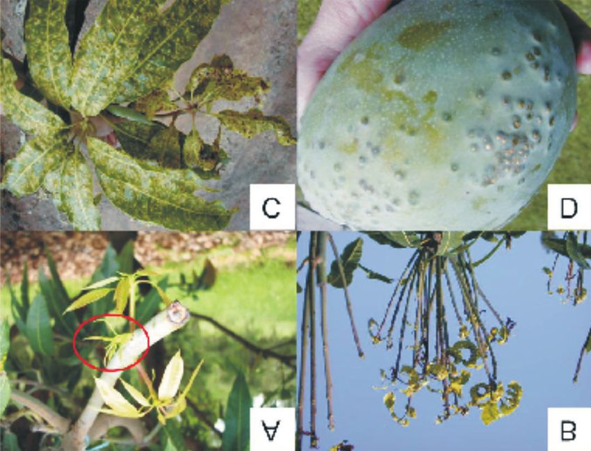
Augustyn et al.: Infestation of Mangifera indica by the mango gall fly, Procontarinia matteiana           81

Table 1. Brief descriptions of morphological features used for distinguishing Procontarinia matteiana from
Chrysonotomyia pulcherimma.

Part of         P. matteiana                                 C. pulcherimma                           Figure reference
organism

Antenna         Clear rounded modular segments               Segments flat and narrow                 Figs 2B and 3B
Eyes            Stacked and slightly elongated eyes          Protrusion extends from each eye         Figs 2C and 3C
Wings           Rounded with a sharp tip                     Round wings displaying fine hairs        Figs 2D and 3D
                                                             on the edges
Legs            No obvious differences and not useful        Figs 2E and 3E
                for identification purposes
Larva           Rounded in shape                             Elongated                                Figs 2F and 3F

emission ä = 490 nm). Digital images were cap-               of the male and female gall fly, as well as the para-
tured using the DXM 1200 camera.                             site, are depicted in Fig. 1. Female gall flies have
                                                             shorter antennae and the genitalia are less
RESULTS AND DISCUSSION                                       pronounced than those of males (Fig. 1A,B). The
                                                             gall fly has a rounded head and a round abdomen.
  Mangifera indica is utilized by over 250 insect            In contrast, the parasite (Fig. 1C) is characterized
species of which about 25 species are gall-inducing          by more angular features that include a sharp
species, with Procontarinia spp. the most prevalent          triangular head and pointed abdomen.
(Raman et al. 2009). In countries where mango is               In many cases, the insect emerging from the gall
indigenous, gall fly numbers are kept under con-             is the parasite, rather than the mature gall fly.
trol by natural enemies. The most prevalent para-            Scanning electron micrographs of emerged insects
site (C. pulcherimma) in South Africa is found on all        simplified subsequent identifications. Figs 2A and
infested cultivars throughout the season, but is             3A are micrographs of the intact insects, while
unable to suppress the gall fly population below             Figs 2B–F and 3B–F represent those of the
the economic threshold (Grové et al. 2003). These            enlarged antennae, eyes, wings and legs of the gall
parasites lay their eggs inside the gall and the             fly and parasite, respectively. The eyes were found
larvae feed on the gall fly while it is still inside the     to be the most useful for identification purposes. A
structure. To investigate the life cycle of the gall fly     summary of the distinguishing features used for
and the effect of insecticides on this cycle, it is          identification is provided in Table 1. Based on
necessary to distinguish the gall fly from other             these morphological characteristics and the eye
insects inhabiting the gall structure such as                structure, the insect displayed in Fig. 4D was
C. pulcherimma. In this study a holistic view of both        therefore identified as the parasite and not the gall
the gall fly and the parasite was adopted. Images            fly.

Fig. 1. Light microscope photographs of adult flies and parasite. A, The female fly has a full abdomen and rounded
posterior with no visible genitalia and short antennae; B, the male fly has a slender abdomen with visible genitalia and
long antennae; C, the parasite of the gall fly. Photographs courtesy of E. Louw, Westfalia.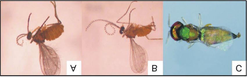
82                                    African Entomology Vol. 21, No. 1, 2013

Fig. 2. Scanning electron micrographs of the mango gall fly and various parts of the insect. A, Intact fly; B, antennae;
C, eye; D, wing; E, leg; F, larva.

  Initially, Schreiner (1990) proposed that the gall         2009). The current SEM studies confirmed that
fly develops only up to the larval stage within the          P. mateiena completes its life cycle within the gall.
leaf, after which it emerges, drops to the ground            Longitudinal and perpendicular sections of galls
and the remainder of the life cycle is completed in          are presented in Figs 4A and 4B, illustrating the
the soil. Some species, such as P. pustulata, have           development of insects inside individual galls. The
been shown to pupate in the soil (Kolesik et al.             fully developed insects depicted in Figs 4C and 4D
Augustyn et al.: Infestation of Mangifera indica by the mango gall fly, Procontarinia matteiana            83

Fig. 3. Scanning electron micrographs of the parasite of the mango gall fly and various parts of the parasite. A, Intact
insect; B, antennae; C, eye; D, wing; E, leg; F, pupa.

were identified as the gall fly and parasite, respec-          The influence of the systemic insecticide thia-
tively, while the larvae of the gall fly and parasite        methoxam on gall and insect development is
are represented in Figs 4E and 4F, respectively.             apparent in Fig. 5. In Fig. 5A, true galls containing
Once again, the rounded shape of the gall fly larva          insects are clearly visible. These galls are of the
and the sharp abdomen of the parasite larva are              same age as the structures depicted in Fig. 5B, a
evident.                                                     micrograph of plant material treated with thia-
84                                      African Entomology Vol. 21, No. 1, 2013

Fig. 4. Scanning electron micrographs illustrating the development of a mango gall fly and parasite within the gall.
A, Longitudinal and perpendicular section of galls with insect larvae visible; B, longitudinal section of galls with larva in
one gall, while the structure of a vacated gall is visible to the left; C, section through a gall fly with eye, antennae and
extremities visible; D, parasite inside a gall; E, gall fly larva in gall; F, parasite larva in gall.

methoxam. Although the insecticide clearly                      insect. The visible damage observed after insecti-
prevented larval development, the gall-like                     cide application can be attributed to earlier infesta-
structure was still consistent with that of true galls,         tion while the leaves were very young and soft.
with randomly ordered parenchyma cells.                         Micrographs thus confirmed the efficacy of
Thickening of the tissue still occurs due to a plant            thiamethoxam as an insecticide to curb gall fly infes-
response following stimulation by the parasitizing              tation in mango orchards. Systemic insecticides are
Augustyn et al.: Infestation of Mangifera indica by the mango gall fly, Procontarinia matteiana            85

Fig. 5. Scanning electron micrographs of sections through galls to verify differences between leaves that were
exposed or not exposed to commercial insecticides. A, Mango (cv. Tommy Atkins) leaves in an organic orchard with
insect development in galls; B, mango (cv. Tommy Atkins) leaves exposed to insecticide – no insects visible.

therefore an effective means of pest management.              associated with the mango and mango insect pests
  Although chemical control offers some relief,               (Githure et al. 1998). Alternative natural com-
non-target organisms, including gall fly parasites,           pound-based solutions that reduce gall fly infesta-
are also killed using this method, thereby eroding            tion, while maintaining parasite populations, are
natural control mechanisms. Organophosphates,                 currently sought by the industry.
for example, have been used to combat gall fly, but             Mango gall flies oviposit on M. indica during the
are not recommended as these pesticides are even              spike stage of flush leaves (encircled in Fig. 6A),
more detrimental to the natural enemy complexes               resulting in the development of galls on leaf

Fig. 6. Photographs of gall fly infestation on leaves and fruit of Mangifera indica. A, flush leaves with the spike stage
circled; B, defoliated branches as a result of severe infestation; C, deformed mature leaves; D, infested fruit. Photo-
graphs courtesy of E. Louw, Westfalia, and D. Le Lagadec, Agri-Science Queensland, Australia.
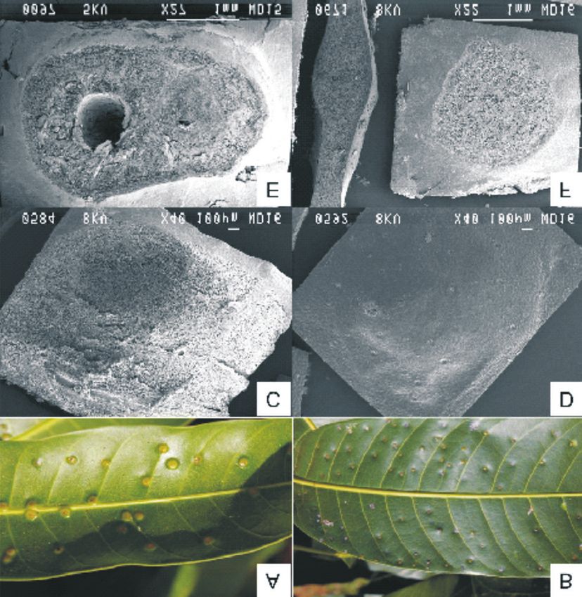
86                                    African Entomology Vol. 21, No. 1, 2013

Fig. 7. Images of true galls and pseudo-galls. A, Photograph of a true gall; B, photograph of a pseudo-gall; C, scanning
electron micrograph of a true gall; D, a longitudinal section through a pseudo-gall; E, scanning electron micrograph of
a pseudo-gall; F, longitudinal section through a true gall.

surfaces. As shown, complete metamorphosis of                ‘Peach’, ‘Zill’, ‘Kensington’, ‘Tommy Atkins’ and
the gall fly takes place within these structures.            ‘Sabre’ develop true galls (Augustyn et al. 2010b);
Colonized leaves appear deformed (Fig. 6C) and               Fig. 7A). In cultivars that present pseudo-galls,
are likely to drop. Severe infestations may result in        such as ‘Keitt’, ‘Kent’ and ‘Irwin’, the presence of
the defoliation of branches (Fig. 6B) thereby                compounds with antibiosis properties deter the
impacting seriously on fruit production and                  development of the pest following oviposition.
photosynthetic abilities of infected trees. In South         Typical shot-hole damage is evident on the leaves
Africa, the incidence of gall fly infestation on fruit       of these cultivars (Githure et al. 1998); Fig. 7B).
is very low, but has been occasionally observed              These marks may be sites where oviposition did
(Fig. 6D).                                                   take place, but the young larvae failed to develop.
  Larval mortality occurs in pseudo-gall-forming             Morphological differences were evident between
cultivars, as observed using microscopy. It is               true galls and pseudo-galls as illustrated in the
thought that secondary host-plant metabolites                micrographs in Fig. 7C–F. True galls (Fig. 7C) have
may contribute to this phenomenon (Augustyn                  a rounded, inflated shape, while pseudo-galls
et al. 2010b). The cultivars, ‘Heidi’, ‘Haden’,              (Fig. 7D) appear deflated. Exudates are emitted by
Augustyn et al.: Infestation of Mangifera indica by the mango gall fly, Procontarinia matteiana      87

Fig. 8. Light microscope images comparing pseudo-galls (A) to galls (B). The fluorescence images at the bottom are
longitudinal (C) and perpendicular (D) cuts through galls stained with natural product.

galls as observed in Fig. 7C; however, no exudates         larval feeding and separated from the rest of the
are present on leaves displaying pseudo-galls. A           gall by a thin wall of sclerenchyma (Stone &
longitudinal section of a true gall (Fig. 7E) revealed     Schönrogge 2003) (Fig. 8B). In the maturation
the gall structure, as well as the cavity in which the     phase, secondary nutritive cells are formed and
insect developed. In contrast, the pseudo-gall             the gall parenchyma cells are lignified. These
(Fig. 7F) contains no insect, no cavity is visible and     secondary cells are the main food source of the
only disorganized plant parenchyma cells can be            larvae (Lalonde & Shorthouse 1985). In Figs 8C
observed.                                                  and 8D, the green fluorescence indicates the pres-
  From the light microscopy of a semi-thin section         ence of phenolic compounds after staining with
of a pseudo-gall (Fig. 8A) the small size of the           natural product reagent. Excessive phenolic com-
cavity can be seen. In true galls (Fig. 8B), the cavity    pounds are produced as a protection mechanism in
and the parenchyma cells surrounding the gall are          response to infestation (Du Plooy et al. 2009).
visible and correspond to the description of gall          Galling insects may award some benefits to their
development by Lalonde & Shorthouse (1985).                hosts as a result of the induction of foliar phenolic
According to these authors, gall development in            defence compounds in the leaves that may indi-
the Canada thistle (Cirsium arvense) consists of           rectly protect against feeding by other herbivores
three phases after infestation with the tephritid fly      (Pascual-Alvarado et al. 2008).
Urophora cardui. In the initiation phase, the insect
takes control of tissue development, whereafter            CONCLUSION
parenchyma cells multiply rapidly, surrounding
the larvae with a thick layer of cells, and primary         Scanning electron microscopy proved that the
nutritive cells appear. This layer is stimulated by        entire larval development of both the mango gall
88                                      African Entomology Vol. 21, No. 1, 2013

fly and that of its parasite takes place within the             micrographs elucidated the structural differences
gall cavity. These findings have contributed to the             between true and pseudo-galls, thereby verifying
entomological data available for P. matteiana, as               that larval development is terminated in the case
well as that of the parasite, C. pulcherimma. Para-             of pseudo-gall-bearing cultivars. Orchard applica-
sites of the gall fly play a role in curbing gall fly           tion of secondary metabolites found to be active in
numbers; however, they never occur in sufficient                larval mortality may be a viable option for gall fly
numbers to eradicate the gall fly problem. Electron             control. This study has provided the necessary
microscopy confirmed that the application of the                tools to investigate the effect of natural compounds
systemic insecticide thiamethoxam WG 250 g/kg                   on larval development and true gall formation.
indeed halted the development of the gall fly. This             Alternatively, cultivars that are good producers of
finding verified that the use of systemic insecti-              antibiotic compounds should be selected and
cides should currently be retained as an integral               cultivated to obviate the need for insecticide
part of pest management in the orchard. In addition,            application.

REFERENCES

AUGUSTYN, W.A., BOTHA, B.M., COMBRINCK, S. &.                   LALONDE, R.G. & SHORTHOUSE, J.D. 1985. Growth
   DU PLOOY, G.W. 2010a. Correlation of volatile pro-              and development of larvae and galls of Urophora
   files of twenty mango cultivars with their susceptibil-         cardui (Diptera, Tephritidae) on Cirsium arvense
   ities to mango gall fly infestation. South African Journal      (Compositae). Oecologia 65: 161–165.
   of Botany 76: 710–716.                                       O’BRIEN, T.P. & McCULLY, M.E. 1981. The Study of Plant
AUGUSTYN, W.A., BOTHA, B.M., REGNIER, T. & DU                      Structures: Principles and Methods. Bradford House
   PLOOY, G.W. 2010b. The role of secondary metabolites            (Pty) Ltd, South Melbourne, Australia.
   in the management of mango gall fly infestations.            PASCUAL -ALVARADO, P., CUEVAS-REYES, P.,
   South African Mango Growers’ Association, Research Jour-        QUESADA, M. & OYAMA, K. 2008. Interactions
   nal 30: 32–39.                                                  between galling insects and leaf-feeding insects: The
DANEEL, M.S., DE JAGER, K., STEYN, W. & HUSSEL-                    role of plant phenolic compounds and their possible
   MAN, J. 2000. Efficacy of different insecticides against        interference with herbivores. Journal of Tropical
   gall fly on mangoes. South African Mango Growers’               Ecology 24: 329–336.
   Association, Research Journal 20: 85–89.                     RAMAN, A., BURCKHARDT, D. & HARRIS, K.M. 2009.
DU PLOOY, G.W., COMBRINCK, S., REGNIER, T. &                       Biology and adaptive radiation in the gall-inducing
   BOTHA, B.M. 2009. Linking lenticel discolouration of            Cecidomyiidae (Insecta Diptera) and Calophyidae
   mango (Mangifera indica L.) fruit to reversed-phase             (Insecta Hemiptera) on Mangifera indica (Anacardia-
   HPLC profiles of phenolic compounds. Journal of                 ceae) in the Indian subcontinent. Tropical Zoology 22:
   Horticultural Science and Biotechnology 84: 421–426.            27–56.
GAGNÉ, R.J., & MEDINA, C. 2004. A new species of                SANKARAN, T. 1988. Recent studies on the mango
   Procontarinia (Diptera: Cecidomyiidae), an important            leaf-gall midge Procontarinia matteiana Kieffer and
   new pest in the Phillipines. Proceedings of the Entomo-         Cecconi (Dip. Cecidomyiidae) and its parasites in
   logical Society of Washington 106: 19–25.                       India and on prospects for biological control of the
GITHURE, G.W., SCHOEMAN, A.S. & McGEOCH, M.A.                      pest in Oman. Acta Horticulturae 231: 587–592.
   1998. Differential susceptibility of mango cultivars in      SAâCO, V.C. 2004. Mango production and world
   South Africa to galling by the mango gall fly,                  market: Current situation and future prospects. Acta
   Procontarinia matteiana Kieffer & Cecconi (Diptera:             Horticulturae 645: 107–116.
   Cecidomyiidae). African Entomology 8: 33–40.                 SCHOEMAN, A.S., McCEOCH, M.A. & GITHURE, W.C.
GROVÉ, T., STEYN, W.P. & DE BEER, M.S. 2003. Biologie              1996. Differential susceptibility of eleven mango
   van galvlieg. South African Mango Growers’ Associa-             cultivars to galling by mango gall fly (Procontarinia
   tion, Research Journal 23: 102–107.                             matteiana Kieffer and Cecconi (Diptera:
HEINRICH, G., PFEIFHOFER, H.W., STABENTHEINER,                     Cecidomyiidae)). South African Mango Growers’ Asso-
   E. & SAWIDIS, T. 2002. Glandular hairs of Sigesbeckia           ciation, Research Journal 16: 23–26.
   jorullensis Kunth (Asteraceae): morphology, histo-           SCHREINER, J.H. 1990. Blotch miner associated with
   chemistr and composition of essential oil. Annals of            mango leaf anthracnose in Micronesia. Plant Disease
   Botany 89: 459–469.                                             74: 253.
KOLESIK, P., RICE, A.D., BELLIS, G.A. & WIRTHEN-                STONE, G.N. & SCHÖNROGGE, K. 2003. The adaptive
   SOHN, M.G. 2009. Procontarinia postulata, a new gall            significance of insect gall morphology. Trends in
   midge species (Diptera: Cecidomyiidae) feeding on               Ecology and Evolution 18: 512–522.
   mango, Mangifera indica (Anarcadiaceae), in northern         TOOKER, J.F. & C. DE MORAES, M. 2008. Gall insects
   Australia and Papua New Guinea. Australian Journal              and indirect plant defences. A case of active manipu-
   of Entomology 48: 310–316.                                      lation? Plant Signal Behaviour 3: 503–507.

                                                                                              Accepted 17 October 2012
You can also read