Is the transperineal ultrasonography approach effective for the diagnosis of rectocele?
←
→
Page content transcription
If your browser does not render page correctly, please read the page content below
Gastroenterology Report, 2021, 1–9

                                                                                doi: 10.1093/gastro/goab019
                                                                                Original Article

ORIGINAL ARTICLE

                                                                                                                                                        Downloaded from https://academic.oup.com/gastro/advance-article/doi/10.1093/gastro/goab019/6288120 by guest on 05 July 2021
Is the transperineal ultrasonography approach
effective for the diagnosis of rectocele?
Yi-Bo Yao1,†, Hao-Qiang Yin2,†, Hai-Jun Wang1, Hong-Tao Liang1, Bo Wang3
and Chen Wang1,*
1
  Department of Proctology, Longhua Hospital affiliated to Shanghai University of TCM, Shanghai, P. R. China,
2
  Department of Ultrasonic, Longhua Hospital affiliated to Shanghai University of TCM, Shanghai, P. R. China;
3
  Shanghai Hao Zhuo Data Service Co. Ltd, Shanghai, P. R. China
†
 These authors should be regarded as joint first authors.
*Corresponding author. Department of Proctology, Longhua Hospital affiliated to Shanghai University of TCM, No. 725 South Wanping Road, Shanghai
200032, P. R. China. Tel: þ86-18917763340; Fax: þ86-21-64698173; Email: wangchen_longhua@163.com

Abstract
Background Transperineal ultrasonography has been used as a diagnostic imaging modality for rectocele for many years.
However, the consistency of ultrasonography and defecography in evaluating the severity of rectocele was not satisfactory.
This study aimed to evaluate the agreement in the measurement of rectocele parameters between the two methods in dif-
ferent positions and provide clinical implications for the diagnosis of rectocele.
Methods In this pilot study, participants were recruited in an outpatient clinic of a tertiary hospital between December 2017
and December 2019. All participants separately underwent defecation proctography at sitting and squatting positions, and
undertook transperineal ultrasonography at left lateral, sitting, and squatting positions. The consistency of ultrasonography
and defecography was evaluated.
Results Thirty female volunteers with rectocele were included in this study. The degree of anorectal angle was significantly
larger at rest and during contraction, maximal Valsalva, and evacuation; the depth of the rectocele was significantly deeper
during maximal Valsalva and evacuation; and the length of the perineum descending was significantly longer during con-
traction and maximal Valsalva in using squatting position compared to the sitting position when performing the defecation
proctography. The degree of anorectal angle, the depth of rectocele, the area of levator hiatus, and the volume of the recto-
cele were significantly different in using squatting, sitting, and left lateral positions when performing the transperineal ul-
trasonography. Bland-Altman semi-quantitative plots showed good consistency in the measurement of the anorectal angle
and the depth of the rectocele between proctography and ultrasonography in both sitting and squatting positions.
Conclusions The findings of our study may be considered as the preliminary evidence to support the use of transperineal ul-
trasonography with sitting and squatting positions as the imaging test of choice for evaluating patients with rectocele.

Key words: transperineal ultrasonography; defecation proctography; rectocele; sitting position; squatting position;
consistency

Submitted: 16 January 2021; Revised: 17 February 2021; Accepted: 20 February 2021
C The Author(s) 2021. Published by Oxford University Press and Sixth Affiliated Hospital of Sun Yat-sen University
V
This is an Open Access article distributed under the terms of the Creative Commons Attribution License (http://creativecommons.org/licenses/by/4.0/),
which permits unrestricted reuse, distribution, and reproduction in any medium, provided the original work is properly cited.

                                                                                                                                               1
2   | Y.-B. Yao et al.

Introduction                                                          [17]. These participants were provided with a Bristol stool chart
                                                                      to describe their daily stool quality [18].
Rectoceles are commonly found in conjunction with obstructed
defecation and pelvic-floor dysfunction. It presents as an out
pocketing of the anterior rectal and posterior vaginal wall into
                                                                      Defecation proctography protocol
the lumen of the vagina, mostly occurring during defecation [1].
Rectocele is a common condition in females. These patients            Defecation proctography was performed by an experienced ra-
usually do not become symptomatic until the fourth or fifth de-       diologist. The rectum was filled with 120 ml diluted barium sul-
cade of life [2]. Clinical diagnosis and evaluation of a woman        fate. The volunteer was examined using a sitting position on a
with rectocele are based on her medical history, physical exami-      radiolucent commode on the first day then the same procedure

                                                                                                                                            Downloaded from https://academic.oup.com/gastro/advance-article/doi/10.1093/gastro/goab019/6288120 by guest on 05 July 2021
nation, and imaging modality [3, 4].                                  was performed using a squatting position on the second day
    To date, the diagnostic gold standard for rectocele is defeca-    (SIEMENS IconosR200 Digital Gastrointestinal System) (Figure 1).
tion proctography, which is used to identify anatomical disor-        Images were recorded in the sagittal plane at rest and during
ders in combination with the need for an extended defecogram,         contraction, maximal Valsalva, and evacuation of contrast.
including contrast in the bladder and vagina as well as occa-
sional peritoneography [5]. However, this approach is relatively
costly with radiation exposure. In recent years, image diagnos-       Transperineal ultrasonography protocol
tic techniques like dynamic magnetic resonance imaging (MRI)          Transperineal ultrasonography was performed by an experi-
and transperineal ultrasonography have been increasingly used         enced sonographer in the left lateral, sitting, and squatting posi-
to diagnose and evaluate defecatory disorders in clinical prac-       tions (Figure 2) with a 1-hour interval with an empty bladder
tice [6, 7]. The advantages of these imaging modalities are the       using a 1.5- to 6-MHz C1-6-D convex array probe and 5.0- to 9-
absence of ionizing radiation, comfort, and non-invasiveness.         MHz RIC-9-D real-time 4D probe (Voluson10 expert, GE
Transperineal ultrasonography can also be used for the diagno-        Healthcare, Milwaukee, WI). The rectum was filled with a 100-
sis of other pelvic-floor disorders linked to rectocele [8].          ml ultrasonographic coupling gel mix with a 0.5-ml suspension
However, the consistency of these methods in the identification       of SF6 microbubbles at each position. The assessment was car-
of rectocele diagnosis compared to defecation proctography            ried out in the midsagittal plane. Volumes were obtained at rest
was not satisfactory [9]. In addition, patients are asked to take a   and during contraction, maximal Valsalva, and evacuation of
sitting position for the defecography test and take a supine po-
                                                                      contrast. Rectocele volume was calculated in contrast-en-
sition and/or left lateral position for the dynamic MRI and ultra-
                                                                      hanced ultrasound 3D mode, the probe scanned, and the vol-
sonography [10, 11].
                                                                      ume was calculated automatically during maximal Valsalva.
    The dynamic MRI method is costlier than the transperineal
ultrasonography and defecation proctography techniques, and
most previous studies have only examined the abnormal defe-
                                                                      Outcome measurement
cate position to scale the severity of the rectocele [12–14].
Therefore, we conducted this pilot study aiming to compare            Data of the anorectal angle and depth of the rectocele of partici-
defecation proctography and transperineal ultrasound in differ-       pants were obtained using both the defecation proctography
ent defecate positions regarding their consistency in the mea-        and transperineal ultrasonography techniques. In addition, the
surement of rectocele parameters.                                     perineum descending was measured when using the defecation
                                                                      proctography method, while the area of the levator hiatus and
                                                                      volume of the rectocele were determined when using the trans-
Materials and methods                                                 perineal ultrasonography method. It is worth noting that all
Study design and study subjects                                       measurements of these two image diagnostic methods were ad-
                                                                      ministered by two different operators.
This prospective observational study was carried out at the
Longhua Hospital affiliated to Shanghai University of
Traditional Chinese Medicine between December 2017 and
                                                                      Statistical analysis
December 2019. This study was approved by the local research
ethics committee of Longhua Hospital (IRB No. 2017LCSY018).           The median and the interquartile range (IQR) were used to de-
All procedures performed in studies involving human partici-          scribe the measurements of anorectal angle, depth of rectocele,
pants were in accordance with the ethical standards of the in-        perineum descending, area of the levator hiatus, and the vol-
stitutional review board at Longhua Hospital affiliated to            ume of the rectocele. The Wilcoxon signed-rank test and re-
Shanghai University of TCM and with the 1964 Helsinki                 peated measures ANOVA were used to compare the difference
Declaration and its later amendments or comparable ethical            in measurements of anorectal angle, depth of rectocele, peri-
standards. Informed consent was obtained from all individual          neum descending, area of the levator hiatus, and volume of the
participants included in the study.                                   rectocele between the defecography and ultrasonography
    Participants meeting the clinical diagnosis of rectocele by       groups, and across different position groups, as appropriate.
digital rectal examination [15] were included in this study. The      Agreement test was used to detect the consistence between def-
exclusion criteria included hemorrhoids, anal fissure, and rectal     ecation proctography and transperineal ultrasonography in sit-
hemorrhage that predict cancer. For all participants, their de-       ting and squatting positions, respectively. A Bland-Altman plot
mographic and clinical characteristics were collected including       was used to show the appearance of differences between the
age, body mass index, reproductive history, favorite defecation       different techniques. Paired t-test was used to compare the time
position, and daily stool quality, obstructed defecation syn-         performed for the two evaluations. The data were analysed us-
drome (ODS) scores [16], and constipation scoring system (CSS)        ing STATA 14.2 and the significant level was set at 0.05.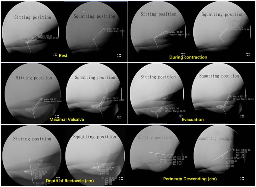
Transperineal ultrasonography and rectocele        |   3

                                                                                                                                                     Downloaded from https://academic.oup.com/gastro/advance-article/doi/10.1093/gastro/goab019/6288120 by guest on 05 July 2021
Figure 1. Different positions for defecation proctography (simulation)

Figure 2. Different positions for transperineal ultrasonography (simulation)

Results                                                                        (P ¼ 0.004); and a longer length of perineum descending during
                                                                               contraction (P ¼ 0.002) and maximal Valsalva (P ¼ 0.022) (Table 1).
Sample characteristics

A total of 30 Chinese female patients were recruited. The aver-                Transperineal ultrasonography
age age of the volunteers was 51.56 6 14.48 years and the aver-
                                                                               The anorectal angle, depth of rectocele, area of levator hiatus,
age body mass index was 22.73 6 2.53 kg/m2. All participants
                                                                               and volume of rectocele images via the transperineal ultraso-
were multipara with one or two eutocie. Nine had a previous                    nography method are shown in Figure 4. There were statisti-
hysterectomy. No patients reported vault prolapse. The median                  cally significant differences among the left lateral, sitting, and
value of the Bristol stool quality among all participants was 5                squatting positions in anorectal angles at rest (P < 0.001); during
(range, 2–6). The average ODS and CSS scores were 12.07 6 5.88                 contraction (P ¼ 0.002), maximal Valsalva (P < 0.001), and evacu-
and 11.67 6 5.03, respectively. Four patients stated that their fa-            ation (P < 0.001); and in depth of rectocele during contraction
vorite defecation position was the squatting position.                         (P ¼ 0.027), maximal Valsalva (P < 0.001), and evacuation
                                                                               (P < 0.001). Significant differences in the area of levator hiatus
Defecation proctography                                                        (P < 0.001) and volume of rectocele (P ¼ 0.002) at maximal
                                                                               Valsalva had also been found among these three positions
The anorectal angle, depth of rectocele, and perineum descend-
                                                                               (Table 2).
ing images via the defecation proctography method are shown in
Figure 3. Compared to the sitting position, participants in the
squatting position showed a larger degree of anorectal angle at                Comparison between the defecography and
rest (P ¼ 0.023) and during contraction (P < 0.001), maximal                   ultrasonography results
Valsalva (P < 0.001), and evacuation (P < 0.001); a deeper depth of            Results from the Bland-Altman semi-quantitative plot showed
rectocele at maximal Valsalva (P < 0.001) and evacuation                       agreement in the measurement of the anorectal angle and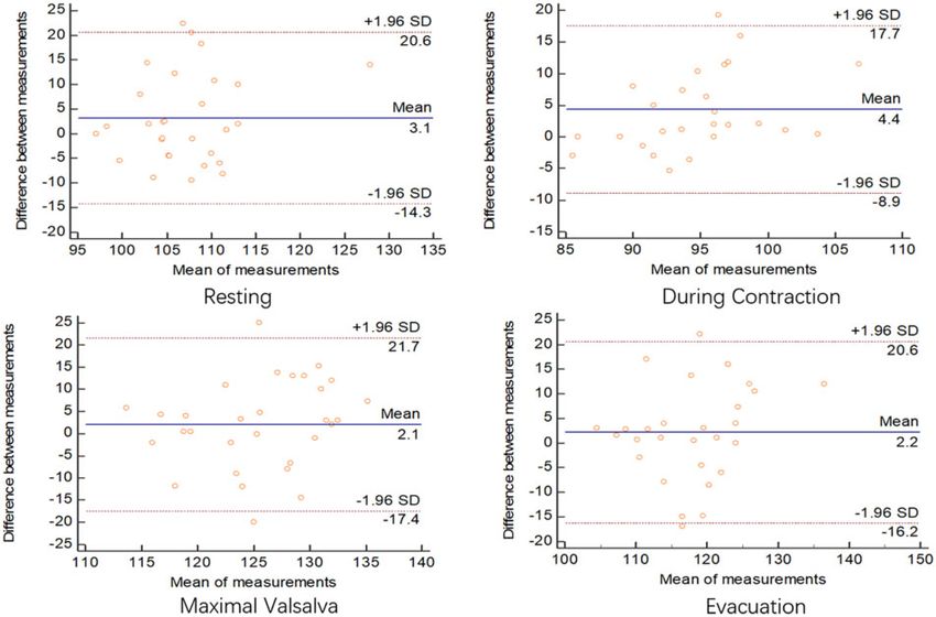
4   | Y.-B. Yao et al.

                                                                                                                                                                      Downloaded from https://academic.oup.com/gastro/advance-article/doi/10.1093/gastro/goab019/6288120 by guest on 05 July 2021
Figure 3. Anorectal angle (at rest and during contraction, maximal Valsalva, and evacuation), depth of rectocele (at maximal Valsalva), and perineum descending (at
maximal Valsalva) by defecation proctography in two positions

Table 1. Results of defecation proctography in two positions

Defecation photography                                                     Sitting position (n ¼ 30)             Squatting position (n ¼ 30)              P-value

Anorectal angle, degree,                 Rest                                   105 (100, 110)                          107 (104, 112)                     0.023
  median (IQR)                           During contraction                      92 (89, 94)                             97 (90, 102)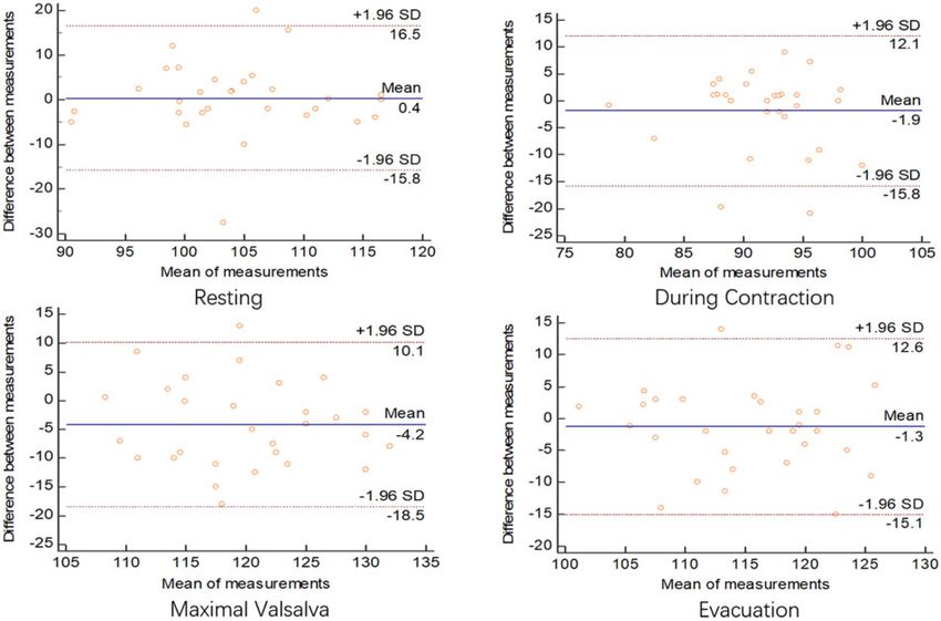
Transperineal ultrasonography and rectocele                    |   5

                                                                                                                                                                           Downloaded from https://academic.oup.com/gastro/advance-article/doi/10.1093/gastro/goab019/6288120 by guest on 05 July 2021
Figure 4. Anorectal angle, depth of rectocele, area of levator hiatus, and volume of rectocele (at maximal Valsalva) by transperineal ultrasonography in three positions

transperineal ultrasonography was significantly longer than                            rectocele between these two methods, previous studies only ex-
that for defecation proctography (55.43 6 26.38 vs 40.69 6                             amined the defecography method in the sitting position and the
18.25 min, P < 0.05).                                                                  ultrasonography method in the left lateral position [9, 19, 20].
                                                                                       Therefore, the position adopted for a patient may play an im-
                                                                                       portant role when determining the diagnosis of rectocele via
Discussion
                                                                                       different image methods in clinical practice.
This study evaluated compared the consistency of the measure-                              Defecography is the conventional image approach for the di-
ments for rectocele diagnosis via defecography and ultrasonog-                         agnosis of rectocele [21], although patients have to receive a
raphy in varied defecate positions. Our study found the                                large dose of ionizing radiation with this approach and it is not
anorectal angle and depth of rectocele between the defecation                          safe for fertile women. As such, healthcare practitioners cau-
proctography and transperineal ultrasonography techniques                              tiously apply defecography to a patient for rectocele and are
were consistent in both sitting and squatting positions.                               seeking a safe and convenient method to replace the defecogra-
Although this finding is not in line with previous studies report-                     phy method [22]. By contrast, transperineal ultrasonography is
ing a significant difference in the anorectal angle and depth of                       a non-invasive approach with a lower cost. This imaging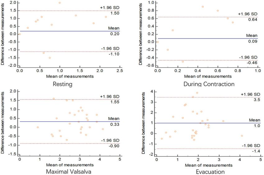
6   | Y.-B. Yao et al.

Table 2. Results of transperineal ultrasonography in three positions

Transperineal ultrasonography                                      Left lateral position          Sitting position        Squatting position            P-value
                                                                          (N ¼ 30)                     (N ¼ 30)                (N ¼ 30)

Anorectal angle, de-               Rest                                 98 (95, 101)               103 (100, 112)            105 (100, 111)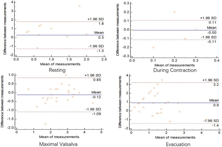
Transperineal ultrasonography and rectocele   |   7

                                                                                                                                                     Downloaded from https://academic.oup.com/gastro/advance-article/doi/10.1093/gastro/goab019/6288120 by guest on 05 July 2021
Figure 6. Consistency of anorectal angle (degree) between defecography and ultrasonography in squatting position

Figure 7. Consistency of depth of rectocele (cm) between defecography and ultrasonography in sitting position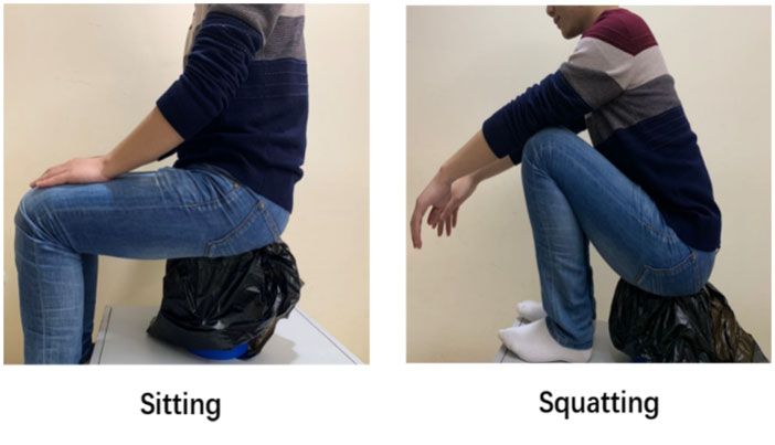
8   | Y.-B. Yao et al.

                                                                                                                                                             Downloaded from https://academic.oup.com/gastro/advance-article/doi/10.1093/gastro/goab019/6288120 by guest on 05 July 2021
Figure 8. Consistency of depth of rectocele (cm) between defecography and ultrasonography in squatting position

Second, the level of the perineum descending was not exam-                          Funding
ined by ultrasonography in this study due to the fact that the
perineum descending can only be obtained when making the                            This study was funded by the National Natural Science
ultrasound probe contact the perineum and keeping pressure                          Foundation of China [81603618, 81603625] and Shanghai
on the perineum. Finally, data were only collected from the vol-                    Municipal Health Commission [2018BR19].
unteers with a rectocele, which did not compare with normal
female volunteers without any pelvic-floor pathology. However,                      Acknowledgements
this is the pilot study exploring the role of the normal defecate
                                                                                    The manuscript was reread carefully by Wenbo Peng from
position for rectocele diagnosis and the consistency of anorectal
angle and depth of rectocele values between the defecography                        the Faculty of Health, University of Technology Sydney.
and ultrasonography methods for the rectocele. An observer-
blinded high-quality clinical trial will be required to investigate                 Conflicts of Interest
the use of transperineal ultrasonography in the wider pelvic-
floor setting in future research. The finding of this study may be                  None declared.
used for the basis of future studies exploring the optimal recto-
cele image diagnostic method.                                                       References
    In conclusion, our results provide preliminary evidence to
support the use of transperineal ultrasonography in sitting and                     1. Wexner SD, Zbar, AP, Pescatori, M, (eds). Complex Anorectal
squatting positions as a safer imaging test option for rectocele                       Disorders: Investigation and Management. New York: Springer,
diagnosis. Transperineal ultrasonography is an alternative that                        2005, p. 446.
correlates satisfactorily with the findings obtained by defecog-                    2. Beck DE, Wexner SD, Rafferty JF, (eds). Gordon and Nivatvongs’
raphy, provides important data in the evaluation of rectoceles,                        Principles and Practice of Surgery for the Colon, Rectum, and Anus,
and offers safety and comfort to patients. Further research with                       4th edn. New York: Thieme, 2019, p. 834.
a larger sample size is needed to explore the physiological ex-                     3. Fazio VW, Church JM, Delaney CP, Kiran CP (eds). Current
amination position in the future.                                                      Therapy in Colon and Rectal Surgery Third Edition. Philadelphia:
                                                                                       Elsevier, 2017, p. 114.
                                                                                    4. Deng Q, Yu KL, Liu ZY et al. Outcomes of a modified Bresler
Authors’ Contributions                                                                 procedure for the treatment of rectocele with rectal intussus-
Study concept, study design, and drafting of the manuscript:                           ception. Gastroenterol Rep (Oxf) 2020;8:457–64.
C.W. Material preparation, data collection, and analysis: Y.B.Y.,                   5. Saclarides TJ, Brubaker LT, Altringer WE et al. Clarifying the
H.Q.Y., H.J.W., and H.T.L. Statistical analysis: B.W. All authors                      technique of four-contrast defecography. Dis Colon Rectum
read and approved the final manuscript.                                                1996;39:826.
Transperineal ultrasonography and rectocele | 9
6. Martin-Martin GP, Garcia-Armengol J, Roig-Vila JV et al. 14. Chevillotte T, Coudert P, Cawley D et al. Influence of posture
Magnetic resonance defecography versus videodefecography on relationships between pelvic parameters and lumbar lor-
in the study of obstructed defecation syndrome: is video defe- dosis: comparison of the standing, seated, and supine posi-
cography still the test of choice after 50 years? Tech Coloproctol tions: a preliminary study. Orthop Traumatol Surg Res 2018;
2017;21:795–802. 104:565–8.
7. Beer-Gabel M, Teshler M, Barzilai N et al. Dynamic transperi- 15. Sultan AH, Monga A, Lee J et al. An International
neal ultrasound in the diagnosis of pelvic floor disorders. Dis Urogynecological Association (IUGA)/International
Colon Rectum 2002;45:239–48. Continence Society (ICS) joint report on the terminology for
8. Kleinübing H, Pinho MSL, Pescatori M, Regadas FSP, Zbar AP female anorectal dysfunction. Int Urogynecol J 2017;28:5–31.
(eds). Transperineal Ultrasonography of Pelvic Floor and Anorectal 16. Altomare DF, Spazzafumo L, Rinaldi M et al. Set-up and statis-
Downloaded from https://academic.oup.com/gastro/advance-article/doi/10.1093/gastro/goab019/6288120 by guest on 05 July 2021
Anatomy: Technique and Images. Milan: Springer, 2008. tical validation of a new scoring system for obstructed defae-
9. Perniola G, Shek C, Chong CCW et al. Defecatioin proctogra- cation syndrome. Colorectal Dis 2008;10:84–8.
phy and translabial ultrasound in the investigation of defeca- 17. Agachan F, Chen T, Pfeifer J et al. Constipation scoring system
tory disorder. Ultrasound Obstet Gynecol 2008;31:567–71. to simplify evaluation and management of constipated
10. Dietz HP, Steensma AB. Posterior compartment prolapse on patients. Dis Colon Rectum 1996;39:681–5.
two-dimensional and tree-dimensional pelvic floor ultra- 18. Lewis SJ, Heaton KW. Stool form scale as a useful guide to in-
sound: the distinction between true rectocele, perineal testinal transit time. Scan J Gastroenterol 1997;32:920–4.
hypermobility and enterocele. Ultrasound Obstet Gynecol 2005; 19. Beer-Gabel M, Teshler M, Schechtman E et al. Dynamic trans-
26:73–7. perineal ultrasound vs. defecography in patients with evac-
11. Broekhuis SR, Kluivers KB, Hendriks JCM et al. POP-Q, dy- uatory difficulty: a pilot study. Int J Colorectal Dis 2004;19:60–7.
namic MR imaging, and perineal ultrasonography: do they 20. Shorvon PJ, McHugh S, Diamant NE et al. Defecography in nor-
agree in the quantification of female pelvic organ prolapse? mal volunteers: results and implications. Gut 1989;30:
Int Urogynecol J 2009;20:541–9. 1737–49.
12. Takano S, Sands DR. Influence of body posture on defecation: 21. Bharucha AE, Dorn SD, Lembo A, American
a prospective study of “The Thinker” position. Tech Coloproctol Gastroenterological Association et al. American
2016;20:117–21. Gastroenterological Association Medical Position Statement
13. Rodriguez-Mias NL, Subramaniam N, Friedman T et al. on Constipation. Gastroenterology 2013;144:211–7.
Prolapse assessment supine and standing: do we need differ- 22. Felt-Bersma RJ, Luth WJ, Janssen JJ et al. Defecography in
ent cutoffs for “significant prolapse”? Int Urogynecol J 2018;29: patients with anorectal disorders: which findings are clini-
685–9. cally relevant? Dis Colon Rectum 1990;33:277–84.You can also read