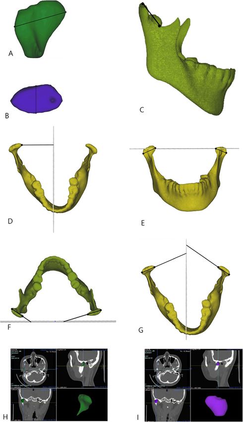
The three-dimensional morphology of mandible and glenoid fossa as contributing factors to menton deviation in facial asymmetry-retrospective study
←
→
Page content transcription
If your browser does not render page correctly, please read the page content below
Oh and Cho Progress in Orthodontics      (2020) 21:33
https://doi.org/10.1186/s40510-020-00335-3

 RESEARCH                                                                                                                                            Open Access

The three-dimensional morphology of
mandible and glenoid fossa as contributing
factors to menton deviation in facial
asymmetry—retrospective study
Min-Hee Oh and Jin-Hyoung Cho*

  Abstract
  Background: The aim of this study is to evaluate whether the three-dimensional (3D) morphology of the
  mandibular condyle, glenoid fossa, and mandible correlated with menton deviation in facial asymmetry.
  Subjects and methods: Thirty adults (15 males and 15 females; mean age, 23.2 ± 3.8 years) with facial asymmetry
  were included. Linear, angular, and volumetric measurements of the 3D morphology of the mandibular condyle,
  glenoid fossa, and mandible were recorded using computed tomography (CT) images. The right/left differences
  were obtained by subtracting the left value from the right value, and an independent t test was used to compare
  the differences between the females and males. Multiple regression analysis was performed to identify the
  correlation between the right/left difference of the 3D morphology and menton deviation.
  Results: The results of the comparative analysis did not show any statistical difference between the females and
  males (P > .05), so the females and males were combined. Multiple regression analysis for the mandibular condyle,
  glenoid fossa, and mandible showed that neck length, ramus length, and frontal ramal inclination had positive
  influences on menton deviation, with 76.5% of explanatory power. The neck length and head volume of the
  mandibular condyle when only the mandibular condyle was considered, and the ramus length and frontal ramal
  inclination when only the mandible was considered had positive influence on menton deviation with 69.9% and
  68.6% explanatory power, respectively. On the other hand, when only considering glenoid fossa, the glenoid fossa
  had little effect on menton deviation with 15.7% of explanatory power.
  Conclusions: In facial asymmetry, the right/left differences in mandibular condyle and mandible have more impact
  on the menton deviation than the right/left differences in glenoid fossa.
  Trial registration: CNUDH, CNUDH-EXP-2017-016. Registered 28 September 2017
  Keywords: Facial asymmetry, Menton deviation, 3D morphology, Computed tomography

* Correspondence: jhcho@jnu.ac.kr
Department of Orthodontics, School of Dentistry, Dental 4D Research
Institute, Dental Science Research Institute, Chonnam National University, 77
Yongbong-ro, Buk-gu, Gwangju 61186, Korea

                                        © The Author(s). 2020 Open Access This article is licensed under a Creative Commons Attribution 4.0 International License,
                                        which permits use, sharing, adaptation, distribution and reproduction in any medium or format, as long as you give
                                        appropriate credit to the original author(s) and the source, provide a link to the Creative Commons licence, and indicate if
                                        changes were made. The images or other third party material in this article are included in the article's Creative Commons
                                        licence, unless indicated otherwise in a credit line to the material. If material is not included in the article's Creative Commons
                                        licence and your intended use is not permitted by statutory regulation or exceeds the permitted use, you will need to obtain
                                        permission directly from the copyright holder. To view a copy of this licence, visit http://creativecommons.org/licenses/by/4.0/.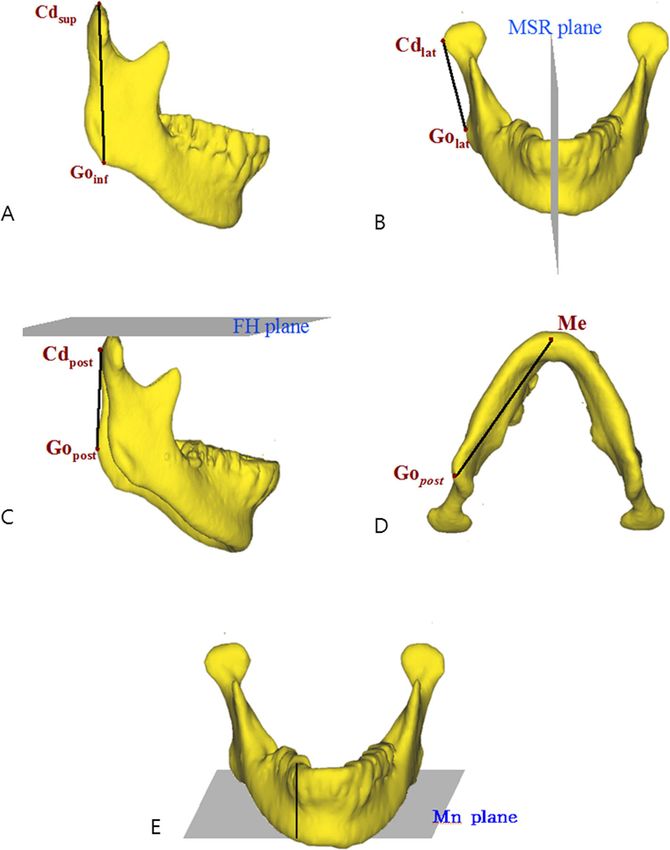
Oh and Cho Progress in Orthodontics   (2020) 21:33                                                                 Page 2 of 9

Background                                                      Methods
Facial asymmetry is influenced more by the lower third          Ethical approval for this retrospective cohort study was
of the face than by the upper and middle thirds of the          obtained (CNUDH-EXP-2017-016). The sample size of
face [1, 2]. The mandibular condyle has been known to           thirty adults was determined using the G*Power 3.1.9.2
affect the asymmetry of the mandible as an important            software (University of Kiel, Kiel, Germany), with an
growth site in the mandible [3, 4]. In addition, mandibular     effect size = 4.92 derived from the preliminary data, α =
asymmetry has been shown to be influenced by other              0.05, and 1-β = 0.95. Based on a previous study [12, 14],
various factors, such as condylar hyperplasia [5], childhood    thirty adults (15 males and 15 females) with skeletal
condylar fractures [6], condylar resorption [7], and internal   class I or III malocclusion were included. The first selec-
derangement of the temporomandibular joint (TMJ) [8].           tion was performed for subjects with menton deviation
However, most studies [5–8] used two-dimensional radio-         on postero-anterior (PA) radiographs exceeding 2° to-
graphs that have several limitations including distortion,      ward the left [16]. Menton deviation was defined as the
magnification, and lack of clarity.                             angle between the vertical reference line drawn from the
  To overcome these limitations, three-dimensional              crista galli to the anterior nasal spine (ANS) and the line
(3D) computed tomography (CT) has been used.                    drawn from the ANS to the menton on PA radiographs
Previous studies [9, 10] have shown that the cranial            [17, 18]. Finally, the amount of menton deviation was
base and the mandible can vary between the deviated             confirmed using CT images. In CT images, menton devi-
and the non-deviated sides. You et al. [11] investigated        ation was defined as the angle between the midsagittal
the mandibular morphology in patients with facial               reference plane (MSR plane) and the line drawn from
asymmetry and mandibular prognathism using CT data              the ANS to the menton on frontal view. The MSR plane
and reported that the length of the mandibular condyle          was defined as the plane perpendicular to Frankfort
and mandibular body were significantly longer on the            horizontal plane (FH plane) passing through the crista
non-deviated side than on the deviated side. Oh et al.          galli and opisthion [14]. The right side was the non-devi-
[12] compared the 3D structure of the mandibular con-           ated side, and the left side was the deviated side because the
dyles between adults with and without facial asymmetry          subjects in this study had menton deviations only toward
and reported that menton deviation was associated with          the left.
right/left differences caused by a smaller condyle, particu-      The inclusion criteria were subjects over 20 years old
larly in condylar neck length and neck and head volume,         with available frontal and lateral cephalograms and CT
on the deviated side. Moreover, Ikeda et al. [13] found that    images acquired before treatment. The exclusion criteria
3D mandibular morphologic asymmetry was associated              were orthodontic treatment, orthognathic surgery,
with condylar movement in patients with mandibular              prosthetic treatment for more than a single crown, TMJ
asymmetry.                                                      morphological changes, signs or symptoms of temporo-
  In addition, several previous studies have suggested that     mandibular disorders, systematic arthritis, facial trauma,
the shape [14] and volume [14, 15] of the glenoid fossa are     craniofacial anomaly, functional discrepancies, functional
necessary to make the diagnosis and administer proper           crossbite, and skeletal class II malocclusion.
treatment in patients with facial asymmetry. Cho et al.           The CT images were acquired using a CT scanner
[14] evaluated the effect of the glenoid fossa on menton        (Light Speed QX/i, GE Medical Systems, Milwaukee,
deviation in facial asymmetry. The vertical position and        WI, USA) before treatment. A more detailed explanation
the depth of the glenoid fossa showed significant               of CT image acquisition can be found in previous stud-
differences between the symmetry and asymmetry groups.          ies [12, 14]. The V-works 4.0 software (CyberMed Inc.,
Kim et al. [15] suggested that the volume of the glenoid        Seoul, Korea) was used to reconstruct the 3D images
fossa, as well as that of the mandibular condyle, should be     from the digital imaging and communication in medi-
considered in facial asymmetry.                                 cine data. Definitions of the landmarks are described in
  A literature review of recent CT [11–15] studies on           Table 1 [12, 14]. The 3D reference planes were con-
the correlation between 3D morphology of the TMJ                structed. The FH plane passed through the right orbitale
and facial asymmetry revealed several measurements              and right and left porions. The MSR plane was the plane
that differed significantly in facial asymmetry. How-           perpendicular to the FH plane passing through the crista
ever, few studies have simultaneously evaluated the             galli and opisthion. The anteroposterior reference plane
contribution of the mandible and glenoid fossa to               (PO plane) was the plane perpendicular to the FH plane
menton deviation. The purpose of this study was to              passing through the right and left porions.
identify the factors contributing to menton deviation             In order to precisely identify the landmarks, the man-
by evaluating the correlation between menton devi-              dible was separated from the whole volume rendering
ation and 3D morphologies of the mandibular condyle,            image by removing the overlapping areas as reported in
glenoid fossa, and mandible in facial asymmetry.                a previous study [12] and exporting it into a selection of
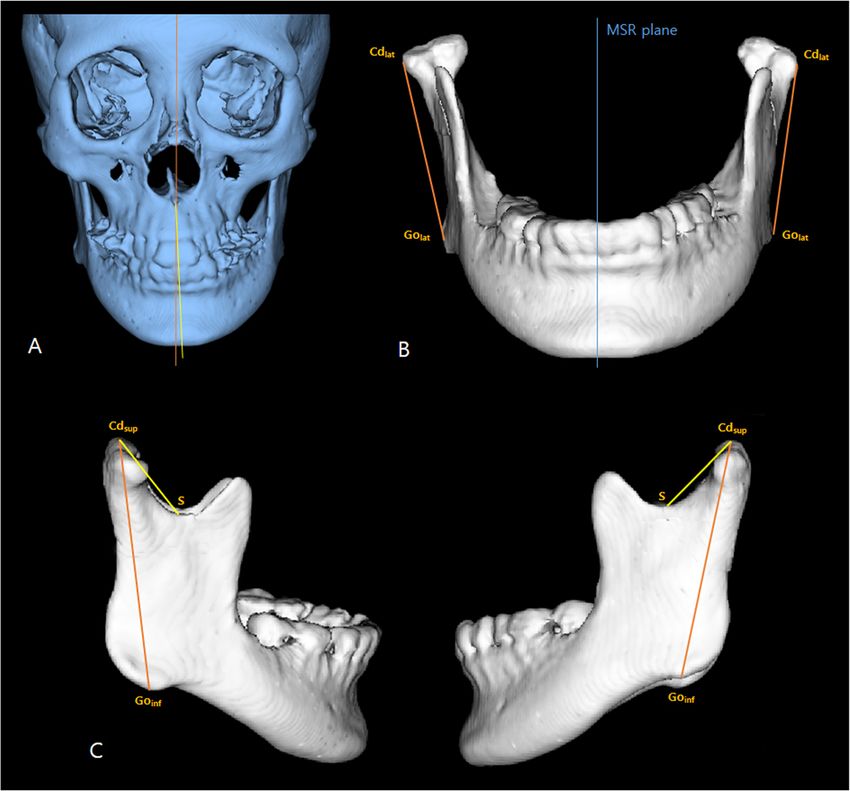
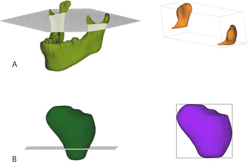
Oh and Cho Progress in Orthodontics          (2020) 21:33                                                                               Page 3 of 9

Table 1 The landmarks used in this study
Landmarks                               Abbreviation                    Description
Crista galli                            Cg                              The most superior point of the crista galli of the ethmoid bone
Opisthion                               Op                              The most posterior point on the posterior margin of the foramen magnum
Porion                                  Po                              The highest point on the roof of the external auditory meatus
Orbitale                                Or                              The deepest point on the infraorbital margin
Condylion superius                      Cdsup                           The most superior point of the condyle head
Condylion medialis                      Cdmed                           The most medial point of the condyle head
Condylion lateralis                     Cdlat                           The most lateral point of the condyle head
Condylion anterius                      Cdant                           The most anterior point of the condyle head
Condylion posterius                     Cdpost                          The most posterior point of the condyle head
Sigmoid notch                           S                               The most inferior point of sigmoid notch
Gonion lateralis                        Golat                           The most lateral point of the gonion area
Gonion posterius                        Gopost                          The most posterior point of the gonion area
Gonion inferius                         Goinf                           The most inferior point of the gonion area
Antegonion                              Ag                              The deepest point of antegonial notch of mandible
Menton                                  Me                              The most inferior point on mandibular symphysis
Roof of glenoid fossa                   RG                              The highest point on the roof of the glenoid fossa
Articular eminence                      AE                              The most inferior point on the articular tubercle

demand (SOD) file. Moreover, the neck SOD file con-                       while parallel to the FH plane (Fig. 1) [12]. The neck
taining the condylar process above the sigmoid notch                      and head SOD files were converted into a 3D surface
was separated by a plane passing through the most infer-                  shaded display (SSD) model to measure the volume of
ior point of sigmoid notch while parallel to the FH                       the 3D object model.
plane. The head SOD file, which included the upper part                     The definition of measurements was described in
of the condylar process, was separated by a plane passing                 Table 2 and Figs. 2 and 3 [12, 14]. All measurements,
through the most contracted part of the condylar neck                     including linear measurements, were measured on a

  Fig. 1 Formation of three-dimensional images. The neck (a) and head (b) selection of demand (SOD) files were separated from the mandible
  SOD file that was separated from the whole volume rendering image by removing the overlapping areas using the sculpt functions of the V-
  works program (CyberMed Inc., Seoul, Korea) (reproduced with permission [12])
Oh and Cho Progress in Orthodontics             (2020) 21:33                                                                         Page 4 of 9

Table 2 Definition of the measurements used in this study                            The right/left differences were obtained by subtracting
Measurements                                  Definition                           the left value from the right value. Thus, if the value on
Mandibular condyle                                                                 the deviated side was smaller than that on the non-
  Mediolateral dimension (mm)                 Cdmed to Cdlat
                                                                                   deviated side, the right/left differences were described as
                                                                                   positive (+), otherwise, they were described as negative
  Anteroposterior dimension (mm)              Cdant to Cdpost
                                                                                   (−). The results of comparative analysis did not show
  Neck length (mm)                            Cdsup to S                           any statistical difference between the females and males
  Mediolateral condylar position (mm)         Cdmed to MSR plane                   (P > .05), so the females and males were combined.
  Condylar angle to FH (°)                    (Cdmed - Cdlat) to FH plane            Multiple regression model was used to determine the
  Condylar angle to PO (°)                    (Cdmed - Cdlat) to PO plane          effect of right/left differences of the measured variables
  Condylar angle to MSR (°)                   (Cdmed - Cdlat) to MSR plane
                                                                                   on the menton deviation. A multiple regression model
                       3                                                           using backward elimination was used to identify the
  Neck volume (mm )                           The volume of condylar neck
                                              above S                              causes of menton deviation. Multiple regression analyses
                                                                                   were performed four times for the different conditions:
  Head volume (mm3)                           The volume of condylar head
                                              above most constriction point        (1) for the mandibular condyle, glenoid fossa, and
                                              of condylar neck                     mandible (n = 30); (2) for the mandibular condyle only
Glenoid fossa                                                                      (n = 30); (3) for the glenoid fossa only (n = 30); and (4)
  Vertical position of RG to FH (mm)          RG to FH plane                       for the mandible only (n = 30).
  Vertical position of AE to FH (mm)          AE to FH plane
                                                                                   Results
  Depth of glenoid fossa (mm)                 AE to RG parallel to MSR plane
                                                                                   The demographic characteristics of the subjects, includ-
  Sagittal position of RG to PO (mm)          RG to PO plane                       ing gender, age, amount of menton deviation, ANB, and
  Sagittal position of AE to PO (mm)          AE to PO plane                       SN-MP are presented in Table 3. The right/left differ-
  Anterior angle of GF to FH (°)              (RG-AE) to FH plane                  ences of all measurements are shown in Table 4.
Mandible                                                                             The regression equation between menton deviation and
                                                                                   the right/left differences of the measurements for all 20
  Ramus length (mm)                           Cdsup - Goinf
                                                                                   variables (n = 30) was as follows: menton = 0.735 • (neck
  Frontal ramal inclination (°)               (Cdlat - Golat) to MSR plane
                                                                                   length) + 0.329 • (ramus length) + 0.331 • (frontal ramal
  Lateral ramal inclination (°)               (Cdpost - Gopost) to FH plane        inclination). The adjusted R2 value was 0.765 (P < .000).
  Body length (mm)                            Gopost - Me                          The regression equation between menton deviation and
  Body height (mm)                            Canine cusp tip to Mandibular        the right/left differences of the nine variables for the man-
                                              plane                                dibular condyle (n = 30) was as follows: menton = 0.903 •
FH Frankfort horizontal plane, PO anteroposterior reference plane, MSR             (neck length) + 0.005 • (head volume). The adjusted R2
midsagittal reference plane, RG roof of glenoid fossa, AE articular eminence, GF
glenoid fossa, Cdmed condylion mediais, Cdlat condylion lateralis, Cdant
                                                                                   value was 0.699 (P < .000). The regression equation for
condylion anterius, Cdpost condylion posterius, Cdsup condylion superius, S        the six variables of the glenoid fossa (n = 30) was as
sigmoid notch, Goinf gonion inferius, Golat gonion lateralis, Gopost gonion        follows: menton = −1.022 • (vertical position of AE to
posterius, Me menton
                                                                                   FH). This equation showed that the adjusted R2 value was
                                                                                   0.157, indicating that the glenoid fossa had almost no
three-dimensional coordinate system, indicating 3D linear,                         effect on menton deviation (P < .05). The regression equa-
angular, and volumetric measurements. The volumes of                               tion for the five variables of the mandible (n = 30) was as
the neck and head were calculated automatically using the                          follows: menton = 0.659 • (ramus length) + 0.407 • (frontal
volume measure function of the V-works program with                                ramal inclination). In this equation, the adjusted R2 value
the SSD model, which is the 3D object model of neck and                            was 0.686 (P < .000, Table 5).
head.
                                                                                   Discussion
Statistical analysis                                                               The degree of recognition of facial asymmetry can be
Statistical analyses were performed using IBM SPSS Statis-                         affected by various factors. Ahn et al. [18] reported that
tics (version 23.0; IBM Co., Armonk, NY, USA). All                                 the degree of asymmetry recognition increased when the
measurements were performed by a single operator (JHC).                            degree of menton deviation increased. Lee et al. [17] also
Twenty images were randomly selected and the measure-                              reported that menton deviation had the greatest effect
ments were performed twice with a 2-week interval between                          on the degree of facial asymmetry recognition. In
the measurements. The intraclass correlation coefficient                           addition, Ferguson [16] evaluated the correlations be-
(ICC) was performed to assess reliability. The ICC (> 0.983)                       tween facial photographs and PA radiographs of patients
indicated excellent intra-observer reliability.                                    with facial asymmetry and reported that the asymmetry
Oh and Cho Progress in Orthodontics (2020) 21:33 Page 5 of 9 Fig. 2 The measurements of the mandibular condyle. a Mediolateral dimension of the condyle. b Anteroposterior dimension of the condyle. c Neck length. d Mediolateral condylar position. e Condylar angle to the Frankfort horizontal plane. f Condylar angle to the anteroposterior reference plane. g Condylar angle to the midsagittal reference plane. h Neck volume. i Head volume (reproduced with permission [12])
Oh and Cho Progress in Orthodontics       (2020) 21:33                                                                                 Page 6 of 9

 Fig. 3 The measurements of the mandible. a Ramus length. b Frontal ramal inclination. c Lateral ramal inclination. d Body length. e Body height
 (reproduced with permission [14])

                                                                           was recognized when menton deviation from the midline
                                                                           was 2° or higher. McAvinchey et al. [19] investigated the
                                                                           perception of facial asymmetry in young adults and re-
Table 3 Description of the subjects (n = 30)
                                                                           ported that the perception of asymmetry was affected by
                                                                           the amount of asymmetry. Menton deviation might be
Demographic characteristic                            % or mean ± SD
                                                                           one of the primary contributing factors for recognition
Gender
                                                                           of facial asymmetry. Thus, it is necessary to evaluate the
  Female (%)                                          50.0                 factors contributing to menton deviation in order to
  Male (%)                                            50.0                 establish treatment plans for facial asymmetry. The
Age (years)                                           23.2 ± 3.8           present study examined the factors contributing to men-
Amount of menton deviation (°)                        5.7 ± 2.5            ton deviation in order to determine what contributed to
ANB (°)                                               −0.5 ± 3.3
                                                                           facial asymmetry.
                                                                             The subjects had skeletal class I or III malocclusion, in
SN-MP (°)                                             34.6 ± 6.3
                                                                           which the ANB angle was lower than 5° on the lateral
Oh and Cho Progress in Orthodontics (2020) 21:33 Page 7 of 9
Table 4 The right/left differences of the measurements (n = 30) Table 5 Multiple linear regression analysis between the
Measurements Mean ± SD menton deviation and the right/left differences of the
Mandibular condyle
measurements (n = 30)
Measurements Coefficient t P value VIF
Mediolateral dimension (mm) 0.71±2.19
For all variablesa
Anteroposterior dimension (mm) 0.38±1.10
Neck length 0.735 3.223 0.003** 2.315
Neck length (mm) 2.43±2.75
Ramus length 0.329 2.082 0.017* 2.346
Mediolateral condylar position (mm) 0.73±2.08
Frontal ramal inclination 0.331 3.031 0.005** 1.430
Condylar angle to FH (°) 2.09±8.13
For the mandibular condyleb
Condylar angle to PO (°) −0.59±7.27
Neck length 0.903 3.698 0.000*** 2.072
Condylar angle to MSR (°) −3.28±13.42
Head volume 0.005 2.625 0.014* 2.072
Neck volume (mm3) 341.99±443.68
For the glenoid fossac
Head volume (mm3) 338.99±379.72
Vertical position of AE to FH −1.022 2.567 0.016* NA
Glenoid Fossa
d
For the mandible
Vertical position of RG to FH (mm) −0.31±2.02
Ramus length 0.659 4.733 0.000*** 1.363
Vertical position of AE to FH (mm) −0.96±2.45
Frontal ramal inclination 0.407 3.305 0.003** 1.363
Depth of glenoid fossa (mm) −0.66±1.35
VIF variance inflation factor, AE articular eminence, FH Frankfort horizontal
Sagittal position of RG to PO (mm) −0.10±1.51 plane, NA not available
a
Adjusted R2 = 0.765, P < 0.000
Sagittal position of AE to PO (mm) 0.21±1.69 b
Adjusted R2 = 0.699, P < 0.000
Anterior angle of GF to FH (°) −3.37±7.34 c
Adjusted R2 = 0.157, P < 0.05
d
Adjusted R2 = 0.686, P < 0.000
Mandible *
P < 0.05
**
Ramus length (mm) 3.27±4.22 P < 0.01
***
P < 0.001
Frontal ramal inclination (°) 3.98±4.53
Lateral ramal inclination (°) 3.25±5.42
size of 200 and 94, respectively, while the present study
Body length (mm) 1.16±3.32 was conducted on only 30 subjects.
Body height (mm) 0.19±1.42 In facial asymmetry, the explanatory power of the re-
SD standard deviation, FH Frankfort horizontal plane, PO anteroposterior gression equation for all 20 variables for the mandibular
reference plane, MSR midsagittal reference plane, RG roof of glenoid fossa, AE
articular eminence, GF glenoid fossa
condyle, glenoid fossa, and mandible was 76.5%, and the
neck length, ramus length, and frontal ramal inclination
cephalogram. Ngan et al. [20] compared the skeletal had positive influences on menton deviation. Multiple
growth changes between class II division 1 and class I regression analysis of the anatomical structures affecting
subjects and reported that the majority of the class II menton deviation showed that the explanatory power of
cases showed mandibular skeletal retrusion or a combin- the regression equation for the mandibular condyle was
ation of horizontal and vertical abnormalities of the 69.9% and the neck length and head volume of the
mandible, rather than maxillary protrusion. Previous mandibular condyle had positive influences on menton
studies [3, 21] compared the position and volume of the deviation. The explanatory power of the regression
condyle in class I, II, and III and reported that the great- equation for the glenoid fossa was 15.7%, indicating that
est condylar decentralization was observed in the class II the glenoid fossa had little effect on menton deviation.
group [21] and significantly lower condylar volume was The explanatory power of the regression equation for
observed in class II subjects compared to those in class I the mandible was 68.6% and the ramus length and
and III [3]. Thus, the present study excluded subjects frontal ramal inclination had positive influences on men-
with skeletal class II malocclusion due to the possible ton deviation (Table 5). The variance inflation factor for
presence of mandibular growth disorders [3, 20, 21]. all variables was less than 3, indicating that there was no
In this study, all measurements, including volumes of multicollinearity. These results suggest that the right/left
condylar neck and head, showed no significant differ- differences of the mandibular condyle and mandible can
ences between males and females. These results are be used to predict menton deviation. Specifically, the
inconsistent with the results of previous studies [3, 4]. right/left differences of neck length and ramus length
Previous studies [3, 4] reported that the condylar volume contributed the most to menton deviation. However, it
was significantly higher in males than in females. This was not possible to predict menton deviation using the
conflicting result is thought to be due to the fact that right/left difference of the glenoid fossa (Fig. 4). These
the previous studies [3, 4] was conducted on a sample results are caused by the mandibular asymmetry isOh and Cho Progress in Orthodontics (2020) 21:33 Page 8 of 9 Fig. 4 A subject consistent with the results of this present study. a Menton is deviated to the left side. b The frontal ramal inclination is greater in the right side (non-deviated side) than the left side (deviated side). c The ramus length and neck length are greater in the right side (non- deviated side) than the left side (deviated side) associated with the condylar growth center, which dir- muscles and bone density in an asymmetrical mandible ectly or indirectly regulates the size of the condyle and and reported that the asymmetrical mandible was also the length of the condylar neck, ramus, and body of associated with asymmetrical distributions of the highest the mandible [22]. In the diagnosis of facial asymmetry mineralized cortical bone and that it was age dependent. patients, the difference between right and left side of Nakano et al. [24] evaluated changes in the calcified mandible including mandibular condyle should be tissue of the mandibular condyle during altered muscle considered and a treatment plan should be established function and showed that both the mandible and the to improve it. condyle modified their shape and size, as well as the The present study had several limitations. Although trabecular bone of the condyle, during the shifting of the the upper and middle thirds of the face have less effect mandible to one side as it closed. Nur et al. [25] found on facial asymmetry than the lower third of the face [17, that soft tissues compensated for hard tissues at the 19], there might be a maxillary asymmetry in facial gonial level. In addition, Kurusu et al. [26] evaluated the asymmetry. The horizontal reference plane, FH plane, relationship between occlusal force and mandibular was defined as a plane passing through the right orbitale condyle morphology and reported that occlusal force and right and left porions to exclude the asymmetry on influenced not only maxillofacial morphology but also orbitale points. Nevertheless, the effects of the maxillary mandibular condyle morphology. Thus, further studies asymmetry could not be completely ruled out, due to are needed to evaluate the effects of soft tissue and func- the use of ANS for measuring the menton deviation. tion to better understand the etiology of facial asymmetry. In addition, menton deviation can be influenced by In the literature, the condylar volume has been also other factors, such as functional adaptation and the related to the type of mandibular divergence [27]. In a surrounding neuromuscular system. Maki et al. [23] study with ninety-four subjects, higher condylar volume evaluated the correlations between the surrounding was a common characteristic of low angle subjects
Oh and Cho Progress in Orthodontics              (2020) 21:33                                                                                             Page 9 of 9

compared to normal and high mandibular plane angle                                  9.    Kim SJ, Lee KJ, Lee SH, Baik HS. Morphologic relationship between the
subjects. In this study, the differences according to the                                 cranial base and the mandible in patients with facial asymmetry and
                                                                                          mandibular prognathism. Am J Orthod Dentofac Orthop. 2013;144:330–40.
type of mandibular divergence were not evaluated. Thus,                             10.   Shibazaki-Yorozuya R, Yamada A, Nagata S, Ueda K, Miller AJ, Maki K. Three-
the role of the neck and ramus length related to the                                      dimensional longitudinal changes in craniofacial growth in untreated
concept of mandibular divergence should be supported                                      hemifacial microsomia patients with cone-beam computed tomography.
                                                                                          Am J Orthod Dentofac Orthop. 2014;145:579–94.
by new studies.                                                                     11.   You KH, Lee KJ, Lee SH, Baik HS. Three-dimensional computed tomography
                                                                                          analysis of mandibular morphology in patients with facial asymmetry and
                                                                                          mandibular prognathism. Am J Orthod Dentofac Orthop. 2010;138:540.e1-8.
Conclusions                                                                         12.   Oh MH, Kang SJ, Cho JH. Comparison of the three-dimensional structures of
In facial asymmetry, the right/left differences in man-                                   mandibular condyles between adults with and without facial asymmetry: a
dibular condyle and mandible have more impact on the                                      retrospective study. Korean J Orthod. 2017;48:73–80.
                                                                                    13.   Ikeda M, Miyamoto JJ, Takada JI, Moriyama K. Association between 3-
menton deviation than the right/left differences in the                                   dimensional mandibular morphology and condylar movement in subjects
glenoid fossa.                                                                            with mandibular asymmetry. Am J Orthod Dentofac Orthop. 2017;151:324–34.
                                                                                    14.   Cho JH, Lee KM, Park HJ, Hwang HS. 3-D CT image study of effect of
Acknowledgements                                                                          glenoid fossa on menton deviation. J Korean Assoc Maxillofac Plast
Not applicable                                                                            Reconstr Surg. 2011;33:337–45.
                                                                                    15.   Kim JY, Kim BJ, Park KH, Huh JK. Comparison of volume and position of the
Authors’ contributions                                                                    temporomandibular joint structures in patients with mandibular asymmetry.
MHO performed the work and wrote the manuscript. JHC designed the                         Oral Surg Oral Med Oral Pathol Oral Radiol. 2016;122:772–80.
study, evaluated the statistical results, and revised the manuscript. All authors   16.   Ferguson JW. Cephalometric interpretation and assessment of facial
read and approved the final manuscript.                                                   asymmetry secondary to congenital torticollis. Int J Oral Maxillofac Surg.
                                                                                          1993;22:7–10.
                                                                                    17.   Lee GH, Cho HK, Hwang HS, Kim JC. Studies of relationship between P-A
Funding
                                                                                          cephalometric measurements and visual facial asymmetry. Korean J Phys
This work was supported by the National Research Foundation of Korea
                                                                                          Anthrop. 1998;11:41–8.
(NRF) grant funded by the Korea government (No. 2019R1G1A1008831 &
                                                                                    18.   Ahn JS, Hwang HS. Relationship between perception of facial asymmetry and
2018R1D1A1B07051057).
                                                                                          posteroanterior cephalometric measurements. Korea J Orthod. 2001;31:489–98.
                                                                                    19.   McAvinchey G, Maxim F, Nix B, Djordjevic J, Linklater R, Landini G. The
Availability of data and materials                                                        perception of facial asymmetry using 3-dimensional simulated images.
Not applicable                                                                            Angle Orthod. 2014;84:957–65.
                                                                                    20.   Ngan PW, Byczek E, Scheick J. Longitudinal evaluation of growth changes in
Ethics approval and consent to participate                                                class II division 1 subjects. Semin Orthod. 1997;3:222–31.
This retrospective study was performed after obtaining approval from the            21.   Fraga MR, Rodrigues AF, Ribeiro LC, Campos MJ, Vitral RW. Anteroposterior
Institutional Review Board of at Chonnam National University Dental Hospital              condylar position: a comparative study between subjects with normal
(CNUDH-EXP-2017-016).                                                                     occlusion and patients with class I, class II division 1, and class III
                                                                                          malocclusions. Med Sci Monit. 2013;19:903–7.
Consent for publication                                                             22.   Erickson GE, Waite DE. Mandibular asymmetry. J Am Dent Assoc. 1974;89:1369–73.
Not applicable                                                                      23.   Maki K, Miller AJ, Okano T, et al. Cortical bone mineral density in
                                                                                          asymmetrical mandibles: a three-dimensional quantitative computed
                                                                                          tomography study. Eur J Orthod. 2001;23:217–32.
Competing interests
                                                                                    24.   Nakano H, Watahiki J, Kubota M, et al. Micro X-ray computed tomography
The authors declare that they have no competing interests.
                                                                                          analysis for the evaluation of asymmetrical condylar growth in the rat.
                                                                                          Orthod Craniofacial Res. 2003;6:168–72.
Received: 1 April 2020 Accepted: 13 August 2020
                                                                                    25.   Nur RB, Çakan DG, Arun T. Evaluation of facial hard and soft tissue
                                                                                          asymmetry using cone-beam computed tomography. Am J Orthod
                                                                                          Dentofac Orthop. 2016;149:225–37.
References                                                                          26.   Kurusu A, Horiuchi M, Soma K. Relationship between occlusal force and
1. Severt TR, Proffit WR. The prevalence of facial asymmetry in the dentofacial           mandibular condyle morphology. Evaluated by limited cone-beam
    deformities population at the University of North Carolina. Int J Adult               computed tomography. Angle Orthod. 2009;79:1063–9.
    Orthodon Orthognath Surg. 1997;12:171–6.                                        27.   Saccucci M, Polimeni A, Festa F, Tecco S. Do skeletal cephalometric
2. Thiesen G, Gribel BF, Freitas MPM, Oliver DR, Kim KB. Mandibular                       characteristics correlate with condylar volume, surface and shape? A 3D
    asymmetries and associated factors in orthodontic and orthognathic                    analysis. Head Face Med. 2012;8:15.
    surgery patients. Angle Orthod. 2018;88:545–51.
3. Saccucci M, D’Attilio M, Rodolfino D, Festa F, Polimeni A, Tecco S. Condylar
    volume and condylar area in class I, class II and class III young adult         Publisher’s Note
    subjects. Head Face Med. 2012;8:34.                                             Springer Nature remains neutral with regard to jurisdictional claims in
4. Nota A, Caruso S, Ehsani S, Baldini A, Tecco S. Three-dimensional volumetric     published maps and institutional affiliations.
    analysis of mandibular condyle changes in growing subjects: a retrospective
    cross-sectional study. Cranio. 2018;24:1–7.
5. Bruce RA, Hayward JR. Condylar hyperplasia and mandibular asymmetry: a
    review. J Oral Surg. 1968;26:281–90.
6. Proffit WR, Vig KW, Turvey TA. Early fracture of the mandibular condyles:
    frequently an unsusepected cause of growth disturbances. Am J Orthod.
    1980;78:1–24.
7. Huang YL, Pogrel MA, Kaban LB. Diagnosis and management of condylar
    resorption. J Oral Maxillofac Surg. 1997;55:114–9.
8. Trpkova B, Major P, Nebbe B, Prasad N. Craniofacial asymmetry and
    temporomandibular joint internal derangement in female adolescents: a
    posteroanterior cephalometric study. Angle Orthod. 2000;70:81–8.
You can also read