ISFRI IAFR - Program & Abstracts - International Society of Forensic Radiology and ...
←
→
Page content transcription
If your browser does not render page correctly, please read the page content below
Program & Abstracts

                          Joint Congress 2017

                ISFRI•IAFR
    6th Congress of the International Society of Forensic Radiology and Imaging
12th Anniversary Meeting of the International Association of Forensic Radiographers

            11-13 May • www.ISFRI2017.com
  University of Southern Denmark • Odense, Denmark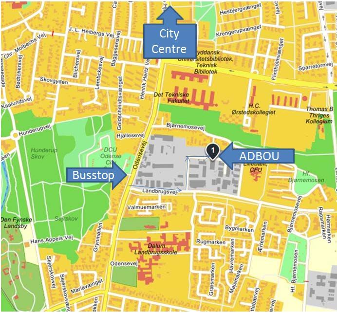
Dear Participants,

It is our great pleasure on behalf of the scientific committee to welcome you to the
joined 6th annual meeting of the International Society for Forensic Radiology and
Imaging (ISFRI) and the 12th Anniversary Meeting of the International Association of
Forensic Radiographers (IAFR), in Odense in Denmark May 11th – 13th 2017.

Forensic imaging plays an increasingly important role in forensic science and
constitutes a rapidly developing research area. The joint ISFRI and IAFR annual
meeting presents an opportunity to hear about the latest developments in this
exciting new field and meet with colleges from all over the world.

The meeting is hosted by the Department of Forensic Medicine at the University of
Southern Denmark in close collaboration with the Departments of Forensic Medicine
at the Universities of Copenhagen and Aarhus.

The University of Southern Denmark is the third largest university in Denmark and has
a close cooperation with Odense University Hospital. This cooperation will be further
strengthened with the opening of a new super hospital in 2022 that will be linked to
the university campus by a new-built faculty of medical science.

Odense is a peaceful and beautiful city situated on the island of Funen in the centre
of Denmark and only 1.5 hours train ride from Copenhagen Airport. The city is named
after Odin, the Norse god of war, poetry and wisdom – a perfect patron for a cultural
and historical hotspot.

As always the spirit of the meeting is one of collegiality and respect. The joined
annual meeting of the ISFRI and IAFR offers the participants the chance to meet old
and new friends, to strengthen bonds and to start collaborations.

Best regards,
Professor Peter Mygind Leth, MD. DMSc. Chair ISFRI
On behalf of the scientific committee
Content
Organizing Committee ……………………………………………………..              2
Venue, Floor plan………..……………………………………………………               3
Program……………………………………………………………………………                     4
Social Program …………………………………………………………………                 8
Oral presentations ……………………………………………………………              12
Poster presentations ………………………………………………………..            14
Abstracts …………….……………………………………………………………                 16

Organizing Committee

Peter Mygind Leth       pleth@health.sdu.dk
Peter Thiis Knudsen     pthiis@health.sdu.dk
Jesper Lier Boldsen     jboldsen@health.sdu.dk
Mette Ramsdal Poulsen mette.ramsdal.poulsen@rsyd.dk
Lene Warner Boel        lwb@forens.au.dk
Lise Loft Nagel         lisenagel@rm.dk
Christina Jacobsen      christina.jacobsen@sund.ku.dk
Niels Lynnerup          n.lynnerup@sund.ku.dk
Chiara Villa            chiara.villa@sund.ku.dk
Lars Oesterhelweg       lars.oesterhelweg@charite.dk
Kim Pelle Christensen   kim.pelle.christensen@rsyd.dk

2
Venue, Floor Plan

SDU - University of Southern Denmark, Campusvej 55, 5230 Odense M

                                                                    3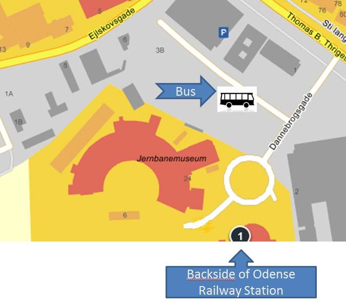
Daily Programs
Wednesday May 10
Pre-Congress meetings

15.30 – 17.00           ISFRI board meeting
                        Library, Department of Forensic Medicine, J.B. Winsløws Vej 17A, 5000 Odense C
                        (closed meeting, by invite only)

18.00 – 20.00           Reception at Odense City Hall

Thursday May 11, 2017

08:00                   Registration, welcome & coffee

09:00                   Opening by Professor Peter Mygind Leth, University of Southern Denmark

09:15 – 11.00           Plenary Session: Forensic imaging in times of terror
                        Chairmen professor Guy Rutty and forensic radiologist Chris O’Donnell

09:15                   Hard data in the face of conflicting narrative
                        Forensic pathologist dr. Alon Krispin. Director of Forensic Imaging Service at the
                        National Center of Forensic Medicine (ICFM), Israel

10:00                   Forensic imaging of the victims from Malaysian Airline Flight MH17
                        Professor Paul Hofman, Department of Radiology, Maastricht University Medical
                        Center

10:45                   Discussion

11:00                   Coffee break and posters

11:45 – 13.00           Scientific session I (6 lectures of 10 minutes)
                        Chairmen associate professor Peter Thiis Knudsen and forensic anthropologist
                        Chiara Villa

13:00 – 14.00           Lunch

14:00 – 15.30           Plenary session: Forensic imaging in times of terror
                        Chairmen professor Guillaume Gorincour and professor Rick R. van Rijn

14:00 - 14:30           Identification of suspects from video-surveillance cameras
                        Forensic Anthropologist Peter K. Larsen, University of Copenhagen, Unit of
                        Biological Anthropology

14:30 - 15:30           The use of forensic radiology for the investigation of the terror attack at Utöy in
                        Norway July 22nd 2011.
                        Senior consultant in forensic pathology and clinical forensic medicine Arne
                        Stray-Pedersen, Department of Forensic Medicine, University of Oslo

4
15:30                 Discussion

15:45                 Coffee break and posters

16:30                 End of day 1

19:00 - 21:00         Casual Dinner at Dept. of Anthropology (ADBOU)

Friday May 12, 2017
08:00                 Registration, welcome & coffee

08:30 – 10.30         Plenary session: Specialized imaging modalities
                      Chairmen professor Jesper Boldsen and professor Lene Boel

08:30                 MicroCT
                      Professor Sarah Hainsworth, Director of ASDEC/Professor of Materials and
                      Forensic Engineering, Department of Engineering, University of Leicester, UK

09:15                 Discussion

09:30                 Contrast enhanced micro-CT; potential forensic applications
                      Dr Robert Stephenson PhD, Marie Curie research fellow, Department of Clinical
                      Medicine, University of Aarhus

10:15                 Discussion

10:30                 Coffee break and posters

11:15 – 13.00         Scientific Session II (6 lectures of 10 minutes)
                      Chairmen forensic radiologist Thomas Ruder & professor Jørgen Lange Thomsen

12:15                 Gil Brogdon Honorary lecture
                      Imaging in archaeology
                      Professor Niels Lynnerup, anthropological laboratory, Department of forensic
                      Medicine, University of Copenhagen.

13:00                 Lunch

14:00                 ISFRI workshops: (all are welcome)

                       1. DVI (Room O98) Moderator: Guy Rutty
                       2. Education (Room O97) Moderator: Guillaume Gorincour
                       3. Image acquisition (Room 96) Moderator: Natalie Adolphi
                       4. Anthropology (Room O94) Moderator: Alison Brough

15:00                 Coffee break and posters

                                                                                                     5
15:30 – 16.45   Scientific session III (6 lectures of 10 minutes)
                Chairmen forensic radiologist Lise Loft Nagel and professor Lars Oesterhelweg

16:45           6th General assembly of the ISFRI

17:30           End of day 2

14:00 – 16:30   Program B: Parallel Program (Room O99)
                12th Annual Meeting of International Association of Forensic Radiographers
                Moderators: M. Viner & J. Kroll

14:00 – 14:10   Welcome by IAFR Chair / Vice-Chair

14:10 – 14:45   Bone Age Guidelines
                E Doyle

14:45 – 15:15   Anthropology - Radiology Assessment of Juvenille remains - MDCT or Digital
                Radiography?
                Amy- Lee Brookes

15:15 – 16:00   Age estimation in forensic osteology
                Jesper Boldsen, ADBOU, Dept. of Anthropology, University of Southern Denmark

20:00           Annual dinner at Hindsgavl Castle

Saturday May 13, 2017
08:00           Registration, welcome & coffee

08:30 – 11.00   Plenary session
                Chairmen professor Krzysztof Wozniak and professor Niels Lynnerup

08:30           The diagnostic accuracy of the triad in “shaken baby syndrome”. Is the evidence
                massive and robust?
                Professor Anders Eriksson and professor Anders Persson, Unit of Forensic
                Medicine, Umeå University and Center for Medical Image Science and
                Visualization (CMIV), Linköping university.

09:30 – 09:45   Discussion

09:45           Coffee break and posters

10:15           Musculoskeletal Ultrasound with Reference to Potential use in Forensics, chief
                physician Michel Bach Hellfritzsch, Aarhus University Hospital

10.45           Discussion

11.00           Coffee break and posters

6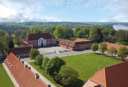
11:30 – 13.00   Scientific session IV (6 lectures of 10 minutes)
                Chairmen professor Michael Thali and associate professor Birgitte Schmidt
                Astrup

12:30 – 13:15   3D Crime Scene reconstruction in bombings
                Senior lecturer Chiara Villa, University of Copenhagen and deputy chief forensic
                pathologist Peter Thiis Knudsen, University of Southern Denmark

13.15           Closing ceremony & Call for ISFRI 2018 Board

13:45           End of ISFRI 2017

10:45 – 12:45   Program B: Parallel Program
                12th Annual Meeting of International Association of Forensic Radiographers
                Moderator: E. Doyle

10:45 - 11:00   Management of a terrorist attack situation
                Paris November 2015
                Philippe Gerson, Hôtel Dieu Paris Hospital, Paris, France

11:00 - 11:45   Terrorist Incidents in London 1990–2005
                Mark Viner, Forensic Institute, Cranfield University, U.K.

11:45 - 12:15   Integrating MDCT into the DVI Process
                Jeroen Kroll, UMC Maastricht, Netherlands

12:15 - 12:45   Forensic Radiography “Down Under”
                Lisa Detleif-Nielson, Dept. of Forensic Medicine, Enmore, Australia

                                                                                               7
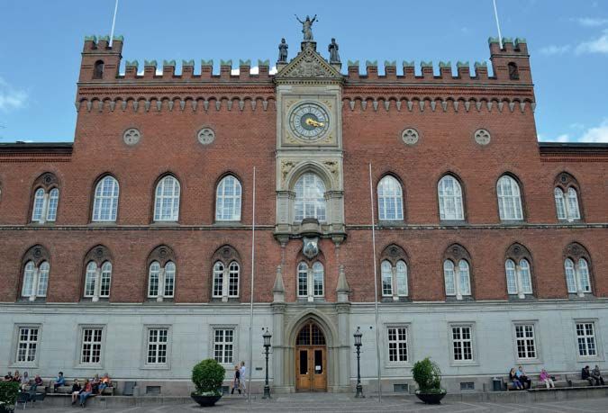
Social Program

Wednesday 10th May, 2017

Welcome reception at Odense City Hall; 18.00 – 20.00

The City of Odense is pleased to invite all delegates to a reception at the beautiful and historic City Hall at the
end of the first day of the Congress.

Odense City Hall is located in the centre of the old part of the city, surrounded by Sct. Knuds Plads, Albani
Torv and Vestergade. The City Hall consists of two different parts, an old and a new one. The part facing
Flakhaven was built in 1881-83 in an Italian-Gothic style. The new part is from 1955 and has been added to
the old building in a very attractive way.

8
Thursday 11th May, 2017
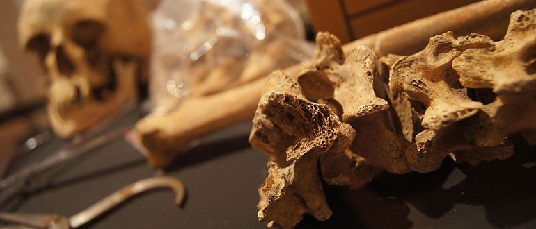
Casual dinner at ADBOU from 19.00-21.00
ADBOU is the Unit of Anthropology at the Department of Forensic Medicine, University of Southern Denmark
in Odense (www.adbou.dk). ADBOU curates the remains of more than 15,000 skeletons excavated from
Viking Age, Medieval and Post Medieval cemeteries. This event is a relaxed, causal evening. Typical Danish
hot sausages, known as Polser, will be served from a typical Danish polsevogn (literally "sausage wagon")
together with draft beer from the small, but popular local Refsvindinge brewery. Some of the skeletons will
be on display (please don’t spill beer on the old bones!) and the units CT-scanner will be demonstrated.
How to get to ADBOU:
5 min by bus from the city centre. Take bus No.
60-62, direction 'Højby'. Busstop 'Odense City
Camp - Landbrugsvej'. 2 min walk from busstop
to ADBOU (1), Lucernemarken 20.
9Friday 12th May, 2017 Congress Dinner at Hindsgavl Castle, Middelfart, 20:00 – 23.00 Venue: Hindsgavl, Middelfart. (30 min from Odense) Departure by bus from the city centre at 19:00. Go to Odense Railway Station, and find tour bus parking on the backside of the building (towards the Railway Museum, Address: Dannebrogsgade). Bus returns to Odense city centre at 23.15. Join your colleagues for the official congress dinner at the beautiful Hindsgavl Castle where you will experience a unique combination of a classic castle, a modern hotel and nuanced Danish cuisine. This charming waterfront castle overlooks the picturesque Fænø Sound and Little Belt Strait, in Middelfart. It is hard not to be overwhelmed when walking in and around Hindsgavl Castle situated at the Little Belt coast of Funen. Already when driving down the castle avenue you get the sense of entering another world and another time. Even Hans Christian Andersen found that the view was the most beautiful in Funen and he did not exaggerate: the garden and the park are outstanding with the pavillons, the streams, the ponds and the Little Belt in the background, and the nearest neighbour is the deer park Hindsgavl Dyrehave with red deer and fallow deer. 10
Oral Presentations

Thursday May 11, 2017 11:45

1          O’Donnell, Chris                 Can torture be inferred on the basis of PMCT
           chris.odonnell@vifm.org          interpretation?

2          Fujimoto, Hideko                 Development of mechanical screening system for
           kyoto.f.o@gmail.com              personal identification

3          Woźniak, Krzysztof et al         Postmortem imaging as a standard in evaluation of
           mpwoznia@cyf-kr.edu.pl           sharp force trauma – overview of recent cases
4          Sieberth, Till                   Interactive Crime Scene Visualisation – The Forensic
           Lars.Ebert@irm.uzh.ch            Holodeck

5          Heimer, Jakob                    Antemortem identification by fusion of MR and CT
           Jakob.Heimer@irm.uzh.ch

6          Schweitzer, Wolf et al           Visualization strategies in forensic imaging &
           Wolf.Schweitzer@irm.uzh.ch       Virtopsy: recommendations based on semiotic
                                            analysis

Friday May 12, 2017 11:15

7          Pelletti, Guido                  Qualitative and quantitative study of false starts on
           guidopelletti@gmail.com          bones through micro-CT. Preliminary results.
8          Borowska-Solonynko, Aleksandra   Occipital condylar fractures – rare or unrecognized
           et al                            injury during traditional autopsy?
           borowska.solonynko@gmail.com
9          Zoelch, Niklaus et al            Non-invasive ethanol quantification in human
           Niklaus.Zoelch@irm.uzh.ch        bodies by in situ magnetic resonance spectroscopy
10         Webb, Bridgette                  Developing an approach to post-mortem MR
           Bridgette.Webb@cfi.lbg.ac.at     angiography (PMMRA): Investigation of vascular
                                            retention of perfusates in ex situ porcine hearts
11         De Tobel, Jannick                The influence of motion artefacts on magnetic
           jannick.detobel@gmail.com        resonance imaging of the clavicles for age
                                            estimation
12         Gascho, Dominic et al            Estimation of body weight from effective radiation
           Thomas.Ruder@irm.uzh.ch          dose of whole-body CT

                                                                                                    11
Friday May 12, 2017 15:30
13 Boglárka, Marcsa et al Forensic Evaluation of Crania Recovered from
toro.klara@med.semmelweis- Archaeological Excavations Exhibiting Evidence of
univ.hu Sharp Force Trauma
14 Rutty, Guy et al Ventilated Post Mortem Computed Tomography
gnr3@leicester.ac.uk (VPMCT). Does it really make a diagnostic
difference?
15 Robinson, C et al Pulmonary Thromboembolism – improving the
claire.robinson@uhl-tr.nhs.uk diagnosis on post-mortem CT (PMCT)
16 Bolster, Ferdia et al Accuracy of PMCT with Death Investigation and
bdaly@umm.edu Toxicology Reports for Cause and Manner of Death
as Determined by Conventional Autopsy
17 Dedouit, Fabrice Death due to aortic dissection or rupture –
Fabrice.Dedouit@chuv.ch comparison of postmortem CT (PMCT) and multi-
phase PMCT angiography (MPMCTA), against
autopsy.
18 Rutty, Guy et al The Leicester Post Graduate Post Mortem Radiology
gnr3@leicester.ac.uk Training Courses; our experience so far.
Saturday May 13, 2017 11:30
19 Pedersen, Dorthe Dangvard et al A possible case of medieval child abuse
dopedersen@health.sdu.dk
20 Stoll, Alexander The Use of Diagnostic Imaging in Forensic
a.stoll@surrey.ac.uk Veterinary Pathology
21 Ebert, Lars C The use of deep learning in forensic medicine – a
Lars.Ebert@irm.uzh.ch feasibility study
22 Jotterand, Morgane New formula for cardiothoracic ratio for the
Morgane.Jotterand@chuv.ch diagnostic of cardiomegaly on post-mortem CT
23 Urschler, Martin Automated Multi-Factorial Age Estimation from
urschler@icg.tu-graz.ac.at Skeletal and Dental MRI
Volumes based on Deep Learning
12Poster Presentations
No. Writer
1 Raquel, Vilarino et al MR Spectroscopy in traumatic death: preliminary
Raquel.Vilarino@hopitalvs.ch results
2 Noor, Mohd et al The value of Postmortem Computed Tomography (PMCT)
mohdsuhani@yahoo.com in differentiating live birth from stillbirth
3 Lee, Heon et al Basal subarachnoid hemorrhage following violence-
acarad@naver.com related minor blunt trauma to the head: Evaluation of
vascular injury with post-mortem CT angiography
4 Hideki, Hyodoh et al An experimental evaluation of intermittent breathing in
hyodohh@yahoo.co.jp the appearance of drowning lung on postmortem CT
5 Makino, Yohsuke et al Evaluation of cervical spinal injuries by post-mortem MRI
ymakino-tky@umin.ac.jp with gradient echo sequences
6 Lundemose, Sissel Post-mortem Hippocampal Measurements in
sissel.lundemose@sund.ku.dk Mentally Ill Individuals
7 Matteo, Nioi et al Optical coherence tomography (OCT) study on
al.chighine@gmail.com reproducibility of
corneal pachymetry map results after death
8 Gomez, Oscar et al Soft Computing and Computer Vision for Comparative
ogomez@decsai.ugr.es Radiography in Forensic Identification
9 Pizzirani, Margherita et al Multi-phase postmortem CT angiography (MPMCTA): first
megpizzirani@gmail.com experiences
of the Forensic Radiology Unit in the University of
Modena (Italy)
10 Pereyra, Jorge et al Conventional Radiology and its Significant contribution
jorgepereyrafernandez@ to Forensic Anthropology
gmail.com
11 Baldoni, F et al Usefulness of PMCT in forensic pathology: our experience
amorico.mariagrazia@policlini
co.mo.it
12 Yoshida, Maiko et al Pseudo Pneumatosis Intestinalis Sign; Postmortem CT
maikichi0711@gmail.com depicted ‘Thaenia saginata (Parasite) ’ in a young-Thai-
man’s intestine.
13 Baron, K. et al Fracture dating: Comparing accuracy between
Katharina.Baron@cfi.lbg.ac.at morphological and quantitative analyses of MR data
14 Noor, Mohd Forensic radiology in the DVI operation for the Wang
mohdsuhani@yahoo.com Kelian clandestine graves in Malaysia
15 Kobayashi, Tomoya Relationship between postmortem MR relaxation time
t.kobayashi1001@gmail.com and body temperature: Is scan parameter optimization
necessary?
16 Beng Ong, Beng et al Diagnosis of venous air embolism with the use of post-
ongbb77@gmail.com mortem CT scan
17 Gascho, Dominic et al Postmortem CT findings of decedents with a short
Dominic.Gascho@irm.uzh.ch postmortem interval and excessive gas- and/or fluid-filled
distension of the bowels
1318 Decker, Summer et 3D Printing Applications for Medicolegal Practice
sdecker@health.usf.edu Purpose
19 Busch, Johannes et al Identifying suspects by matching hand photographs with
johannes.busch@sund.ku.dk video evidence
20 Chatzaraki, Vasiliki Unexpected brain finding in pre-autopsy postmortem CT
Vasiliki.Chatzaraki@irm.uzh.ch
21 Carballeira Álvarez, Ana et al Value of un-enhanced post-mortem computed
aacarballeira@gmail.com tomography in the detection of traumatic abdominal
injuries
22 Tashiro, Kazuya Relaxation time of the skeletal muscles in postmortem
k_tashiro0219@yahoo.co.jp MR imaging of adult humans
23 Hindsø, Louise et al Epicardial adipose tissue estimation by computed
louise.hindsoe@sund.ku.dk tomography of eviscerated hearts – A forensic method
study
24 Leipner, Anja et al Reconstruction of reflective surfaces such as vehicle
Anja.Leipner@irm.uzh.ch mirrors based on 3D scan data for visibility simulation
25 Robinson, C et al Evaluating a new service using PMCT to replace autopsy
claire.robinson@uhl-tr.nhs.uk in natural death
26 Fonseca-Pinto, Ana Carolina B. Comparison of computed tomography brain and soft
C. et al tissue windows to evaluate frozen artifacts in the liver
anacarol@usp.br and gallbladder of animal cadavers
27 De Matteis, Maria Post-mortem CT in criminal disposal of homicide
maria.dematteis@icloud.com victims. A ten-year retrospective study in Padua
28 Adolphi, Natalie Non-invasive Temperature Determination by Post-
NAdolphi@salud.unm.edu Mortem MR
29 Schweitzer, Wolf et al Very Affordable Immersion Pump for Post Mortem CT
Wolf.Schweitzer@irm.uzh.ch Angiography in Forensic Pathology: First 10 Cases
30 Jeanson, Alizé Lacoste Can we predict nutritional status from the skeleton?
alize.lacoste.jeanson@gmail.c Inputs from forensic radiology.
om
31 Boel, Lene Warner Thorup et Death from displacement of airway stents
al
lwb@forens.au.dk
32 Boel, Lene Warner Thorup et Distribution of skull and facial fractures according to
al circumstances of death
lwb@forens.au.dk
14Non-invasive Temperature Determination by Post-Mortem MR
Adolphi, Natalie; Martinez Barrera, Julio; Weisand, Jordan; Gerrard, Chandra
Center for Forensic Imaging, University of New Mexico School of Medicine
To determine which MR tissue parameters correlate strongly with temperature, but not post-
mortem interval, such that these parameters may be useful for determining subject temperature
non-invasively.
Using mammalian tissues from several species and a spin echo pulse sequence, T1 and T2 were
measured as a function of A) temperature, PMI < 48 hours, and B) post-mortem interval (PMI), at
constant temperature (4, 19, or 35 oC). Both the temperature- and PMI-dependence of the
apparent diffusion coefficient (ADC) of brain tissue were also investigated.
After controlling for temperature, the ADC of brain tissue showed a robust dependence on
temperature and no dependence on PMI over many days. In the body, the T1 values for cardiac
muscle, skeletal muscle, and lung parenchyma showed the strongest dependence on temperature;
however, of these, only the skeletal muscle T1 showed no PMI-dependence.
Measurements performed in this study, which accounted for both temperature and PMI,
demonstrate that our previous report of a PMI-dependence of the ADC of brain tissue was
incorrect.
The ADC of brain tissue and the T1 of skeletal muscle are good candidates for use as non-invasive
thermometers, based on their strong temperature-dependence and minimal PMI-dependence.
Keywords: PMMR, non-invasive thermometer
15Usefulness of PMCT in forensic pathology: our experience Baldoni F.1, Pizzirani M.2, Procicchiani D.1, Todaro R.3, Amorico M.G.1, Vecchio S.2, Santunione A.L.2, Tata C.1, Torricelli P.1, Silingardi E.2 1 Department of Diagnostic and Clinical Medicine and Public Health, Section of Diagnostic Imaging, University of Modena and Reggio Emilia, Modena, Italy. 2Department of Diagnostic and Clinical Medicine and Public Health, Section of Legal Medicine, University of Modena and Reggio Emilia, Modena, Italy. 3Radiodiagnostic and Radiotherapy Unit, University Hospital “Policlinico-Vittorio Emanuele”, Via Santa Sofia 78, Catania 95123, Italy. To evaluate the usefulness of PMCT in assessing cause of death and its diagnostic aid to the forensic pathologist before conventional autopsy. Define the advantages and limitations of the technique. From 2006-2016, 115 subjects, divided by different forensic type underwent CT. 103 of these were subjected to conventional autopsy, the remaining 12, victims crushed in the 2012 earthquake, were subjected only to TC. The study was performed with a 64-row CT-unit (GE Milwaukee USA) from head to foot, using the following scan parameters: slice thickness 1 mm and interval of reconstruction 1 mm. 2D and 3D reconstructions were performed on all. The reporting was done by two radiologists with specific experience in the field. We compared autopsy findings with those of PMCT by highlighting findings of forensic interest. They were divided into: basic, very relevant, useful, not important. Compared to conventional / virtual autopsy, resulted significantly useful in skeletal findings (98%), and not essential but important in the remaining areas. PMCT carried out before traditional autopsy greatly facilitates the work of the forensic pathologist in preliminarily identifying the most significant findings, but does not entirely replace traditional autopsy. PMCT provides an important support to post-mortem examinations used in court due to the immediacy of the images to be used in the reconstruction of events, as a means of consultation and of objective and repeatable proof. Keywords: PMCT, Autopsy, Forensic radiology 16
Fracture dating: Comparing accuracy between morphological and quantitative
analyses of MR data
K. Baron, T. Widek, S. Ferk, T. Ehammer, S. Heinze, E. Scheurer.

Ludwig Boltzmann Institute for Clinical-Forensic Imaging, Graz, Austria and Institute of Forensic Medicine,
Medical University Graz, Austria

To evaluate the accuracy in fracture dating using qualitative and quantitative MRI data and the
comparison of both approaches.

During a longitudinal study examining healing fractures, 75 MR datasets of 35 test subjects were
aquired and analysed. Two blinded radiologists (one being also a medical examiner, one a clinical
radiologist with forensic experience) determined the age of fractures based on qualitative MRI data.
A third blinded examiner with basic training used quantitative MRI data to analyse 10 fractures by
comparing T1 and T2 relaxation times to reference values. Both approaches were compared
regarding their accuracy in dating fractures.

The agreement between both radiologists regarding qualitative data was moderate with an ICC of
0.4. However, both radiologists correctly determined fracture age, with an accuracy of up to 39%.
The preliminary quantitative analysis showed an accuracy of 30% (3 out of 10), however, after
including information regarding the sex of the test subject the accuracy increased towards 60%.

The results show that MRI can add valuable information concerning the

Both approaches seem equivalently accurate in their current state, however the quantitative
approach may have more potential to increase the accuracy when compared to radiological
examinations.

Keywords: Fracture analysis, qMRI

                                                                                                              17
Death from displacement of airway stents Lene Warner Thorup Boel, Lars Uhrenholt, Marianne Rohde Department of Forensic Medicine, Aarhus University Case reports illustrating the importance of post mortem CT for documentation of displacement of airway stents Two cases where the deceased had a silicone stent in the trachea. Post mortem CT was performed before autopsy. Case 1 was a woman who died the day after having a replacement of a tracheal stent. Case 2 was a man, handicapped from a traffic accident with brain injury and having a tracheal stent, who was found dead. In case 1, the stent was found displaced to the tracheal bifurcature. The cause of death was presumed to be suffocation due to inadequate air passage in the trachea. In case 2, the stent was shifted to the left and compressed due to a large benign tumor of the thyroid gland. The cause of death was presumed to be suffocation from a compressed trachea possibly in association with an epileptic seizure Tracheal stents have been used in the past decades for treatment of various conditions involving the airways. Stent migration is a well-known complication, but death associated with tracheal stents is rare and usually caused by hemoptysis or related to the surgical procedure Post mortem CT serves as important documentation that displacement of a tracheal stent contributed to death Keywords: Tracheal stent, airway stent, post mortem CT 18
Distribution of skull and facial fractures according to circumstances of death

Lene Warner Thorup Boel, Lars Uhrenholt, Alice Svensson

Department of Forensic Medicine, Aarhus University

To describe the distribution of skull and facial fractures according to the cause of death, manner of
death and mechanism of trauma

Deceased autopsied at the Department of Forensic Medicine, Aarhus University in the period 2008-
2015 in which a fracture to the face and/or skull were detected and a post-mortem CT was
performed

A total of 211 deceased were included in this study, consisting of 167 males and 44 females. The
most common cause of death was traumatic lesions to the brain and nervous system (54,5%). The
most common manner of death was accident (61,1%) of which the most common mechanism of
trauma was a traffic crash. The commonest facial fracture among females was maxilla (45,5%) and
zygoma (37,7%) among males. The commonest skull fracture among females was parietal (52,3%)
and temporal (59,9%) among males.

The findings revealed that facial fractures were common in relation to natural death and active
blunt trauma. Skull fractures were present in all suicides. Males were shown to commit suicide
more violently than females. Interestingly, one in five females with a skull or facial fracture had
been killed.

Skull and facial fractures varies within a forensic postmortem population

Keywords: Cranial fracture, post mortem CT scanning

                                                                                                      19
Accuracy of PMCT with Death Investigation and Toxicology Re-ports for Cause and Manner of Death as Determined by Conven-tional Autopsy Bolster, Ferdia MD; Ali, Zabiullah MD; Sheroke, Amanda; Daly, Barry MD; Fowler, David MD Office of the Chief Medical Examiner and Radiology Department, Unversity of Maryland, Baltimore, U.S.A. To compare the accuracy of PMCT with both death investigation and toxicology reports (PMCTit), for cause of death (COD) using conventional autopsy (CA) as standard of reference. A second-ary aim was to determine accuracy of PMCTit for the manner of death (MOD). Two readers interpreted PMCTs in 303 consecutive decedents who underwent PMCT and CA as part of death investigation over a period of 7 months. The COD and MOD were determined based on the combination of PMCT findings, the scene investi-gation, medical reports, and toxicology results. Sensitivity and positive predictive value (PPV) with 95% confidence-intervals (95%CI) were calculated. Observed agreement between [COD PMCTit vs COD Autopsy], and [MOD PMCTit vs MOD Autopsy] were calculated. These were 207 males (68.3%) and 96 females (31.7%). Age range was 0-94 years (mean 37.2 years). PMCTit accurately determined the COD in 89.4% of cases, and MOD in 89.1% of cases. Observed agreement between [COD PMCTit] vs. [COD Autopsy] was 100% concordant for drowning, gunshot related deaths, sharp force injuries and asphyxia, and excellent (>80%) for trauma (97%), infant (87.5%), and drug overdose-related deaths (87%). Observed agreement between [COD PMCT it] vs. [COD Autopsy] was good (74.2 %) for natural deaths. When interpreted in combination with death scene investigation, medical reports, and toxicology results, PMCT correlates favora-bly for COD and MOD as determined by CA In selected cases PMCT can play an important role as a triage tool, to determine when conventional autopsy may not be re-quired. Keywords Postmortem CT, Autopsy, Cause of Death, Manner of Death 20
Occipital condylar fractures – rare or unrecognized injury during traditional
autopsy?

Aleksandra Borowska-Solonynko1, Agnieszka Dąbkowska1, Dorota Samojłowicz1, Jarosław Żyłkowski 2
1
    Department of Forensic Medicine, Medical University of Warsaw
2
    Second Department of Clinical Radiology, Medical University of Warsaw

Occipital condylar fractures (OCF) practically are not examined during traditional autopsy due to
anatomical location, therefore are considered as rare injury. The aim of this study is to determine
the true frequency of OCF based on postmortem computed tomography examination (PMCT)
conducted in traumatic cases.

438 PMCT studies performed in Departement of Forensic Medicine Medical University in Warsaw
between November 2014 and December 2016 on victims of: traffic accidents, falls from height,
batteries and low energy head injuries with known circumstances were analyzed retrospectively.
The cases with OCF were divided into three subgroups. Type I-an impaction-type fracture, with a
comminution of the condyle, type II-as part of a basio-occipital fracture and III-an avulsion type.
Statistical analysis of all data has been performed. Further, more detailed analysis of cases with OCF
type I and III-the most useful for the reconstruction purpose, including data from the autopsy
protocols will be conducted.

OCF was present in 22,6% cases (n=99), the most often occurred in cases of hitting by train (48,5%,
n=17), falls from height (26,6%, n=29), in cyclists (24%, n=6) and pedestrians hit by a car (22,5%,
n=29). There were no OCF in fatal battery cases and low energy head injuries. Isolated OCF were
found in 5,5% of cases, the most often in cyclists (12%, n=3) and pedestrians hit by a car (9,3%,
n=12).

PMCT revealed that OCF is quite common in deaths caused by high-energy mechanical injuries and
can be useful for the reconstruction purpose

Keywords: postmortem computed tomography, occipital condylar fractures

                                                                                                      21
Identifying suspects by matching hand photographs with video evidence Busch, Johannes, Lynnerup, Niels Department of Forensic Pathology, Institute of Forensic Medicine, University of Copenhagen Reports by minors of sexual relations against their will are not rare, and in some cases pornographic photography is a part of the abuse. Such material can be used to help identify the perpetrator. We examined the efficacy of visual comparison between high resolution photography and low resolution video image stills of the hand. We obtained single blinded still images from video recordings and high quality camera images of the back of the right hand from 51 Caucasian male volunteers. The images were compared in pairs (2601 combinations) and a judgment was made about whether they were a highly likely, possible or unlikely match, using an algorithm based on several types of anatomical features as markers for comparison. All 51 high quality images were correctly matched with the video image from the same person, though in some cases there was up to 5 other samples that could not be excluded as possible matches. In total there were no false positive “highly possible” matches, but there were 50 false positive “possible” matches. Visual comparison of the back of the hand is a valuable addition to the burden of evidence in a judicial setting, but should not be used as a standalone method to establish proof of identity. The applicability of the method is dependent on the existence of a database of reference images; the degree of certainty of the identification is directly correlated to the size of the database. Keywords: Forensic image comparison; Hand photography; Forensic anthropology; Photographic evidence 22
Value of un-enhanced post-mortem computed tomography in the detection of
traumatic abdominal injuries
Carballeira Álvarez, Ana1 , Mancini, Julien2,3, Tuchtan-Torrents, Lucile3, Bartoli, Christophe 3, Desfeux,
Jacques3, Piercecchi, Marie-Dominique3, Gorincour, Guillaume3

1
  Hospital Universitario Donostia, Donostia/San Sebastián, Spain
2
  Aix-Marseille University, INSERM, IRD, UMR912 SESSTIM, Marseille, France
3
  CHU La Timone, Marseille, France

To determine the accuracy of unenhanced post-mortem computed tomography (PMCT) in detecting
traumatic abdominal lesions

The population was collected retrospectively from the “virtopsy” database in a period of 5 years in a
single institution. Traumatic deaths who benefited from both PMCT and classical autopsy were
included, excluding cases of gunshot injuries. Liver, spleen, pancreas and kidney injuries and
hemoperitoneum were searched. Sensitivity, specificity, negative (NPV) and positive (PPV)
predictive values of the PMCT as a whole and for each finding were estimated using the autopsy
report as gold standard.

For the 71 cases included from victims of a traumatic event (52 males, 19 females, median age of
38,6 years), PMCT as a whole has shown to have a low sensitivity (80%) and a high specificity (94%),
with a PPV of 98% and a NPV of 59%. The highest sensitivity was obtained for the detection of
hepatic lesions (69%) and the lowest for pancreatic lesions (12%). PMCT was very specific (100%) for
the detection of hemoperitoneum, with a PPV of 100%. A high NPV was found for the detection of
perihepatic hematomas (98%).

Major studies reporting the accuracy of PMCT for traumatic abdominal lesions are limited. The low
sensitivity and NPV showed in our study discards the PMCT as an alternative to the conventional
autopsy to diagnose and rule out traumatic abdominal lesions. Nevertheless, it remains a helpful
tool and its accuracy would be increased by the use of PMCT angiography.

Keywords: Un-enhanced post-mortem computed tomography, polytrauma, abdominal lesions,
abdominal injuries, accuracy

                                                                                                             23
Unexpected brain finding in pre-autopsy postmortem CT Chatzaraki Vasiliki Institute of Forensic Medicine, University of Zurich Forensic pathologists often face great danger from spreading infections during autopsies. Pre- autopsy postmortem imaging can reveal potentially infectious foci and thus enable forensic pathologists to enforce protective measures during autopsies. Pre-autopsy imaging is also helpful for early differential diagnosis. Combining imaging and autopsy findings improves the quality of forensic investigations. A man was found dead on his bed and was delivered to Institute of Forensic Medicine for further investigation. Pre-autopsy imaging performed on a CT scanner and images were evaluated on a multimodality workstation. Then, autopsy and microbiological sample collection took place. Postmortem CT (PMCT) revealed a hypodense lesion in the frontal brain lobe, compatible with abcess or necrotic tumor. In addition, PMCT revealed frontal sinus opacification and a small osseous defect in the frontal bone adjacent to the lesion. The autopsy and microbiological culture confirmed the brain abscess diagnosis. This case highlights how PMCT is useful for early differential diagnosis and enables forensic pathologists to protect their own health by wearing appropriate clothing during autopsy. Complementary use of imaging, autopsy and microbiology set the final diagnosis. The abscess originated from a chronic frontal sinusitis which had spread by continuous expansion through the eroded posterior wall of the frontal sinus. PMCT enables forensic pathologists to adapt their autopsy approach by wearing protective clothing. It is of primary importance to match and complement PMCT with other methods’ findings to improve the quality of forensic investigations and set final diagnosis. Keywords: Postmortem computed tomography; brain abscess; Streptococcus anginosus; sinusitis; autopsy 24
3D Printing Applications for Medicolegal Practice

Decker, Summer; Ford, Jonathan

Department of Radiology, Morsani College of Medicine, University of South Florida

The purpose of this presentation is to provide examples of how 3D printing can assist forensic
practitioners in criminal reconstruction and documentation for long term evidence preservation.

3D reconstructions from CT, MRI and laser scanning technologies have been used in the
documentation of pathologies, human identification and even in child abuse investigations. In the
past, 3D printing was cost prohibitive but more consumer friendly desktop printers are making the
process affordable.

Applications of 3D printing will be demonstrated with some of the strengths and pitfalls of the
technology highlighted through specific case examples. With a variety of printers and print
materials avaiable, the intended application (be it expeimental, demonstration etc.) is only limited
by printer capability and model complexity.

3D printing allows for a tangible representation of evidence that can be utilized in court for juries
and also provide the opportunity for subsequent analyses after a body has been released or after a
surviving victim has healed. 3D prints allow for the display of evidence in a non-prejudicial manner.
It has been shown that juries respond better to 3D visual aids as opposed to gory autopsy or trauma
photos. 3D printing is a viable tool available to forensic practitioners for evidence documentation,
preservation of evidenced, real world analyses and useful in court room demonstrations.

Keywords: 3D Printing, Forensic, Reconstruction, Child Abuse

                                                                                                   25
Death due to aortic dissection or rupture – comparison of postmortem CT (PMCT) and multi-phase PMCT angiography (MPMCTA), against autopsy. DEDOUIT Fabrice, MICHAUD Katarzyna, BRUGUIER Christine, DUCROT Kewin, GRABHERR Silke Unit of Forensic and Anthropological Imaging, University Center of Legal Medicine Lausanne-Geneva, Switzerland We analysed the performance of postmortem computed tomography (PMCT) and multi-phase PMCT angiography (MPMCTA) compared to medicolegal autopsies in cases of sudden death associated with aortic pathology, this includes sudden: natural (ND) or unnatural death (UND), directly related to aortic dissection and/or rupture. We retrospectively selected all cases concerning aortic dissection or rupture autopsied at our centre between 2013 and 2014. Autopsy was preceded by PMCT, and where possible MPMCTA, as per the established protocol developed by Grabherr et al. Results of 3 imaging items (aortic lesion presence, it’s location, and pericardial fluid presence) were compared against autopsy findings. Our study of 35 cases underwent PMCT (ND:12; UND:23), with 21 cases undergoing MPMCTA (ND:11; UND:10). For all three items studied, and especially so for the identification and localisation of aortic lesions, MPMCTA was superior to PMCT. PMCT showed false negative results for the presence or location of an aortic lesion in 10/14 cases (71.4%) against 3/21 (14.3%) for MPMCTA. MPMCTA was falsely positive for the presence or location of aortic lesions in 1/14 cases (4.8%). Pericardial fluid was missed in 1/35 (7.5%) by PMCT, however was falsely positive in 2/14 cases (14.3%) and MPMCTA in 1/21 (4.8%). PMCT and less often MPMCTA, are already incorporated into routine work at numerous forensic institutes. In cases of death due to aortic pathology, they give useful information on the presence of aortic lesions and pericardial fluid, and lesion location. MPMCTA was superior to PMCT for items studied, although autopsy remains the gold standard. Keywords: Forensic imaging; Postmortem computed tomography (PMCT); Multi Phase PMCTA – angiography; Sudden aortic death; Autopsy 26
The use of deep learning in forensic medicine – a feasibility study

Ebert, Lars C.; Schweitzer, Wolf; Thali, Michael J.; Ampanozi, Garyfalia

University of Zurich, Institute of Forensic Medicine

The amount of data generated by modern medical scanners can be huge, especially in a forensic
setting, were the entire body is documented in high resolution. For a forensic case, reading of
images can therefore easily take several hours. A solution these issues could the use of deep
learning techniques..
We hypothesize that deep learning techniques might help in automatically detecting and
segmenting cases of hemopericardium in PMCT.

We used the ViDi Suite 2.0 as a deep learning image analysis software. 28 cases (20 male, 8 female)
of hemopericardium were selected retrospectively. For each dataset, one predefined slice was
extracted, windowed and converted to the PNG format. We tested the performance of the software
to classify and segment a hemopericardium by using 50% of the input data for training and the
other 50% for validation. Training was repeated 20 times, randomly selecting different images for
training and validation.

For classification, the software achieved an average f-score of 0.79 ±0.1 ( max 0.96, min 0.54). The f-
score for the correct segmentation of the hemopericardium was 0.8 ±0.02 (max 0.84, min 0.78)
compared to the manual segmentation serving as reference.

The deep learning software was able to detect and segment a hemopericardium with reasonable
success considering the limitations. While this technique is not yet feasible for routine use, we could
demonstrate that deep learning might be a suitable tool to solve the problem of large datasets in
forensic medicine in the future.

Keywords: Deep Learning, PMCT, hemopericardium, image analysis

                                                                                                    27
Comparison of computed tomography brain and soft tissue windows to evaluate frozen artifacts in the liver and gallbladder of animal cadavers Fonseca-Pinto, Ana Carolina B. C.1; Heng, Hock Gan2; Massad, Mara Rita Rodrigues3; Lim, Chee Kin2; Ribas, Laila Massad3; Baroni, Carina Outi1; Ferrante, Bruno1; Muramoto, Caterina1; Rocha, Noeme Sousa3 1 School of Veterinary Medicine and Animal Science – University of São Paulo 2 College of Veterinary Medicine – Purdue University 3 School of Veterinary Medicine and Animal Science – São Paulo State University The purpose of this study was to compare the computed tomography (CT) brain window (BW) and the soft tissue window (STW) in identifying frozen artifacts (FA) and to characterize the appearance and Hounsfield units (HU) of these FA in the liver and gallbladder of inadequately thawed cadavers. Fourteen animal cadavers of different species were frozen and thawed at the room temperature (15-35˚C) for one to two days. Three investigators evaluated the transverse post mortem CT images. Soft tissue (WL 40/WW 350) and brain (WL 50/WW 100) windows were used to detect and characterize the FA in the liver and gallbladder, and the HU were recorded. Compared to the STW, the BW allowed better delineation and identification of FA in the liver parenchyma. Only 47% of the liver FA identified using BW was noted in STW. Commonly identified liver FA include the ‘crescent line’ appearance and centrally hypoattenuating ‘frozen’ regions. The mean HU of the liver FA was 33 HU and the presumed normal liver parenchyma have higher tissue attenuation up to approximately 70 HU. The gallbladder was better visualized using STW than BW. Common gallbladder FA include ‘icicles’ and ‘sherbet’ appearance within the lumen. The mean HU of the gallbladder frozen content was -22 HU. Different detected appearances of FA became more conspicuous with the BW showing the relevance of its use in frozen cadavers. The addition of the BW to the conventional STW is important to identify and characterize FA present in the liver and gallbladder of animal cadavers. Keywords: PMCT, brain window, soft tissue window, frozen artifact, animal 28
Development of mechanical screening system for personal identification

Fujimoto, Hideko1,2, Hayashi, Takeshi2, Iino, Morio2
1
Kyoto Forensic Odontology Center, Fujimoto Clinic for Oral and Maxillofacial Surgery, 2Division of Legal
Medicine, Tottori University Faculty of Medicine

We presented our study ”Development of a new personal identification method” at the 4th ISFRI.
This method is performed using Procrustes analysis between two images. This time we have
improved the method which can refine the same person candidates by semi-automatic mechanical
operation using Procrustes analysis .

For 252 CT panoramic images and panoramic X-ray images, the x and y coordinates of the
landmarks on the tooth sockets were recorded. Each image was divided into six regions and the
overall Procrustes distance (d) was calculated. All combinations that can be taken among the 252
cases were calculated by Procrustes analysis.
Considering the alveolar lack portion, d / √k divided by the square root of the total number
landmarks (k) was verified for differences between the same person group and non-identical person
group.
Distribution on the histogram was somewhat asymmetrical, so we converted it into d '= ln (1000 × d
/ √k) in order to make it closer to the normal distribution.

The average d’ for the same person group (n=341) was 4.59 and the standard deviation was 0.18.,
which were 5.10 and 0.18 for the non-idential person group (n=31,285 ).

The smaller d / √k indicates the closer the coordinate distribution on the image.The results
suggested that candidates could be easily refined to 70 % or less with a probability of 1.5% using
this method.

This method proved that it was an useful screening system for personal identification.

Keywords: Personal identification, Panoramic CT, Panoramic X-ray, Procrustes analysis, Forensic
odontology

                                                                                                           29
Estimation of body weight from effective radiation dose of whole-body CT Purpose Gascho, Dominic et al Department of Forensic Medicine and Imaging, Institute of Forensic Medicine, University of Zurich, Wintherthurerstrasse 190/52, 8057 Zurich, Switzerland Documentation of the visual appearance is required in any forensic report, including body weight of the decedent. Due to a failure of our in-house, floor-embedded weighting scale, we developed a method for body weight estimation based on postmortem computed tomography (PMCT) using automated dose modulation, as each case underwent PMCT in our institute. The aim of this study was to evaluate the correlation between effective milliampere second (mAseff) values, based on automatic exposure control, and body weight for the purpose of determining body weight just by the means of PMCT. The study population comprised 349 (114 female, 234 male) decedents. In a retrospective evaluation the previously listed weight measurements, using the embedded weighting scale, were correlated with the PMCT mAseff value of the automatically provided patient protocol. All data were statistically analyzed. An excellent correlation between mAseff values and body weight for the whole study population was revealed, which allows for calculate a conversion factor. The present method based on computed tomography using dose modulation techniques is an accurate and quick possibility to determine body weight of decedents and shows potential for further application areas, e.g. in pediatric radiology for weight-based application of contrast media during emergency computed tomography treatment. 30
Postmortem CT findings of decedents with a short postmortem interval and
excessive gas- and/or fluid-filled distension of the bowels
Dominic Gascho1, Sarah Schaerli1, Lucile Tuchtan-Torrents2,3, Michael J Thali1, Guillaume Gorincour4
1
 Department of Forensic Medicine and Imaging, Institute of Forensic Medicine, University of Zurich,
Wintherthurerstrasse 190/52, 8057 Zurich, Switzerland
2
 Department of Forensic Pathology APHM, CHU Timone, 264, rue Saint-Pierre, 13385 Cedex 5, Marseille,
France
3
 Aix-Marseille Université, CNRS, EFS, ADES UMR 7268, 13916, Marseille, France
4
 Department of Pediatric Imaging, APHM, CHU Timone, 264, rue Saint-Pierre, 13385 Marseille, France

The aim of this small case study was to assess postmortem CT findings in non-traumatic cases with a
postmortem interval of less than three days displaying excessive bowel distension.

9 adult decedents with excessive bowel distension underwent postmortem CT. CT findings have
been assessed regarding autopsy findings and bacteriological analyses.

In 6/9 cases CT findings were confirmed by autopsy and in 2/9 cases autopsy did not reveal further
conclusion. The cause of excessive bowel distension was identified by means of CT in 6/9 cases and
was related to bowel obstruction (n=5) or paralytic ileus (n=1). One case showed a strong indication
for bowel obstruction causing bowel distension; however, autopsy did not confirm bowel
obstruction or bowel injuries. In 6/9 cases cardiac failure due to septic shock was determined as
cause of death using bacteriological analyses (bowel obstruction: n=4, paralytic ileus n=1, unknown
origin: n=1).

CT indicates a high correlation with autopsy (8/9 cases) and shows high sensitivity for the detection
of bowel obstruction. Bowel obstruction is mostly related to cardiac failure due to septic shock.

Keywords: bowel distension, bowel obstruction, septic shock

                                                                                                       31
Soft Computing and Computer Vision for Comparative Radiography in Forensic Identification Gomez, Oscar and Ibáñez, Oscar and Valsecchi, Andrea and Cordón, Oscar and Kahana, Tzipi University of Granada Comparative radiography traditionally involves the comparison of consistencies and inconsistencies of ante-mortem (AM) and post-mortem (PM) radiographs, taken trying to simulate the AM’s in scope and projection. However it is based on manual comparisons and thus it is a time consuming and error prone visual inspection process. The objective of this work is to describe and validate a novel computer-aided automatic paradigm based on a 3D bone -2D radiograph super-imposition process. The proposed 3D-2D superimposition approach is based on a Computer Vision technique called Image Registration which automatically, objectively, and pre-cisely searchs for the AM radiograph (2D) acquisition parameters using an opti-mizer. A simulated radiograph is obtained applying the latter acquisition param-eters to the 3D model and projected over the AM radiograph. A matching de-gree of the superimposed radiographs is calculated according to a given similari-ty metric considering the external contour (bone shape) of the target bone in both radiographs (AM and simulated PM). With the goal of validating the capability of our method to precisely perform 3D-2D radiograph superimposition we used 30 CTs (from which we extracted the following 3D models: 10 frontal sinuses, 10 clavicles, and 10 patellas) provided by the Hospital de Castilla la Mancha, and obtained 5 simulated radiographs from each 3D model (50 frontal sinuses, 50 clavicles and 50 patellas). From each of the 150 radiographs we generated three additional virtual x-rays with increasing degree of occlusion of the target bone up to 45% in order to model bone contour occlusion in x- rays. Promising result has been found with an average overlapping error around 0,05%. We have managed to automatize the comparative radiograph technique while providing reproducibility, objectivity, and higher precision. Keywords: Forensic Identification, Forensic Radiology, Comparative Radiography, 3D-2D bone superimposition, Soft computing, Computer Vision. 32
Antemortem identification by fusion of MR and CT

Heimer, Jakob

Institut für Rechtsmedizin – Zurich, Switzerland

Computed tomography (CT) has become a valuable addi-tion to radiologic identification while
magnetic-resonance-imaging (MR) has only rarely been used for this purpose. In our case,
identification was facilitated by fusion of MR- and CT-imaging in a living victim of assault.

A man was hospitalized with disfiguring injury that rendered establishment of his identity
impossible. Head-CT was per-formed and the PACS featured an earlier MR of the brain. We were
tasked to confirm the presumed identity by radio-logical identification. Images were processed with
syn-go.via (SIEMENS).

Unenhanced comparison of MR and CT imaging of the frontal sinuses did not allow for
identification. In order to allow for identification, MR and CT data had to be adapted in contrast,
windowing, and color. Subsequent fusion of the images allowed for differentiation of the two
modalities and enabled identification.

Due to familiar differences in bone tissue contrast, the di-rect visual comparison of CT and MR
imaging is challeng-ing. The syngo.via fusion tool enables intermodal identifica-tion. This is a first
step towards objective rather than sub-jective identification. The case also reports on the rare
occasion of antemortem radiologic identification.

Intermodal radiologic identification by comparison of CT to MR imaging is feasible despite
differences in tissue contrasts.

Keywords radiological identification, computed tomography, magnetic resonance
imaging, paranasal sinuses

                                                                                                         33
Epicardial adipose tissue estimation by computed tomography of eviscerated hearts – A forensic method study Hindsø, Louise Jakobsen; Lykke Schrøder; Jacobsen, Christina ;Lynnerup, Niels; Banner, Jytte Department of Forensic Medicine, University of Copenhagen The purpose of this study was to investigate the ability to estimate epicardial adipose tissue (EAT) volume by computed tomography (CT) of eviscerated hearts during autopsy. Furthermore, we wanted to test the correlation between total heart volume (HV) measured by CT and heart weight (HW). We included 144 individuals who underwent a medicolegal autopsy and did a CT-scan of the eviscerated hearts. 12 subjects were excluded due to inadequate CT-scan. Of the remaining 132 subjects included in the results, 74 (56%) were males. Mean age was 53 years (range: 22-94 years). Using the software Mimics® we determined EAT and myocardial volumes. HW was measured during autopsy. Intra- and interobserver analyses of the CT measurements were performed on 10 randomly chosen subjects. The median HW was 405 g (range: 249-838g), median HV 354 mL (range: 209-787 mL) and median EAT volume 71 mL (range: 10-296 mL), which corresponded to 20% of the HV. HV measured by CT correlated with HW (R2=89%). Mean intraobserver differences of HV and EAT were -0.5 mL and -1.1 mL, respectively. Mean interobserver differences were 11.5 mL and 1.5 mL, respectively. HV measured by CT of eviscerated hearts during autopsy highly correlated with HW. It was possible to estimate EAT volume using CT on eviscerated hearts. The EAT-HV-ratio corresponded with values published on data from former autopsy studies using manual dissection. We conclude that CT of eviscerated hearts may be a useful method to determine EAT at autopsy. We expect to apply this method in future research. Keywords: Epicardial fat tissue, Computed tomography, Autopsy, Method study 34
You can also read