A Retrospective Study of Histopathology of Psoriasis Vulgaris in a Tertiary Hospital - Journal of Medical ...
←
→
Page content transcription
If your browser does not render page correctly, please read the page content below
JMSCR Vol||07||Issue||06||Page 33-38||June                                                  2019
                                                                              www.jmscr.igmpublication.org
                                                                              Index Copernicus Value: 79.54
                                                                  ISSN (e)-2347-176x ISSN (p) 2455-0450
                                                              DOI: https://dx.doi.org/10.18535/jmscr/v7i6.07

 Research Article
 A Retrospective Study of Histopathology of Psoriasis Vulgaris in a Tertiary
                                 Hospital
                                                  Authors
         Renuka Satish Ashtekar , Anup Phadke2, Pramila Patil3, Yogesh M Shah4
                                       1
                 1
                    Professor & HOD, Department of Dermatology, Venereology & Leprosy
                    2
                      Senior Resident, Department of Dermatology, Venereology & Leprosy
                                     3
                                       Ex-Professor, Department of Pathology
                      4
                        Ex- Professor, Department of Dermatology, Venereology & Leprosy
          In Bharati Vdyapeeth (Deemed to be University) Medical College & Hospital, Sangli, India
                                              Corresponding Author
                                            Renuka Satish Ashtekar
                   Professor & HOD, Department of Dermatology, Venereology & Leprosy
          In Bharati Vdyapeeth (Deemed to be University) Medical College & Hospital, Sangli, India
 Abstract
 Introduction: Psoriasis affects about 1.5-3% of world's population. There are other papulosquamous
 dermatoses mimicking psoriasis morphologically which can be differentiated histopathologically. The
 histologic changes in psoriasis vulgaris (plaque psoriasis) vary considerably depending on the stage of the
 lesion.
 Aims and Objectives: This study was done to evaluate the occurence of Munro micro-abscess and the
 spongiform pustule of Kogoj in psoriasis vulgaris and to evaluate various histologic changes in psoriasis
 vulgaris.
 Materials & Methods: A retrospective, cross sectional study done in Dept of Pathology & Dept. of DVL,
 for a period of 6 months, in which Histopathology slides and Clinical notes in the records of patients with
 diagnosis of Psoriasis from Jan 2012 to Dec 2016.
 Statistical analysis: Percentages
 Results: A total of 44 diagnosed cases of psoriasis were studied, male to female ratio was 1.09: 1 and
 maximum patients were in 3rd & 4th decade. Out of 44 cases, 40 (90.90%) cases had erythematous scaly
 plaques f/b hyperpigmented plaques in 4 (9.10%) and limbs was commonest site of involvement f/b trunk,
 scalp & face. Histopathological findings of Acanthosis, Parakeratosis, Suprapapillary thining,
 Hypogranulosis & Dilated capillaries were seen in maximum cases. Munro’s micro abscesses &
 Spongiform pustule of Kogoj were seen in 38 (86.36%) & 10 (22.72%) respectively.
 Conclusion: Psoriasis has varied clinical presentations. So, biopsy of a lesion at different stages of
 presentation in same patient will differ. Presence of Munro’s microabscess & Spongiform Pustule of Kogoj
 is seen in early lesion of psoriasis, these may not be seen in long – standing lesions. Other histological
 features alongwith clinical correlation may help in diagnosis of psoriasis even in absence of Munro’s
 microabscess & Spongiform pustule. So, histopathology serves as a diagnostic tool and rules out other
 lesions that mimic psoriasis.
 Keywords: Psoriasis, Munro’s microabscesses, Spongiform Pustule of Kogoj.
 Renuka Satish Ashtekar et al JMSCR Volume 07 Issue 06 June 2019                                    Page 33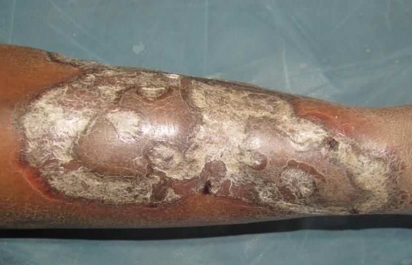
JMSCR Vol||07||Issue||06||Page 33-38||June                                                 2019
 Introduction                                           Materials and Methods
 Psoriasis (Greek. Psora, the itch)1 is a common,       A Retrospective, cross sectional study was
 chronic, relapsing, papulosquamous dermatitis,         conducted in Department of Pathology &
 characterized by an epidermis covered by silvery       Department of Dermatology, Venereology &
 scales.2 Papulosquamous dermatitis comprises a         Leprosy (DVL) at Bharati Vidyapeeth (Deemed to
 group of dermatoses that have distinct                 be University) Medical College & Hospital,
 morphologic features. Psoriasis affects about 1.5-     Sangli from January 2012 to 2016 for a period of
 3%       of    world's      population.4.5     Other   6 months, in which Histopathology slides and
 papulosquamous dermatoses mimicking psoriasis          Clinical notes in the records of patients with
 morphologically are Pityriasis rosea, Lichen           diagnosis of psoriasis were enrolled, studied and
 planus, Seborrheic dermatitis, Pityriasis rubra        analyzed. There were no exclusion criteria’s. All
 pilaris, Parapsoriasis, Drug eruptions, Tinea          the histology slides were reviewed to confirm the
 corporis, and Secondary syphilis.3 The typical         clinical    diagnosis    and     to    study     the
 histological changes seen in Psoriasis are regular     histopathological and clinical features in psoriasis
 acanthosis,     papillomatosis,      thinning     of   vulgaris. The findings were tabulated. Statistical
 suprapapillary plates of epidermis, diminished or      analysis was done with percentages. The study
 absent granular layer, confluent parakeratoses,        was carried out after approval from Institutional
 munro microabscess and the spongiform pustule          Ethical Committee.
 of Kogoj,4,5 and dilated & tortuous capillaries with
 perivascular lymphocytic infiltrate. Munro’s           Observations and Results
 microabscesses are collections of neutrophils and      Total 44 diagnosed cases of psoriasis were studied
 pyknotic nuclei of neutrophils in mounds of            from January 2012 to December 2016, out of
 parakeratoses.5,7 It is observed only in early         which total males were 23 (52.27%) and females
 scaling papule and near the margin of the              were 21 (47.73%). The male to female ratio been
 advancing plaques.5 Sometimes it may not be seen       1.09: 1. The maximum patients were in 3rd & 4th
 because of the sampling error.7The Spongiform          decade. Clinically 40 (90.90%) patients had
 pustule of Kogoj is a micropustule of neutrophils      erythematous scaly plaques (Fig 1,2) followed by
 in a sponge-like network formed by degenerated         hyperpigmented plaques (Fig 3) in 4 (9.10%).
 and thinned epidermal cells.5 It is considered to be   Limbs was commonest site of involvement
 truly diagnostic and specific for psoriasis however    followed by trunk, scalp (Fig 4), palms (Fig 5),
 is not always seen in long-standing lesions.5,6        soles & face. Itching was present in 32 cases
                                                        (72.72%), Auspitz’s sign 15 cases (34.09%) &
 Aims and Objectives                                    Koebner’s phenomenon in 6 (13.63%).
 To evaluate the occurence of Munro’s micro
 abscess and the Spongiform pustule of Kogoj in
 psoriasis vulgaris and various histologic changes
 in psoriasis vulgaris. To study the usefulness of
 histologic features like Munro’s microabscess and
 the Spongiform pustule of Kogoj in the diagnosis
 of psoriasis and to explore the alternative
 histologic features for the diagnosis of psoriasis
 vulgaris.

                                                        Fig 1: Well defined erythematous scaly plaques
                                                        on both lower extremities.

 Renuka Satish Ashtekar et al JMSCR Volume 07 Issue 06 June 2019                                   Page 34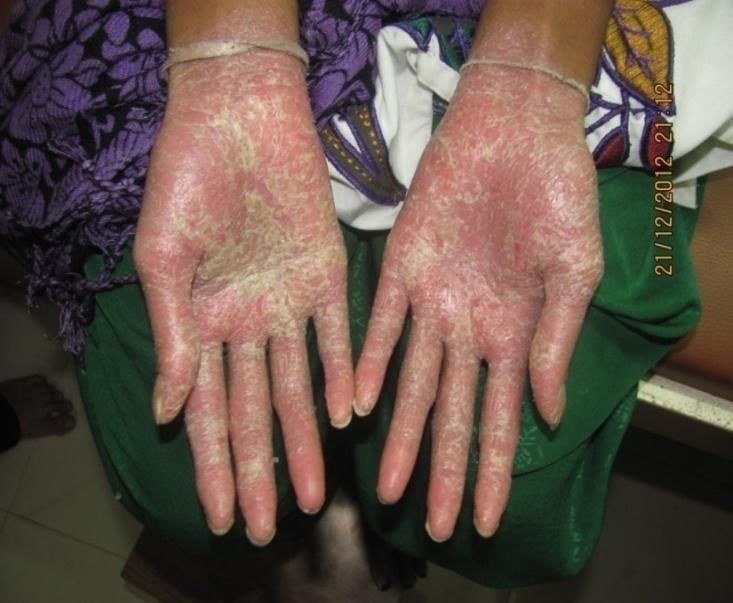
JMSCR Vol||07||Issue||06||Page 33-38||June                                                  2019

                                                              Fig 5: Scaly plaques on palms.
 Fig 2: Well defined erythematous scaly plaques
 on lower back.
                                                    Table 1: Histopathological findings of psoriasis
                                                    of skin
                                                      Histological features      Number      Percentages
                                                                                 of cases        %
                                                                                 (n =44)
                                                      Acanthosis                    42          95.45
                                                      Hyperkeratosis                18          40.90
                                                      Parakeratosis                 41          93.18
                                                      Munro’s                       38          86.36
                                                      Microabscesses
                                                      Elongated Rete Ridges        38           86.36
                                                      Suprapapillary Thining       40           90.90
                                                      Hypogranulosis               40           90.90
                                                      Spongiosis                   18           40.90
                                                      Spongiform Pustule of        10           22.72
                                                      Kogoj
                                                      Dilated Capillaries          42           95.45
                                                      Lymphocytic Infiltrate       40           90.90
 Fig 3: Hyperpigmented scaly plaque on lower leg      Mononuclear         cell      4            9.09
                                                      infiltrate
                                                    Table 1 shows that the above histopathological
                                                    findings seen in slides of 44 patients.
                                                    The histopathological findings seen were
                                                    Acanthosis (95.45%), Hyperkeratosis (18%),
                                                    Parakeratosis (41%), Munro’s micro abscesses
                                                    (86.36%), Elongated rete ridges (86.36%),
                                                    Suprapapillary thining (90.90%), Hypogranulosis
                                                    (90.90%), Spongiosis (40.90%), Spongiform
                                                    pustule of Kogoj (22.72%), Dilated capillaries
                                                    (95.45%), Lymphocytic infiltrate (90.90%) and
                                                    Mononuclear         infiltrate    (9.09%).     The
                                                    histopathological     findings     of   Acanthosis,
                                                    Parakeratosis,         Suprapapillary      thining,
                                                    Hypogranulosis & Dilated capillaries were seen in
          Fig 4: Scaly plaques on scalp.            maximum cases. Munro’s micro abscesses &

 Renuka Satish Ashtekar et al JMSCR Volume 07 Issue 06 June 2019                                   Page 35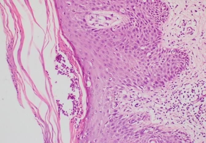
JMSCR Vol||07||Issue||06||Page 33-38||June 2019
Spongiform pustule of Kogoj were seen in 38
(86.36%) & 10 (22.72%) respectively. (Fig 6 to
9).
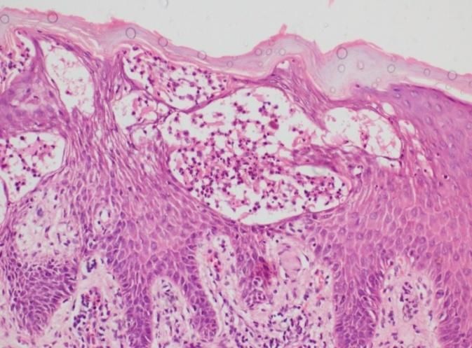
Fig 9: shows Spongiform pustule of Kogoj
Discussion
Psoriasis has many different clinical variants and
Fig 6: shows hyperkeratosis, suprapapilary can resemble other skin diseases such as
thining, elongated rete ridges and dilated secondary syphilis, dyshidrotic eczema,
capillaries in papillary dermis. seborrheic dermatitis, pityriasis rosea,
psoriasiform drug rash, and parapsoriasis.3
Besides, the same patient can present at different
times with a different clinical presentation or
variant.3 The recurrent nature and prognosis of
psoriasis differs, so further highlighting the
importance of reaching the correct diagnosis.3
Typical histologic picture of Psoriasis is not
always found even if biopsy is taken from a
clinically typical lesion.9 The diagnosis of
psoriasis in a classical case, can be made on
clinical grounds and histopathology is only
supportive.6
Fig 7: shows acanthosis and parakeratosis
Table 2 shows Present study’s histopathological
features in comparison to other studies
Histoplogical features Gordon and Pandit Present
Johnson 11 GA12 study
(n = 100 ) (n = 42 ) (n = 44 )
Acanthosis 100 41 42
Hyperkeratosis 28 10 18
Parakeratosis 97 42 41
Munro’s Microabscesses 75 35 38
Elongated Rete Ridges - 36 38
Suprapapillary Thining 98 40 40
Hypogranulosis 75 39 40
Spongiosis 84 40 38
Spongiform Pustule of 31 5 10
Kogoj
Fig 8: shows parakeratosis and Munro’s micro Dilated Capillaries 96 41 42
Lymphocytic Infiltrate 95 42 40
abscesses Mononuclear cell infiltrate - - 4
Renuka Satish Ashtekar et al JMSCR Volume 07 Issue 06 June 2019 Page 36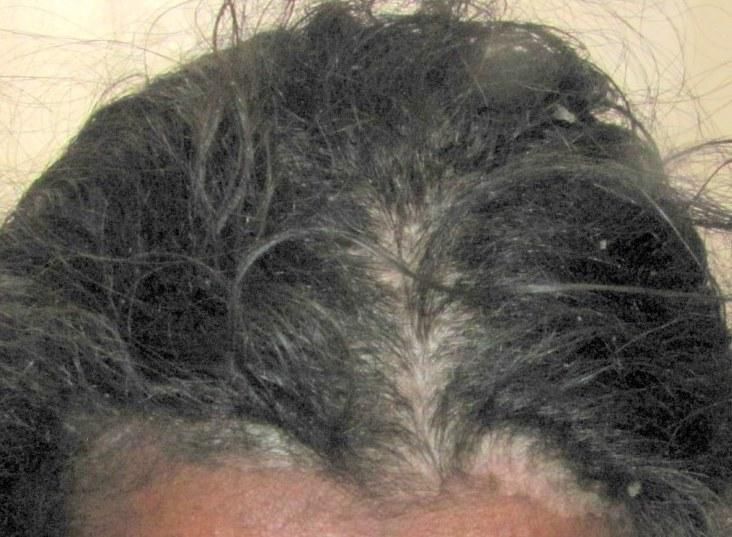
JMSCR Vol||07||Issue||06||Page 33-38||June                                                  2019
 The present study was comparable with studies           Spongiform Pustule of Kogoj is seen in early
 done by Gordan & Jonnson et al11 and Pandit GA          lesion of psoriasis, these may not be seen in long –
 et al.12 The       histopathological findings of        standing lesion. Other histological features like
 acanthosis, pararkeratosis, suprapapillary thining,     Acanthosis,      Hyperkeratosis,      Suprapapillary
 dilated capillaries and lymphocytic infiltrated         thining,     Dilated     capillaries,   Perivascular
 around the capillaries were seenin all 3 studies in     lymphocytic infiltrate along with clinical
 maximum cases. The finding of Munro’s                   correlation may help in diagnosis of psoriasis even
 microabscesses in Pandit GA et al was almost            in absence of Munro’s microabscess &
 same as in present study, also finding of               Spongiform pustule. So, histopathology serves as
 Spongiform pustule of Kogoj was in comparison           a diagnostic tool and rules out other lesions that
 to other studies.                                       mimic psoriasis. The most accurate diagnosis is
 The histopatholgical findings vary in every patient     the one that most closely correlates with clinical
 of psoriasis as it depends on the type of lesion        outcome and helps to direct the most appropriate
 which is taken for biopsy, duration of psoriasis        clinical intervention.
 since when the patient is having.
 Table 3 shows Clinical Features In Comparison to        References
 Other Study                                                1. Fry L. Psoriasis. Br J Dermatol
    Clinical Features      Pandit GA11   Present Study         1988;119:445-61.
                             (n=42)         (n=44)
    No. of males               24             23            2. De Rosa G, Mignogna C. The
    No. of females             18             21               histopathology of psoriasis. Reumatismo
    Age            group   20-40 years    20-40 years
    presentation                                               2007;59 Suppl 1:46-.
    Scaly plaques              39             40            3. Mehta S, Singal A, Singh N, Bhattacharya
    Limbs                      35             41
    Trunk                      20             34               SN. A study of clinicohistopathological
    Scalp                      17             16               correlation in patients of psoriasis and
    Face                       10             13
    Itching                    35             32
                                                               psoriasiform dermatitis. Indian J Dermatol
    Auspitz’s sign             27             15               Venereol        Leprol        2009;75:100.
    Koebner’s                  5               6
    Phenomenon
                                                               [PUBMED]
                                                            4. Krueger GG, Duvic M. Epidemiology of
 Table 3 shows commonest age group presentation                psoriasis: Clinical issues. J Invest
 is between 20 to 40 years in present study and                Dermatol 1994;102:14S-8.
 study done by Pandit GA et al11. The maximum               5. Mobini N, Toussaint S, Kamino H,
 patients had scaly plaques, the commonest site                Noninfectious erythematous, papular and
 involved were limbs followed by trunk, scalp &                squamous diseases In: Elder DE (Ed in
 face on the which are comparable in both studies.             Chief) Lever’s Histopathology of the Skin.
 Itching was present in both studies. Auspitz’s sign           9th ed Lippincott Williams & Wilkins,
 and Koebners’s phenomenon was seen in                         Philadelphia 2005:pp185-186.
 comparable cases in both studies.                          6. Grover C. Psoriasis. In: Sacchidananand S
                                                               (Chief Ed). IADVL Textbook of
 Conclusion                                                    Dermatology 4rded Bhalani Publishing
 In present study it was concluded that Psoriasis              House, Mumbai, India 2015: pp1044-
 has varied clinical presentations. Besides, the               1045.
 same patient may present with different types of           7. James WD, Berger TG, Elston DM.
 lesions of psoriasis. So, biopsy of a lesion at               Andrews’ Diseases of the Skin Clinical
 different stages of presentation in same patient              Dermatology 11th ed. Saunders Elsevier,
 will differ. Presence of Munro’s microabscess &               Publication, US 2011:pp.194

 Renuka Satish Ashtekar et al JMSCR Volume 07 Issue 06 June 2019                                    Page 37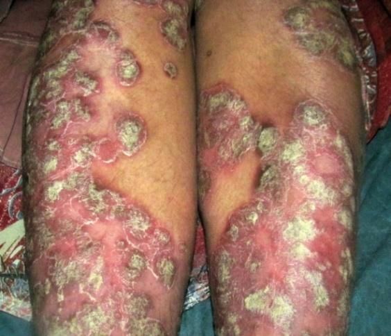
JMSCR Vol||07||Issue||06||Page 33-38||June                         2019
    8. Rupec M. Zur ultrastruktur der
        spongiformen pustel. Arch Kli Exp
        Dermatol 1979; 239:30.
    9. Cox AH, Watson W. Histologic variations
        in    lesions    of    psoriasis.    Arch
        Dermatol1972; 106:503.
    10. Abel EA. Papulosquamous disorders. In:
        Dale DC, Federman DD, editors.
        Dermatology. Sec. II. Ontario: ACP
        (American College of Physicians)
        Medicine Principles and Practice; 2007. p.
        1-9.
    11. Gordon M, Johnson WC. Histopathology
        and histochemistry of psoriasis. I. The
        active lesion and clinically normal skin.
        Arch Dermatol 1967;95:402-7.
    12. Pandit GA, Narayankar SL. Significance
        of clinicopathological correlation in
        psoriasis. Med J DY Patil Univ [serial
        online] 2015 [cited 2017 Sep 5];8:481-5.
        Available
        from: http://www.mjdrdypu.org/text.asp?2
        015/8/4/481/160789.

 Renuka Satish Ashtekar et al JMSCR Volume 07 Issue 06 June 2019          Page 38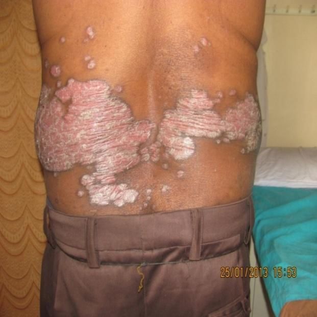
You can also read