Long-term Follow-up of Optic Disc Pit Maculopathy Treated with Laser Photocoagulation: A Case Report
←
→
Page content transcription
If your browser does not render page correctly, please read the page content below
DOI:10.14744/bej.2021.83723
           Beyoglu Eye J 2021; 6(2): 151-154
                                                                                                            Case Report

Long-term Follow-up of Optic Disc Pit Maculopathy
Treated with Laser Photocoagulation: A Case Report

  Sergios Taliantzis, Asli Perente, Christina Mitsi,                             Eirini Kanella Panagiotopoulou,
  Ioannıs Fotiadis, Doukas Dardabounis
Department of Opthalmology, University Hospital of Alexandroupolis, Alexandroupolis, Greece

           Abstract
           This report describes a case of optic disc pit maculopathy treated successfully with laser photocoagulation. Optical co-
           herence tomography (OCT) was used to assess the optic nerve and the macular area. Green laser photocoagulation was
           performed in an arcuate pattern to manage the macular edema. Eighteen months later, OCT showed complete regression
           of the macular edema. Fifty months after the laser treatment, visual acuity remained 20/20. The results of an Amsler grid
           test were negative and the macular area was visualized as normal. Slit-lamp laser photocoagulation is a minimally invasive
           technique that should be considered as a first-line treatment option in patients with optic pit maculopathy who retain
           their visual capacity.
           Keywords: Green laser, metamorphopsia, minimal invasive treatment, optical coherence tomography, optic disc pit.

Introduction                                                                 serous detachment, cystic degeneration, and changes to the
                                                                             retinal pigment epithelium, and is characterized by the accu-
Optic disc pit (ODP) is a rare, congenital anomaly with an
                                                                             mulation of intraretinal and/or subretinal fluid (3).
incidence of 1 in 11.000 individuals that typically appears as a
circumscribed, discolored (often gray), oval-shaped depression                   Several theories have been proposed to explain the origin
of the optic disc (1). It is thought to arise due to an incomplete           of the fluid seen in ODP-M. Possibilities reported include the
closure of the fetal fissures affecting the lamina cribrosa; a her-          vitreous body, the choroid, cerebrospinal fluid, and leakage
niation of dysplastic retinal tissue becomes a collagen-rich ex-             from blood vessels at the site of the ODP (1).
cavation extending toward the subarachnoid space (2). Most                       There are no universally accepted guidelines for the treat-
cases are asymptomatic, though significant visual deterioration              ment of ODP-M; however, laser photocoagulation, vitrecto-
may be observed when maculopathy occurs, a complication                      my, gas tamponade and/or inner limiting membrane (ILM)
detected in 25% to 75% of patients with ODP (3). ODP mac-                    peeling, macular buckling surgery, and recently, autologous
ulopathy (ODP-M) is a clinical entity, which presents with                   platelet injection after pars plana vitrectomy, are among the

   How to cite this article: Taliantzis S, Perente A, Mitsi C, Panagiotopoulou EK, Fotiadis I, Dardabounis D. Long-term Follow-up of Optic Disc Pit
   Maculopathy Treated with Laser Photocoagulation: A Case Report. Beyoglu Eye J 2021; 6(2): 151-154.

  Address for correspondence: Asli Perente, MD. Alexandroupolis Universite Hastanesi, Goz Hastaliklari Bolumu, Alexandroupolis, Yunanistan
                                     Phone: +306951577205 E-mail: asli_perende-90@hotmail.com
                   Submitted Date: January 02, 2021 Accepted Date: March 14, 2021 Available Online Date: June 08, 2021
                  Copyright 2021 by Beyoglu Eye Training and Research Hospital - Available online at www.beyoglueye.com
                   ©

             OPEN ACCESS This work is licensed under a Creative Commons Attribution-NonCommercial 4.0 International License.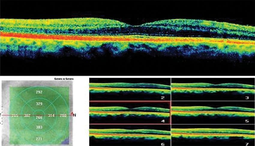
152                                                                                                       Taliantzis et al., Optic Disc Pit Maculopathy

therapeutic interventions used in clinical practice (4).                    a
   This case of ODP-M was successfully managed with laser
photocoagulation.
Case Report
A 66-year-old male patient presented at the outpatient ser-
vice complaining of blurred vision in his right eye. A full
ophthalmological examination was performed. His uncor-
rected visual acuity (UVA) was 20/20 in both eyes, but the
results of an Amsler grid test were positive in the right eye.
The intraocular pressure (IOP) was 35 mmHg in the right
eye (OD) and 15 mmHg in the left eye (OS). No abnormal-
ities were observed in the anterior segment. A dilated fun-
dus examination disclosed a glaucomatous optic disc with                    b
an estimated cup to disc ratio of 0.8 and retinal elevation
from the optic disc to the foveola in the OD. Optical co-
herence tomography (OCT) revealed an optic pit with se-
rous detachment of the retina in the OD. Treatment with a
prostaglandin analogue to be administered in a dose of one
drop once daily was introduced and a 6-month follow-up
program was suggested. On re-examination 6 months later,
the UVA was found to be unchanged and the IOP in the
OD was 13 mmHg. However, the patient mentioned a sig-
nificant worsening of the metamorphopsia symptoms that
affected his daily life activities. OCT showed a progression
of intraretinal fluid toward the foveola. To address the de-
                                                                           Figure 2. (a) Infrared fundus photograph of the right eye before laser
terioration of the clinical picture, 14 burns with a green                 photocoagulation. (b) Infrared fundus photograph of the right eye after
laser were performed in an arcuate pattern (60–27 mW of                    laser photocoagulation showing the laser scars on the temporal side of
energy and spot size of 50μm) (Fig. 1) at the temporal side                the optic disc (green arrows).

Figure 1. Optical coherence tomography images of the right eye showing fluid in the outer nuclear
layer of the retina and a color fundus photograph of the right optic disc indicating the presence of an
optic disc pit.
Taliantzis et al., Optic Disc Pit Maculopathy                                                                                   153

of the right optic disc (Fig. 2). Eight months later, it was          of approximately 50% (5). Pneumatic retinopexy with an in-
observed that the macular edema had improved, and 18                  travitreal gas tamponade with or without laser application is
months after the treatment, no fluid was detected on OCT.             another therapeutic option with encouraging outcomes (7).
Fifty months after the laser treatment, the UVA remained              Scleral buckling is a newer surgical approach with satisfacto-
20/20, an Amsler test was negative, there were no subjec-             ry long-term visual and anatomical outcomes proposed by
tive complaints of metamorphopsia, the IOP was normal,                Theodossiadis et al. (8).
and complete regression of the macular edema was visible                  Slit-lamp laser photocoagulation is an initial, mini-
on OCT (Fig. 3).                                                      mally invasive treatment options for the management of
                                                                      ODP-M. Laser spots applied to the temporal side of the
Discussion                                                            peripapillary area are used to create chorioretinal adhe-
ODP is one of several congenital cavitary anomalies with a            sion, which interrupts circulation of the fluid into the sub-
similar impact on visual capacity. ODP is characterized by            retinal space (9).
isolated cavitations, usually located on the temporal side of             According to the available literature data, the efficacy
the optic disc. Related maculopathy occurs in 2 steps: intra-         of laser treatment is controversial; some published reports
retinal pooling of fluid and consequent retinoschisis, and ex-        demonstrate full absorption of fluid and reattachment of
tension of the fluid in the subretinal layers through an outer        the retina after laser photocoagulation, while other reports
break (3,5).                                                          note low success rates and severe perimetrical defects (9).
    Although spontaneous resolution has been reported in                  In our case, the macular edema completely resolved fol-
approximately 25% of cases, the general visual prognosis              lowing laser treatment, and the disturbing metamorphop-
is poor. Steel et al. (6) conducted a retrospective study of          sia was eliminated. The patient’s visual capacity remained
134 eyes to evaluate the risk factors associated with poor            consistent for the follow-up period of 50 months, with
vision in patients with ODP-M. The researchers found that             no signs of macular edema. Inferotemporally, there was a
extension of the edema beyond the vascular arcades and                significant retinal nerve fiber layer (RNFL) loss detected
the presence of both subretinal fluid and outer retinal layer         during follow-up. However, temporally, there was no loss of
fluid were among the primary determinants for poor visual             RNFL seen where laser photocoagulation was applied. The
acuity.                                                               inferotemporal RNFL loss may have been a result of ad-
    Different treatment options for ODP-M have been pro-              vanced glaucoma (Fig. 4). In conclusion, given that slit-lamp
posed; however, the efficacy of each therapeutic interven-            laser photocoagulation is a minimally invasive technique,
tion seems to be controversial. Pars plana vitrectomy with            it should be considered as a first-line option, particularly
laser treatment, gas tamponade, and/or ILM peeling is the             in patients who have maintained their visual acuity despite
most used surgical procedure, with a reported success rate            macular edema.

Figure 3. Optical coherence tomography images of the right eye 50 months after the laser photoco-
agulation treatment.
154 Taliantzis et al., Optic Disc Pit Maculopathy
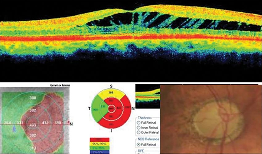
Figure 4. Nerve fiber optic nerve head/ganglion cell complex analysis of the right eye.
GCC: Ganglion cell complex; NDB: Normative database; RNFL: Retinal nerve fiber layer
Disclosures 4. Shah SD, Yee KK, Fortun JA, Albini T. Optic disc pit maculop-
Informed consent: Written informed consent was obtained athy: a review and update on imaging and treatment. Int Oph-
from the patient for the publication of the case report and the thalmol Clin 2014;54:61–78. [CrossRef]
accompanying images.
5. Chatziralli I, Theodossiadis P, Theodossiadis GP. Optic disk pit
Peer-review: Externally peer-reviewed.
maculopathy: current management strategies. Clin Ophthalmol
Conflict of Interest: None declared.
2018;12:1417–22. [CrossRef]
Authorship Contributions: Involved in design and conduct of 6. Steel DH, Williamson TH, Laidlaw DA, Sharma P, Matthews C,
the study (ST); preparation and review of the study (DD, IP, AP,
Rees J, et al. Extent and location of intraretinal and subretinal
CM, EKP); data collection (EKP, CM, AP).
fluid as prognostic factors for the outcome of patients with
References optic disk pit maculopathy. Retina 2016;36:110–8. [CrossRef]
7. Wan R, Chang A. Optic disc pit maculopathy: a review of diag-
1. Moisseiev E, Moisseiev J, Loewenstein A. Optic disc pit maculop-
athy: when and how to treat? A review of the pathogenesis and nosis and treatment. Clin Exp Optom 2020;103:425–9. [CrossRef]
treatment options. Int J Retina Vitreous 2015;1:13. [CrossRef] 8. Theodossiadis GP, Chatziralli IP, Theodossiadis PG. Macular
2. Ferry AP. Macular detachment associated with congenital pit buckling in optic disc pit maculopathy in association with the
of the optic nerve head. Pathologic findings in two cases sim- origin of macular elevation: 13-year mean postoperative results.
ulating malignant melanoma of the choroid. Arch ophthalmol Eur J Ophthalmol 2015;25:241–8. [CrossRef]
1963;70:346–57. [CrossRef] 9. Kalogeropoulos D, Ch'ng SW, Lee R, Elaraoud I, Purohit M,
3. Georgalas I, Ladas I, Georgopoulos G, Petrou P. Optic disc pit: a Felicida V, et al. Optic Disc Pit Maculopathy: A Review. Asia Pac
review. Graefes Arch Clin Exp Ophthalmol 2011;249:1113–22. J Ophthalmol (Phila) 2019;8:247–55.You can also read