MiR-513b-5p inhibits the proliferation and promotes apoptosis of retinoblastoma cells by targeting TRIB1
←
→
Page content transcription
If your browser does not render page correctly, please read the page content below
Open Medicine 2021; 16: 1364–1371
Research Article
Li-Juan Zhang, Fang Wang, Pei-Yan Qi, Wei-Yan Zhou, Bing Wang*
miR-513b-5p inhibits the proliferation and
promotes apoptosis of retinoblastoma cells
by targeting TRIB1
https://doi.org/10.1515/med-2021-0343
received March 23, 2021; accepted August 12, 2021
1 Introduction
Abstract: MicroRNAs are involved in the pathogenesis Retinoblastoma (RB) is the most common intraocular malig-
of various human malignant tumors. This study aims to nant tumor in infants, which seriously endangers their visual
explore the role of miR-513b-5p in the malignant prolifera- function and life. It accounts for 2.5–4% of malignant tumors
tion of retinoblastoma (RB) cells and its potential molecular in children, with a prevalence of 1/20,000 to 1/15,000 [1,2]. In
mechanisms. The function-gain and function-loss experi- recent years, with the continuous improvement of the diag-
ments were performed in Weri-RB1 cells using miR-513b-5 nosis and treatment methods, the prognosis and survival rate
mimics and inhibitors. miR-513b-5p mimics inhibited the of RB patients have been improved. However, the pathogenic
proliferation and clone formation and promoted apoptosis factors and molecular mechanisms of the disease have not
of Weri-RB1 cells. In contrast, the miR-513b-5p inhibitor been fully studied. Therefore, further research and explora-
promoted the proliferation and clone formation of Weri- tion of genes and action mechanism that related to the occur-
RB1 cells and inhibited cell apoptosis. miR-513b-5p can rence and development of RB are of great significance [3].
directly bind to the 3′UTR region of TRIB1 mRNA, and MicroRNAs (miRNAs) are a group of endogenous non-
inhibit its protein expression. Overexpression of TRIB1 pro- coding RNA molecules with a length of 20–22 nucleotides
moted the proliferation and cloning of Weri-RB1 cells but [4]. They can directly interact with the 3′-non-translation
inhibited their apoptosis. The knockdown of TRIB1 inhibited regions (3′UTR) of the target mRNA, leading to mRNA
the proliferation and clone formation of Weri-RB1 cells and
degradation or translation inhibition, thereby negatively
promoted cell apoptosis. In addition, miR-513b-5p mimics
regulating the expression of the target genes [5]. miRNAs
neutralized the effects of TRIB1 overexpression on the
have an important relationship with the occurrence and
proliferation and apoptosis of Weri-RB1 cells. Finally,
development of cancer [6]. Many different miRNA abnorm-
miR-513b-5p can inhibit the phosphorylation level of AKT,
alities have been identified in RB. Their abnormal expression
mTOR, and p70, while TRIB1 played the opposite role. miR-
plays an important role in various processes of cancer,
513b-5p inhibits the malignant proliferation of Weri-RB1
including cell proliferation, metastasis, apoptosis, invasion,
cells by repressing the expression of TRIB1. miR-513b-5p
epithelial–mesenchymal transformation, and angiogenesis [7].
and TRIB1 may be the biomarkers and/or key targets for
Previous studies have shown that miR-513b-5p can
clinical diagnosis and treatment of RB.
play a unique role in different types of solid tumors. For
Keywords: miR-513b-5p, TRIB1, Weri-RB1 cells, prolifera- example, the expression of miR-513b-5p is significantly
tion, apoptosis, retinoblastoma downregulated in non-small cell lung cancer (NSCLC)
[8,9] and gastric cancer [10]. Its exogenous overexpres-
sion can significantly inhibit the proliferation, invasion,
* Corresponding author: Bing Wang, Department of Ophthalmology,
migration, and promote apoptosis of NSCLC [8,9] and
Shandong Provincial Hospital Affiliated to Shandong First Medical gastric cancer cells [10]. However, some studies have
University, Jinan 250021, Shandong, China, also reported that miR-513b-5p plays an oncogene in cer-
e-mail: wang2001bing@163.com vical cancer [11] and ovarian cancer [12]. In addition,
Li-Juan Zhang, Fang Wang, Wei-Yan Zhou: Department of
miR-513b-5p has also been identified to play an important
Ophthalmology, Shandong Provincial Hospital Affiliated to
Shandong First Medical University, Jinan 250021, Shandong, China
role in testicular development and male sexual matura-
Pei-Yan Qi: Guangzhou International Travel Health Care Center, tion [13,14]. However, the role of mir-513b-5p in RB gene-
Guangzhou 510000, Guangdong, China ration has not been reported.
Open Access. © 2021 Li-Juan Zhang et al., published by De Gruyter. This work is licensed under the Creative Commons Attribution 4.0
International License.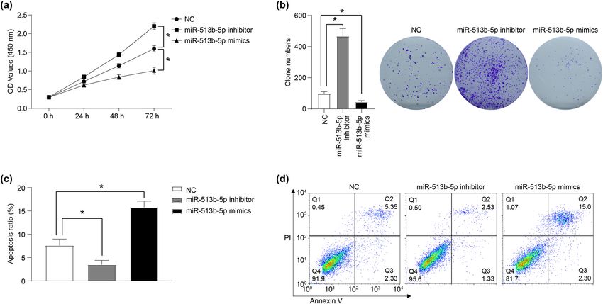
miR-513b-5p inhibits retinoblastoma progression 1365
The aim of this study was to investigate the effect of medium was removed, and the cells were carefully washed
miR-513b-5p overexpression and downexpression on the twice with 2 mL of PBS. After the PBS was air-dried, the
proliferation and apoptosis of RB cells and its possible clones were fixed with 4% paraformaldehyde for 30 min at
mechanism. The results may provide some new insights room temperature and then stained with 0.1% crystal
into the pathogenesis of RB and help to explore new violet dye for 30 min at room temperature. After dyeing,
treatments. the dye was cleaned and then the number of clones was
counted.
2 Materials and methods
2.5 Flow cytometric analysis of apoptosis
2.1 Cell culture
The apoptosis of Weri-RB1 cells was evaluated by using
an Annexin V-FITC Apoptosis Detection Kit (BD Biosciences)
The retinoblastoma cell line Weri-RB-1 and 293 T cell were
and PI staining according to the manufacturer’s instructions.
obtained from the American Type Culture Collection (ATCC),
After 48 h of transfection, the cells were digested with
and cultured in the Dulbecco’s modified Eagle’s medium
trypsin without EDTA, and centrifuged at 3,000g for 5 min
(DMEM; Gibco; Thermo Fisher Scientific, Inc.) with 10%
at room temperature. About 3 mL PBS was added to resus-
FBS, 100 U/mL penicillin, and 0.1 mg/mL streptomycin at
pend and wash the cell precipitation. Subsequently, the cells
37°C with the atmosphere of 5% CO2.
were centrifuged at 3,000g for another 5 min at room tem-
perature. 1× binding buffer was added to resuspend the cells,
and the cell density was adjusted to 1–5 × 10⁶/mL. About
2.2 Cell transfection 100 μL of the cell suspension was added into a 5 mL flow
cytometry tube, and mixed with 5 μL Annexin V/FITC, and
miR-513b-5p mimics, miR-513b-5p inhibitor, siRNA speci- incubated in the dark for 5 min at room temperature. Then,
fically targeting TRIB1 (TRIB1-KD), TRIB1 overexpression 10 μL of the PI dye solution and 400 μL of PBS was added,
plasmid (TRIB1-OE), and their respective negative control and the apoptosis of Weri-RB1 cells in each group was ana-
(NC) were obtained from GenePharma Co. Ltd. (Shanghai, lyzed by flow cytometer (FACS Calibur; BD Biosciences,
China). Transfection was performed according to the Franklin Lakes, NJ). FlowJo software was used to analyze
instructions of the Lipofectamine2000 transfection kit the flow cytometry outcomes.
(Invitrogen; Thermo Fisher Scientific, Inc.).
2.6 Western blot analysis
2.3 Cell Counting Kit‑8 (CCK-8) assay
After 48 h of transfection, the cells were lysed with RIPA
The transfected cells were seeded into a 96-well plate at a lysate (Pierce, USA) in ice for 10 min, and the total protein
concentration of 1 × 10³ cells per well. After culturing for was extracted. The concentration of protein was mea-
24, 48, and 72 h, the old medium in each well was sured with the BCA protein assay kit (Pierce, USA). The
removed, and fresh DMEM containing 10% CCK8 reagent protein samples were separated by SDS polyacrylamide
(Beyotime Institute of Biotechnology) was added and gel electrophoresis and transferred into polyvinylidene
incubated at 37°C for 2 h. Subsequently, the absorbance difluoride membranes (PVDF; Millipore, Billerica, MA).
value (OD) of each well was detected with a microplate The membranes were blocked with 5% skim milk for
reader at a wavelength of 450 nm. 1 h at room temperature and incubated overnight at 4°C
with primary antibodies. The primary antibodies are as
follows: anti-Bax (50599-2-Ig), anti-Bcl2 (12789-1-AP),
anti-caspase3 (19677-1-AP), anti-p53 (60283-2-Ig), anti-
2.4 Clone formation assay GAPDH (60004-1-Ig), anti-AKT (10176-2-AP), anti-p-AKT
(Ser473, 66444-1-Ig), anti-mTOR (66888-1-Ig), and anti-
The transfected cells were seeded in a 6-well plate and p-mTOR (Ser2448, 66888-1-Ig) were obtained from PTG,
cultured for 1–2 weeks until the clones were visible. The and anti-p70 (ab184551), anti-p-p70 (Ser371, ab109393),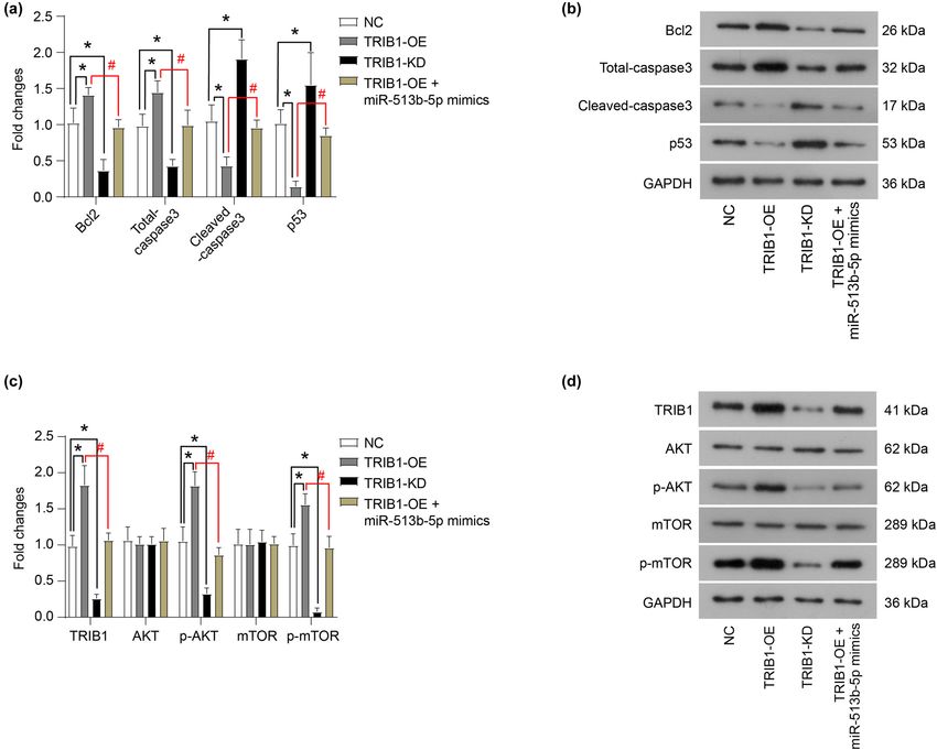
1366 Li-Juan Zhang et al. and anti-TRIB1 (ab137717) were obtained from Abcam. 2.8 Statistical analysis Following this, the secondary antibody (HRP-conjugated Goat Anti-Rabbit IgG, SA00001-2; and HRP-conjugated GraphPad Prism 6.0 was used for data analysis (GraphPad Goat Anti-Mouse IgG, SA00001-1, PTG) was applied and Software Inc., San Diego, CA). All data were presented as incubated with the membrane for 1 h at room tempera- the mean ± standard error of the mean (SEM). The differ- ture. Finally, the proteins were visualized with enhanced ences were evaluated using one-way ANOVA followed by chemiluminescence reagents (Pierce, USA). The QUANT- Tukey’s post hoc test for multiple comparisons and the ITY ONE software (Bio-Rad Laboratories, Inc.) was used Mann–Whitney U-test for pair-wise comparisons. A P-value to scan and calculate the relative expression quantity of less than 0.05 was considered to indicate a statistically sig- each protein. nificant difference. 2.7 Double luciferase assay 3 Results The TRIB1 3′UTR region was cloned into the pmiR GLO vector, and the binding region of TRIB1 3′UTR to miR- 3.1 miR-513b-5p inhibits the proliferation of 513b-5p was mutated. TRIB1-3′UTR-WT and TRIB1-3′UTR- Weri-RB1 cells in vitro MUT plasmids were constructed and co-transfected into 293T cells with a miR-513b-5p mimics or negative control In order to investigate the role of miR-513b-5p on the (NC) using Lipofectamine 2000 (Invitrogen), respectively. proliferation of Weri-RB1 cells, CCK-8 and clone forma- After 48 h, the Dual Luciferase Assay System Kit (Promega, tion assays were performed. The results of the CCK-8 WI, USA) was employed to analyze the activity of the assay showed that compared with the NC group, miR- luciferase. 513b-5p mimics inhibited the proliferation of Weri-RB1 Figure 1: miR-513b-5p inhibits the proliferation and promotes the apoptosis of Weri-RB1 cells in vitro. The role of miR-513b-5p mimics and inhibitors in the proliferation, clone formation, and apoptosis of Weri-RB1 cells was detected by CCK-8 (a), clone formation (b), and flow cytometry (c and d) assays. The differences were evaluated using the Mann–Whitney U-test for pair-wise comparisons. *P < 0.05. Each group of a includes six samples, each group of b–d includes three samples. All experiments were repeated three times independently, and the individual experiments were performed 1 week apart.
miR-513b-5p inhibits retinoblastoma progression 1367
cells, while the miR-513b-5p inhibitor promoted cell prolif- miR-513b-5p inhibitor group was decreased (Figure 1c and d,
eration (Figure 1a, P < 0.05). In addition, the results of the P < 0.05). In addition, after miR-513b-5p mimics were
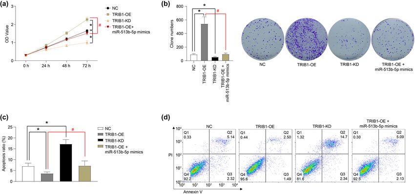
clone formation experiment were consistent with the results transfected, the protein level of anti-apoptotic protein
of the CCK-8 assay. The number of cell clones formed in the Bcl2 was decreased, while the protein level of pro-apop-
miR-513b-5p mimics group was significantly lower than that totic protein Bax, cleaved-caspase3, and p53 was increased
in the NC group, while the clone numbers in the miR-513b-5p (Figure 2a and b, P < 0.05). Furthermore, after transfection
inhibitor group were decreased (Figure 1b, P < 0.05). of miR-513b-5p inhibitor, the expression of anti-apoptotic
protein Bcl2 increased, and the expression of pro-apop-
totic protein Bax, cleaved-caspase3, and p53 decreased
(Figure 2a and b, P < 0.05).
3.2 miR-513b-5p promotes the apoptosis of
Weri-RB1 cells in vitro
The role of miR-513b-5p on the apoptosis of Weri-RB1 3.3 miR-513b-5p inhibits the activation of
cells was detected using flow cytometry and western the AKT/mTOR signaling pathway
blot. The results showed that the apoptosis ratio in the
miR-513b-5p mimics group was significantly higher than The effect of miR-513b-5p on the AKT/mTOR signaling
that in the NC group, while the apoptosis ratio in the pathway activation was determined by western blot.
Figure 2: miR-513b-5p inhibits the activation of the AKT/mTOR signaling pathway. (a and b) The protein expression of key proteins related to
apoptosis was measured using a western blot. (c and d) The protein expression of key proteins in the AKT/mTOR signaling pathway was
measured using western blot. The differences were evaluated using the Mann–Whitney U-test for pair-wise comparisons. *P < 0.05. Each
group of (b and d) includes three samples. All experiments were repeated three times independently, and the individual experiments were
performed 1 week apart.1368 Li-Juan Zhang et al.
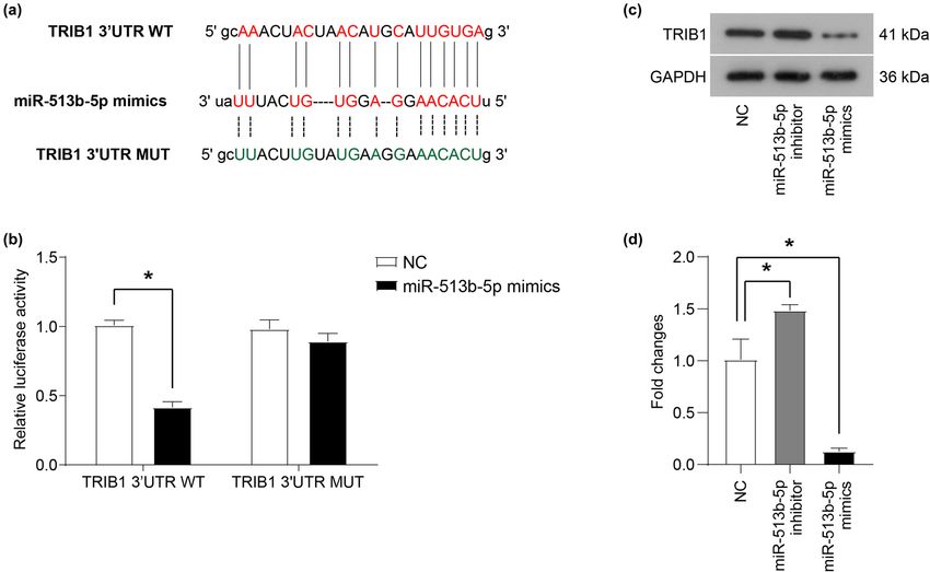
The results showed that miR-513b-5p mimics inhibited UTR-MUT recombinant plasmid did not significantly affect
the phosphorylation of AKT, mTOR, and p70 (Figure 2c the level of luciferase. This result revealed that miR-513b-
and d, P < 0.05). In contrast, the miR-513b-5p inhibitor 5p could directly bind to the 3′UTR region of the TRIB1
increased the phosphorylation levels of AKT, mTOR, and mRNA through the predicted site. In addition, the miR-
p70 (Figure 2c and d, P < 0.05). 513b-5p mimic inhibited the protein expression of TRIB1,
while the miR-513b-5p inhibitor up-regulated the expres-
sion of the TRIB1 protein (Figure 3c and d, P < 0.05). In
summary, miR-513b-5p can bind to the 3′UTR region of
3.4 miR-513b-5p can bind to TRIB1 mRNA TRIB1 mRNA and regulate its protein expression.
and regulate its protein expression
The TargetScan software predicted the direct binding site
of miR-513b-5p and 3′UTR region of TRIB1 mRNA (Figure 3a). 3.5 TRIB1 promotes the proliferation and
Subsequently, the direct binding of miR-513b-5p to the inhibits the apoptosis of Weri-RB1 cells
3′UTR region of TRIB1 mRNA was determined by using in vitro
a double-luciferase assay. As shown in Figure 3b, the
co-transfection of miR-513b-5p mimics and pmiR-GLO- The effect of TRIB1 on the proliferation and apoptosis of Weri-
TRIB1-3′UTR-WT recombinant plasmid significantly RB1 cells was determined. As shown in Figure 4a and b,
reduced the expression of luciferase, while the co-trans- the overexpression of TRIB1 (TRIB1-OE) promoted the pro-
fection of miR-513b-5p mimics and pmiR-GLO-TRIB1-3′ liferation and clone formation of Weri-RB1 cells, while the
Figure 3: miR-513b-5p can bind to TRIB1 mRNA and regulate its protein expression. (a) The TargetScan software predicted the direct binding
site of miR-513b-5p and the 3′UTR region of TRIB1 mRNA. (b) The relative expression of luciferase and direct binding of miR-513b-5p to the
3′UTR region of TRIB1 mRNA was determined by using a double-luciferase assay. (c and d) The protein expression of TRIB1 was measured
using a western blot. The differences of (b) were evaluated using one-way ANOVA followed by Tukey’s post hoc test, and the Mann–Whitney
U-test for (d). *P < 0.05. Each group of (b and d) includes three samples. All experiments were repeated three times independently, and the
individual experiments were performed 1 week apart.miR-513b-5p inhibits retinoblastoma progression 1369
knockdown of TRIB1 (TRIB1-KD) inhibited the proliferation 5% of children’s blindness is caused by RB [16]. There-
and clone formation of Weri-RB1 cells. In addition, the fore, it is of great significance to investigate the patho-
overexpression of TRIB1 inhibited the apoptosis of Weri- genesis and therapeutic targets of RB.
RB1 cells (Figure 4c and d), upregulated the protein expres- It is well known that miRNAs are involved in the
sion of Bcl2, and downregulated the protein expression pathogenesis of various human malignancies. In this
of cleaved-caspase3 and p53 (Figure 5a and b). The study, we found that miR-513b-5p inhibited the prolifera-
knockdown of TRIB1 induced apoptosis of Weri-RB1 cells tion and promoted apoptosis of Weri-RB1 cells in vitro.
(Figure 4c and d), downregulated the protein expression of The results of this research are consistent with previous
Bcl2 and upregulated the protein expression of cleaved- studies on the role of miR-513b-5p in NSCLC [8], embryonic
caspase3 and p53 (Figure 5a and b). Furthermore, the testicular cancer [13], gastric cancer [10], and osteosar-
miR-513b-5p mimic could neutralize the role of TRIB1 over- coma [17] cells, but are contrary to the role of miR-513b-
expression on the proliferation and apoptosis of Weri-rb1 5p in cervical cancer [11] and ovarian cancer [12]. This may
cells (Figures 4a–5b, P < 0.05). be due to the uniqueness of the tumor type.
In addition, the overexpression of TRIB1 promoted the TRIB1 (tribbles homologue 1) is a member of the
phosphorylation of AKT and mTOR, while the knockdown Tribbles family, which is initially found to modulate
of TRIB1 decreased the phosphorylation levels of AKT and string/cdc25 in Drosophila morphogenesis [18]. TRIB1
mTOR (Figure 5c and d, P < 0.05). Furthermore, miR-513b- has been reported to be associated with the occurrence
5p can counteract the effect of TRIB1 overexpression on the and development of a variety of tumors, including leukemia
AKT and mTOR phosphorylation (Figure 5c and d, P < 0.05). [19], hepatocellular carcinoma [20], colorectal cancer [21],
and prostate cancer [22]. It has been revealed that TRIB1 is
highly expressed in prostate cancer and follicular thyroid
cancer, and promotes the proliferation, survival, and
4 Discussion tumor growth of cancer cells. In this article, we determined
that TRIB1 is the direct binding target of miR-513b-5p and
RB is a malignant ocular tumor in infants with a high detected that its overexpression contributed to the prolif-
blindness rate. In China, approximately 1,000 children eration and cloning of Weri-RB11 cells, but inhibited their
are diagnosed with RB every year [15]. Moreover, about apoptosis. Our findings are consistent with prevenient
Figure 4: TRIB1 promotes proliferation and inhibits the apoptosis of Weri-RB1 cells in vitro. The role of TRIB1 overexpression (TRIB1-OE) and
TRIB1 knockdown (TRIB1-KD) in the proliferation, clone formation, and apoptosis of Weri-RB1 cells was detected by CCK-8 (a), clone formation
(b), and flow cytometry (c and d) assays. The differences were evaluated using the Mann–Whitney U-test for pair-wise comparisons. *P < 0.05,
compared to NC; #P < 0.05, compared to TRIB1-OE. Each group of (a) includes six samples, each group of (b and d) includes three samples. All
experiments were repeated three times independently, and the individual experiments were performed 1 week apart.1370 Li-Juan Zhang et al.
Figure 5: TRIB1 promotes the activation of the AKT/mTOR signaling pathway. (a and b) The protein expression of key proteins related to
apoptosis was measured using western blot. (c and d) The protein expression of key proteins in the AKT/mTOR signaling pathway was
measured using western blot. The differences were evaluated using the Mann–Whitney U-test for pair-wise comparisons. *P < 0.05,
compared to NC; #P < 0.05, compared to TRIB1-OE. Each group of (b and d) includes three samples. All experiments were repeated three
times independently, and the individual experiments were performed 1 week apart.
conclusions about the role of TRIB1 in malignancies, sug- In conclusion, our study suggested that miR-513b-5p
gesting that TRIB1 may be an oncogene. inhibited the expression of TRIB1 and exerted a tumor
The dual-luciferase assay confirmed that TRIB1 is the suppressor gene in Weri-RB1 cells. We demonstrated
direct binding target of miR-513b-5p. In addition, the for the first time the role and the interaction of miR-
results of western blot showed that miR-513b-5p inhibited 513b-5p and TRIB1 in Weri-RB1 cells. It is speculated
the protein expression of TRIB1. Meanwhile, this study that miR-513b-5p and TRIB1 may be the biomarkers
also clarified that miR-513b-5p/TRIB1 can regulate the and/or key targets for clinical diagnosis and treatment
phosphorylation of the PI3K signaling pathway. The over- of RB in the future.
expression of miR-513b-5p inhibited the phosphorylation
of AKT, mTOR, and p70, while TRIB1 played the opposite Funding information: This article has received financial
role. However, there are many shortcomings in our experi- support from the Jinan City Clinical Medicine Science
ment, such as the lack of in vivo experiments and the and Technology Innovation Plan (201907078) and the
relatively single target and action mechanism. Further stu- Shandong Medicine and Health Science and Technology
dies will be conducted. Development Plan (2019WS499).miR-513b-5p inhibits retinoblastoma progression 1371
Conflict of interest: The authors declare that they have no 3 protein in gastric cancer. Tumour Biol. 2014;35(11):11081–9.
competing interests, and all authors should confirm its doi: 10.1007/s13277-014-2405-z.
accuracy. [11] Liu H, Zhang L, Ding X, Sui X. LINC00861 inhibits the pro-
gression of cervical cancer cells by functioning as a
ceRNA for miR513b5p and regulating the PTEN/AKT/
Data availability statements: The datasets used and/or mTOR signaling pathway. Mol Med Rep. 2021;23(1):1.
analyzed during the current study are available from the doi: 10.3892/mmr.2020.11662.
corresponding author on reasonable request. [12] Lin W, Ye H, You K, Chen L. Up-regulation of circ_LARP4 sup-
presses cell proliferation and migration in ovarian cancer by
regulating miR-513b-5p/LARP4 axis. Cancer Cell Int.
2020;20:5. doi: 10.1186/s12935-019-1071-z.
[13] Wang X, Zhang X, Wang G, Wang L, Lin Y, Sun F. Hsa-miR-513b-
References 5p suppresses cell proliferation and promotes P53 expression
by targeting IRF2 in testicular embryonal carcinoma cells.
[1] McEvoy JD, Dyer MA. Genetic and epigenetic discoveries in Gene. 2017;626:344–53. doi: 10.1016/j.gene.2017.05.033.
human retinoblastoma. Crit Rev Oncog. 2015;20(3–4):217–25. [14] Sun Z, Zhang Y, Zhang R, Qi X, Su B. Functional divergence of
doi: 10.1615/critrevoncog.2015013711. the rapidly evolving miR-513 subfamily in primates. BMC Evol
[2] Mendoza PR, Grossniklaus HE. The biology of retinoblastoma. Biol. 2013;13:255. doi: 10.1186/1471-2148-13-255.
Prog Mol Biol Transl Sci. 2015;134:503–16. doi: 10.1016/ [15] Shields CL, Shields JA. Basic understanding of current classi-
bs.pmbts.2015.06.012. fication and management of retinoblastoma. Curr Opin
[3] Abramson DH, Shields CL, Munier FL, Chantada GL. Treatment Ophthalmol. 2006;17(3):228–34. doi: 10.1097/
of retinoblastoma in 2015: agreement and disagreement. 01.icu.0000193079.55240.18.
JAMA Ophthalmol. 2015;133(11):1341–7. doi: 10.1001/ [16] Donaldson SS, Smith LM. Retinoblastoma: biology, presenta-
jamaophthalmol.2015.3108. tion, and current management. Oncology (Williston Park).
[4] Pan JH, Abernathy B, Kim YJ, Lee JH, Kim JH, Shin EC, et al. 1989;3(4):45–51. discussion-2.
Cruciferous vegetables and colorectal cancer prevention [17] Zhang Z, Wu X, Han Q, Huang Z. Downregulation of long non-
through microRNA regulation: a review. Crit Rev Food Sci coding RNA UCA1 represses tumorigenesis and metastasis of
Nutr. 2018;58(12):2026–38. doi: 10.1080/10408398. osteosarcoma via miR-513b-5p/E2F5 axis. Anticancer Drugs.
2017.1300134. 2021;32(6):602–13. doi: 10.1097/CAD.0000000000001034.
[5] Guarnieri DJ, DiLeone RJ. MicroRNAs: a new class of gene [18] Seher TC, Leptin M. Tribbles, a cell-cycle brake that coordi-
regulators. Ann Med. 2008;40(3):197–208. doi: 10.1080/ nates proliferation and morphogenesis during Drosophila
07853890701771823. gastrulation. Curr Biol. 2000;10(11):623–9. doi: 10.1016/
[6] Rupaimoole R, Slack FJ. MicroRNA therapeutics: towards a new s0960-9822(00)00502-9.
era for the management of cancer and other diseases. [19] Yoshida A, Kato JY, Nakamae I, Yoneda-Kato N. COP1 targets C/
Nat Rev Drug Discov. 2017;16(3):203–22. doi: 10.1038/ EBPalpha for degradation and induces acute myeloid leukemia
nrd.2016.246. via Trib1. Blood. 2013;122(10):1750–60. doi: 10.1182/blood-
[7] Delsin LEA, Salomao KB, Pezuk JA, Brassesco MS. Expression 2012-12-476101.
profiles and prognostic value of miRNAs in retinoblastoma. [20] Ye Y, Wang G, Wang G, Zhuang J, He S, Song Y, et al. The
J Cancer Res Clin Oncol. 2019;145(1):1–10. doi: 10.1007/ oncogenic role of tribbles 1 in hepatocellular carcinoma
s00432-018-2773-7. is mediated by a feedback loop involving microRNA-23a
[8] Wang J, Sheng Z, Cai Y. Effects of microRNA-513b on cell pro- and p53. Front Physiol. 2017;8:789. doi: 10.3389/
liferation, apoptosis, invasion, and migration by targeting fphys.2017.00789.
HMGB3 through regulation of mTOR signaling pathway in non- [21] Wang Y, Wu N, Pang B, Tong D, Sun D, Sun H, et al. TRIB1
small-cell lung cancer. J Cell Physiol. 2019;234(7):10934–41. promotes colorectal cancer cell migration and invasion
doi: 10.1002/jcp.27921. through activation MMP-2 via FAK/Src and ERK pathways.
[9] Cai Y, Wu Q, Liu Y, Wang J. AZIN1-AS1, Novel A oncogenic Oncotarget. 2017;8(29):47931–42. doi: 10.18632/
LncRNA, promotes the progression of non-small cell lung oncotarget.18201.
cancer by regulating miR-513b-5p and DUSP11. Onco Targets [22] Mashima T, Soma-Nagae T, Migita T, Kinoshita R, Iwamoto A,
Ther. 2020;13:9667–78. doi: 10.2147/OTT.S261497. Yuasa T, et al. TRIB1 supports prostate tumorigenesis and
[10] Chen X, Zhao G, Wang F, Gao F, Luo H, Wang Y, et al. tumor-propagating cell survival by regulation of endoplasmic
Upregulation of miR-513b inhibits cell proliferation, migration, reticulum chaperone expression. Cancer Res.
and promotes apoptosis by targeting high mobility group-box 2014;74(17):4888–97. doi: 10.1158/0008-5472.CAN-13-3718.You can also read