MOLECULAR-GENETIC DIAGNOSTICS OF ANGELMAN SYNDROME - THE BULGARIAN EXPERIENCE - Sciendo
←
→
Page content transcription
If your browser does not render page correctly, please read the page content below
10.2478/AMB-2020-0002
MOLECULAR-GENETIC DIAGNOSTICS OF ANGELMAN
SYNDROME – THE BULGARIAN EXPERIENCE
B. Georgieva1, S. Atemin1, A. Todorova1,2, T. Todorov2, A. Miteva3, D. Avdjieva-Tzavella4, V. Mitev1
1
Department of Medical Chemistry and Biochemistry, Medical Faculty, Medical University – Sofia, Bulgaria
2
AND Genetic Medico-Diagnostic Laboratory “Genica” – Sofia, Bulgaria
3
Department of Medical Ethics and Law, Faculty of Public Health, Medical University – Sofia, Bulgaria
4
Children’s Hospital “Prof. Ivan Mitev MD”, Department of Endocrinological and Genetic Diseases, Medical
Faculty, Medical University – Sofia, Bulgaria
Abstract. Objective: The aim of the study was to determine the molecular mechanisms
of mutagenesis in Bulgarian patients with Angelman syndrome (AS). AS is a severe neu-
rodevelopmental disorder caused by loss of expression in brain of the maternally inherited
UBE3A gene as a result of various 15q11.2-q13 alterations. Material and Methods: In total
24 patients (11 boys, 13 girls) from 22 unrelated families with suspected clinical diagnosis
AS were analysed. We used methylation specific PCR, multiplex ligation-dependent probe
amplification, methylation sensitive MLPA, and direct sequencing of the UBE3A gene. Re-
sults: In 9 families (41%) pathogenic mutations were detected, which confirmed the clinical
diagnosis on а molecular-genetic level. In 4 (44%) of these families we found 15q11-q13
region deletion with breakpoints BP1-BP3 or BP2-BP3. In 1 (11%) of the families we found
imprinting defect: deletion of the AS-SRO regulatory region (part of the PWS-AS imprinting
center). In 1 (11%) of the families we detected a rare finding paternal uniparental disomy
of chromosome 15. In 3 (33%) of the families different point mutations in the UBE3A gene
were detected: two novel missence mutations c.488T > C; p.Leu163Ser and c.1832A > T;
p.Gln611Leu, and one known frameshift mutation c.2576_2579delAAGA; p.Lys859Argfs*4.
Conclusion: The obtained results helped us to develop a systematic diagnostic algorithm
in order to provide proper diagnosis for the patients with AS. Combining excellent knowl-
edge of the molecular mechanisms of mutagenesis and proper molecular-genetic testing
approaches is a cornerstone in the management of AS patients, ensuring AS families would
receive both adequate genetic counseling and prophylaxis of the disease in the future.
Key words: Angelman syndrome, UBE3A, imprinting center, CpG methylation, 15q11.2-q13, paternal UPD
Corresponding author: Bilyana Georgieva, PhD, Department of Medical Chemistry and Biochemis-
try, Medical Faculty, Medical University, 2 Zdrave Street, 1431 Sofia, Bulgaria, tel: +359 2/9172684;
+359 887967177, e-mail: gueorguievab@yahoo.com
INTRODUCTION delay (evident by 6-12 months of age), motor dysfunc-
tion (patients do not walk until 3-4 years of age or do
A
ngelman syndrome (OMIM#105830) is a rare not achieve ambulation), severe intellectual and learn-
and severe neurodevelopmental disorder, with ing disability, speech impairment (none or minimal use
prevalence of between 1:10 000 and 1:40 000 of words), specific behavioural phenotype (waving
[1]. Clinically AS is characterized by developmental movements, hypermotoric state, excessive laughter
Acta Medica Bulgarica, Vol. XLVII, 2020, № 1 // Original article 9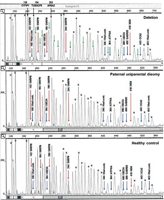
and happy grimacing), epileptic seizures of various                     cases) [1, 10]. In 90% of the cases the deletion spans
types (developed at 1-3 years of age), ataxia of gait                   the region between the breakpoins BP1-BP3 ot BP2-
and/or tremulous movement of the limbs, dysmorphic                      BP3, and rarely reaches BP4 or BP5 [11, 12]. 2) Pa-
facies (prominent chin, deep set eyes, wide mouth                       ternal uniparental disomy of chromosome 15, where-
with protruding tongue), microcephaly with a flat oc-                    in one inherits both chromosomes 15 from the father
ciput (by the age of 3), sleep disturbances (night time                 (in 1-3% of the cases) [1, 10]. 3) Imprinting center
awakenings, diminished total sleep time) [1, 2].                        defects resulting from small deletions or disruption of
AS results from loss of expression of the UBE3A gene                    DNA methylation in the imprinting center (in 3-6% of
(MIM 601623), which encodes human papillomavirus                        the cases) [13]. 4) UBE3A gene mutations (in 5-10%
E6-assocoated protein (Е6-АP) that functions as both                    of the cases) [14].
an ubiquitin-protein E3 ligase in the ubiquitin proteasome              The molecular-genetic diagnostics of AS requires ap-
pathway and as a transcriptional coactivator [3, 4].                    plication of different analytical methods [10, 15]. The
The gene UBE3A is located in 15q11.2-q13 chro-                          methylation pattern within 15q11-q13 region could be
mosomal region where several imprinted genes are                        assessed using methylation specific PCR (MS-PCR).
clustered (Fig. 1). Prader–Willi syndrome (OMIM                         Changes in the copy number of numerous genes
176270) is caused by loss of function of paternally                     within the target region could be detected by multi-
expressed genes, whereas Angelman syndrome                              plex ligation-dependent probe amplification (MLPA).
is caused by loss of function of the maternally ex-                     The simultaneous assessment of methylation status
pressed gene UBE3A in neuronal cells [5].                               and changes in copy numbers across the 15q11-q13
                                                                        region could be done by methylation sensitive multi-
Methylation and gene expression in the imprinted                        plex ligation-dependent probe amplification (MS-ML-
region is regulated in cis by a bipartite imprinting                    PA). Direct sequencing for pathogenic variants in the
centre located in the small nuclear ribonucleoprotein-                  UBE3A gene could be used as well.
associated protein N (SNRPN) upstream region [6].
Imprinted expression of UBE3A is regulated indirect-                    The aim of the current work was to confirm on molec-
ly by small nucleolar RNA host gene 14 (SNHG14,                         ular-genetic level the suspected clinical diagnosis AS
formerly known as UBE3AATS) whose product is a                          in Bulgarian patients in order to reveal the molecular
non-coding antisense transcript that is initiated at                    mechanisms of mutagenesis in our sample and to
the paternal SNRPN promoter. Normally in neuronal                       develop a systematic diagnostic algorithm for proper
cells the paternal region lacks methylation, resulting                  diagnosis, providing opportunities for adequate ge-
in transcription of SNHG14 and thus UBE3A gene is                       netic counseling and prophylaxis of the disease in the
not expressed. The maternal region is methylated,                       affected AS families.
SNHG14 is not expressed, resulting in UBE3A gene
transcription [7, 8].
                                                                                         MATERIAL AND METHODS
AS can result from various molecular mechanisms by
which UBE3A can be disrupted including: 1) Dele-                        In total 24 patients (11 boys, 13 girls) from 22 unrelat-
tions of the 15q11.2-q13 region (in 70%-75% of the                      ed families with suspected clinical diagnosis AS were

Fig. 1. Chromosomal region 15q11.2-q13. Paternally expressed genes are represented in blue, maternally expressed genes are marked
in red, genes expressed from both alleles are given in grey. Transcription orientation is pointed by with arrows. AS-IC is the Angelman syn-
drome imprinting center. PWS-IC is the Prader-Willi syndrome imprinting center. BP1 to BP5 are the deletion breakpoints. CH3 indicates
methylation [adapted from 9]

10                                                                                        B. Georgieva, S. Atemin, A. Todorova et al.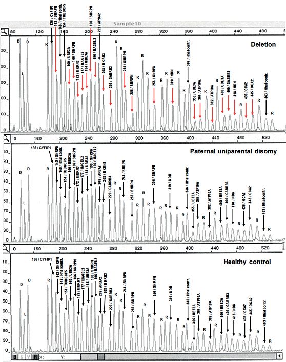
analysed, and where possible their parents were in- formed on ABI Prism 3130 Sequence Genetic An- cluded in the research as well. DNA samples were alyzer (Applied Biosystems) with polymer POP7 obtained from peripheral blood after written informed (Applied Biosystems). The electrophoretic data were consent was obtained. The study was approved by automatically analyzed with ABI3130 Data Collec- the Ethics Committee of Sofia Medical University. tion Software (Applied Biosystems, Foster City, CA) In all patients we accessed the methylation pattern for the direct sequencing or GeneMapper Software within the imprinted 15q11-q13 region by methylation v.4.0 (Applied Biosystems, Foster City, CA) for the specific PCR (MS-PCR). Two different primer pairs fragment analysis. specific for differentially methylated sites within the SNRPN exon 1/promoter regions were used, accord- RESULTS ing to the protocol of Kosaki et al. [16]. The DNA tem- plate for MS-PCR was chemically modified with so- In 9 of the analyzed families (41%; 9/22) genetic ab- dium bisulfite, resulting in conversion of cytosine, but normalities in the 15q11-q13 region were found, with not 5-methyl-cytosine, to uracil, which permits to dis- which the clinically suspected diagnosis of AS was tinguish successfully the methylated maternal from confirmed. the unmethylated paternal allele in the subsequent In 6 families the MS-PCR showed loss of the mater- PCR. For the sodium bisulfite treatment we used EZ nally methylated copy of the allele, but that test can’t DNA Methylation–Gold Kit (Zymo Research, USA) distinguish if AS is due to deletion of the 15q11-q13 following the instructions of the manufacturer. Normal region, paternal uniparental disomy or an imprinting individuals have unmethylated paternal and a meth- defect (Fig. 2, Lines 2, 3 and 5). To determine the ylated maternal allele. In AS patients with deletion of mechanisms of mutagenesis in these patients we 15q11-q13 region, paternal uniparental disomy or an proceeded further with MLPA and MS-MLPA. MS- imprinting defect, only a paternal unmethylated allele PCR cannot prove the diagnosis of AS in patients can be detected. with UBE3A gene mutation, because they have To access changes in the copy number in some pa- the same methylation profile as the normal control tients we used MLPA with SALSA MLPA kit P245- samples (Fig. 2, Lines 4 and 6). In cases of normal A2 Microdeletion-1 probemix with 49 MLPA probes MS-PCR result UBE3A gene sequencing was per- (MRC-Holland), which can detect 20 different micro- formed. deletion syndromes, including AS. MLPA and MS-MLPA results showed: 1) De novo To access simultaneously the methylation status and deletion of the maternally inherited 15q11.2-q13 re- changes in the copy number of some of the genes gion in 4 of the 9 genetically confirmed AS cases within the 15q11-q13 region we used SALSA MS- (44%) (Fig. 3 and Fig. 4). Of note, mothers were MLPA ME028-А1 PWS/AS probemix (MRC-Holland) not deletion carriers. 2) Paternal uniparental disomy with 43 MLPA probes or SALSA MS-MLPA ME028-В2 in 1 of the 9 confirmed AS cases (11%) (Fig. 3 and PWS/AS probemix (MRC-Holland) with 48 MLPA Fig. 4). The parental chromosomes were normal. 3) probes. The protocols for SALSA MLPA and SALSA Imprinting defect (deletion of the AS-SRO regulatory MS-MLPA kits were performed strictly following the region in AS/PWS imprinting center) in 1 out of the 9 instructions of the manufacturer. confirmed AS cases (11%) (data not shown), where In patients with normal methylation status and no the mother was a carrier of the mutation and was changes in the copy number within the 15q11-q13 symptoms free. region we proceeded with direct sequencing of the In the profile of the patient with deletion the red ar- UBE3A gene in order to screen for pathological vari- rows indicate the fragments with half of the intensity ants by BigDye Terminator v3.1 Cycle Sequencing compared to the normal sample, which shows dele- Kit (Applied Biosystems). The nucleotide changes tion of MGRN3, MAGEL2, NDN, SNRPN, UBE3A, in the UBE3A gene were described according to the ATP10A, GABRB3 and ОСА2 genes (TUBGCP5, coding DNA reference sequence of the longest iso- CYFIP1 and АРВА genes are not included in the de- form of the protein (UBE3A isoform II), where +1 nu- letion, because the corresponding fragments are with cleotide is the base adenine of the ATG start codon the same intensity compared to the normal sample). in the middle of exon 4 of the gene (https://databas- In the profile of the patient with paternal uniparen- es.lovd.nl/shared/refseq/UBE3A_NM_000462.3_ tal disomy all fragments are with the same intensity codingDNA.html). compared to the normal sample, which shows that The electrophoretic separation of the MLPA, MS- each of the genes in the 15q11-q13 region are pre- MLPA and direct sequencing fragments was per- sented in 2 allele copies. Molecular-genetic diagnostics of Angelman syndrome... 11
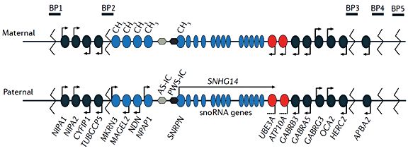
Fig. 2. Agarose gel electrophoresis of the fragments amplified by MS-PCR using two different primer pairs of primers in a multiplex reac-
tion. Line 1 – 100 bp. ladder, Line 2 – patient with 15q11.2-q13 deletion, Line 3 – patient with paternal uniparental disomy, Line 4 – patient
with UBE3A gene mutation, Line 5 – patient with imprinting defect, Line 6 – normal control sample. Normal individuals have unmethylated
paternal allele (164 bp.) and a methylated maternal allele (131 bp.). In AS patients with deletion of 15q11-q13, paternal uniparental disomy
or an imprinting defect only the paternal unmethylated allele (164 bp.) can be detected, while the maternal methylated allele is missing

Fig. 3. MLPA results of a patient with 15q11-q13 deletion with breakpoints BP2-BP3, patient with paternal uniparental disomy, and healthy
control sample with kit SALSA MS-MLPA ME028-А1. The length (in base pairs) of all specific fragments is provided with numbers. The
control fragments “D” (two denaturation controls), “L” (one ligation control), “R” (15 reference probes on 10 chromosomes) are marked with
letters. Black arrows indicate all fragments with normal intensity corresponding to 2 alleles (no deletion)

12                                                                                         B. Georgieva, S. Atemin, A. Todorova et al.
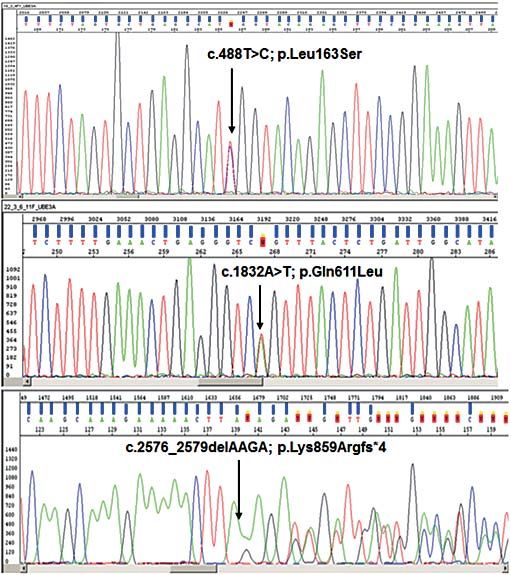
Fig. 4. MS-MLPA results of a patient with 15q11-q13 deletion with breakpoints BP2-BP3, patient with paternal uniparental disomy, and
healthy control sample with kit SALSA MS-MLPA ME028-А1. The length (in base pairs) of some of the fragments is provided in num-
bers. The control fragments “D” (two denaturation controls), “L” (one ligation control), “R” (15 reference probes on 10 chromosomes) are
marked by letters. Blue arrows indicate the eight fragments which obligatory are unmethylated, therefore cut by HhaI restrictase and the
corresponding peaks are missing. Red arrows indicate the five methylation-specific sensitive to HhaI digestion fragments in CpG islands
in SNRPN and NDN genes, which are missing in the patients either because of deletion of the maternal methylated copy of the 15q11-q13
region or because no maternal chromosomal 15q11-q13 15 region is inherited. In the profile of the patient with deletion the green arrows
indicate the fragments with reduced intensity compared to the normal sample (corresponding to deletion of MGRN3, MAGEL2, NDN, SN-
RPN, UBE3A, ATP10A, GABRB3 and ОСА2 genes), and with black arrows are marked the fragments with the same intensity compared
to the normal sample (meaning the genes TUBGCP5, CYFIP1 and АРВА are not included in the deletion). In the profile of the patient
with paternal uniparental disomy the fragments are with the same intensity compared to the normal sample, which shows that each of the
genes in the 15q11-q13 region is present in 2 alleles, but both of them are unmethylated and inherited from the father

UBE3A gene sequencing results showed three                            carrier and had no clinical symptoms, while in the
different point mutations in 3 of the 9 geneti-                        rest two families the mutations were de novo.
cally confirmed AS cases (33%) – two previ-
ously unpublished missense mutations (c. 488T                                                  DISCUSSION
> C; p.Leu163Ser in exon 7 and c.1832А > Т;
p.Gln611Leu in exon 9), and one known frameshift                      The current study revealed that all known mecha-
mutation c.2576_2579delAAGА; p.Lys859Argfs*4                          nisms of mutagenesis (four types of mechanisms) re-
in exon 14 (Fig. 5). In one of the families with                      sulting in AS were seen among 9 Bulgarian patients
UBE3A gene mutation the mother was mutation                           with AS.

Molecular-genetic diagnostics of Angelman syndrome...                                                                               13
Fig. 5. Sequencing results of the UBE3A gene mutations c.488T > C; p.Leu163Ser in exon 7, c.1832А > Т; p.Gln611Leu in exon 9, and
c.2576_2579delAAGА; p.Lys859Argfs*4 in exon 14 in three AS cases

Fig. 6. Location of the Bulgarian point mutations along the UBE3A gene. The UBE3A gene exons are schematically represented by
numbered rectangles – 1 to 14. The translation of the longest isoform of the protein (UBE3A isoform II composed of 875 amino acid resi-
dues) is initiated in exon 4 (the ATG starting codon). The stop codon (TAA) in exon 14 is marked. Part of exon 7 to exon 14 encodes the
conservative catalytic HECT (homologous to the E6-AP carboxyl terminus) ubiquitine ligase domain which constitutes the last 350 amino
acid residues of the protein. The three pathological variants found in our patients are marked with arrows above the affected exon. The
two novel missense mutations c.488T > C; p.Leu163Ser in exon 7 and c.1832А > Т; p.Gln611Leu in exon 9 are marked with asterisks

14                                                                                     B. Georgieva, S. Atemin, A. Todorova et al.
The most common mechanism of mutagenesis in acid residues), a large number of small deletions and
our sample was deletion of the maternally inherited insertions were reported there [17].
15q11.2-q13 region – 44%, which is much lower Adequate genetic counseling requires proper assess-
than the percent published in the literature (70-75% ment of the recurrence risk after molecular-genetic
of the cases) [1, 10]. The deletions described in our testing of all family members. In cases with uniparen-
study fall between the most common breakpoints tal disomy, when parental chromosomes are normal,
BP1-BP3 or BP2-BP3, and none of them spans to the recurrence risk is very low (< 1%). In cases with
BP4 or BP5, which corresponds to the published de novo mutation (deletion, imprinting center muta-
data [11, 12]. tion, or UBE3A gene mutation) the recurrence risk is
The imprinting center defect in our AS patients was very low (< 1%), but germ line mosaicism should be
detected once, but having in mind our cohort was considered (about 10%). In cases when the mother is
very small, this presumably resulted in a falsely high a carrier of a UBE3A gene mutation, there is a 50%
percent 11%, which is twice as common compared recurrence risk, that is a 50% risk that the mother
to the literature (3-6%) [13]. The detected mutation could have another child with AS.
belongs to the most common type of the imprinting
center defects microdeletion of the AS-SRO regula- CONCLUSION
tory region in AS/PWS imprinting center resulting in
loss of methylation in the SNRPN exon 1/promoter Our results helped us to develop a systematic di-
regions and inhibition of the UBE3A gene transcrip- agnostic algorithm for genetic evaluation of patients
tion [13]. with AS in Bulgaria. Combining excellent knowledge
We had the unique chance to find paternal unipa- of the molecular mechanisms of mutagenesis and
rental disomy of chromosome 15 where the patient proper molecular-genetic testing approaches is
does not inherit UBE3A gene from the mother (one a cornerstone in the management of AS patients,
patient), which is considered to be the most rare ensuring AS families would receive both adequate
mechanism of mutagenesis in AS (1-3% of the cas- genetic counseling and prophylaxis of the disease
es) [1, 10]. in the future.
The percentage of UBE3A gene point mutations in Conflict of interests: The authors declare no conflict of
our AS patients (33%) was more than three times interests.
higher than that published in the literature (5-10%) Acknowledgements: Part of the work was supported by
[14]. Our finding of two novel missense mutations grant No D-56/03.05.2018, Medical University of Sofia.
(c.488T > C; p.Leu163Ser in exon 7 and c.1832А >
Disclosure Summary: The authors have nothing to disclose.
Т; p.Gln611Leu in exon 9) enriches the spectrum of
missense mutation in AS. According to the published
data, most of the mutations are insertions, deletions REFERENCES
and nonsense mutations, while missense mutations
1. Clayton-Smith J, Laan L. Angelman syndrome: a review of the
are less common [14, 17].
clinical and genetic aspects. J Med Genet. 2003; 40: 87-95.
The novel missense mutations c.488T > C; 2. Williams CA, Beaudet AL, Clayton-Smith J et al. Angelman
p.Leu163Ser in exon 7 is not a surprising finding, syndrome 2005: updated consensus for diagnostic criteria.
considering the large size of that exon (encodes 415 Am J Med Genet A. 2006; 140(5):413-418.
out of totally 875 amino acid residues) where about 3. Huibregtse JM, Scheffner M, Howley PM. Cloning and ex-
pression of the cDNA for E6-AP, a protein that mediates the
half of all UBE3A gene mutations are reported.
interaction of the human papillomavirus E6 oncoprotein with
Our novel missense mutation in exon 9, togeth- p53. Molec Cell Biol. 1993; 13:775-784.
er with the already published frameshift mutation 4. Kishino T, Lalande M, Wagstaff J. UBE3A/E6AP mutations
c.2576_2579delAAGА; p.Lys859Argfs*4 in exon 14, cause Angelman syndrome. Nat Genet. 1997;15:70-73.
which we also detected in one patient, fall in the high- 5. Vu T H, Hoffman AR. Imprinting of the Angelman syndrome
gene, UBE3A, is restricted to brain. Nat. Genet. 1997;
ly conservative catalytic HECT (homologous to the
17(1):12-13.
E6-AP carboxyl terminus) ubiquitine ligase domain of
6. Sutcliffe JS, Nakao M, Christian S et al. Deletions of a
the protein, probably resulting in severe impairment differentially methylated CpG island at the SNRPN gene
of the protein function (Fig. 6). Although exon 14 is define a putative imprinting control region. Nat. Genet.
10 times smaller than exon 7 (encodes only 39 amino 1994;8(1):52-58.
Molecular-genetic diagnostics of Angelman syndrome... 157. Rougeulle C, Cardoso C, Fontés M et al. An imprinted an- 13. Bürger J, Buiting K, Dittrich B et al. Different mechanisms and
tisense RNA overlaps UBE3A and a second maternally ex- recurrence risks of imprinting defects in Angelman syndrome.
pressed transcript. Nat Genet. 1998;19(1):15-16. Am J Hum Genet. 1997;61(1):88-93.
8. LaSalle JM, Reiter LT, Chamberlain SJ. Epigenetic regulation 14. Sadikovic B, Fernandes P, Zhang VW et al. Mutation Up-
of UBE3A and roles in human neurodevelopmental disorders. date for UBE3A variants in Angelman syndrome. Hum Mutat.
Epigenomics. 2015; 7(7):1213-1228. 2014;35(12):1407-1417.
9. Buiting K, Williams C, Horsthemke B. Angelman syndrome 15. Ramsden SC, Clayton-Smith J, Birch R et al. Practice guide-
– insights into a rare neurogenetic disorder. Nat Rev Neu- lines for the molecular analysis of Prader-Willi and Angelman
rol. 2016;12(10):584-593. syndromes. BMC Med Genet. 2010;11:70.
10. Buiting K, Clayton-Smith J, Driscoll DJ et al. Clinical utility 16. Kosaki K, McGinniss MJ, Veraksa AN et al. Prader-Willi
gene card for: Angelman Syndrome. Eur J Hum Genet. 2015; and Angelman syndromes: diagnosis with a bisulfite-treated
23(2). doi: 10.1038/ejhg.2014.93. methylation-specific PCR method. Am J Med Genet. 1997;
11. Amos-Landgraf JM, Ji Y, GottliebW et al. Chromosome 73(3):308-313.
breakage in the Prader-Willi andAngelman syndromes in- 17. Camprubí C, Guitart M, Gabau E et al. Novel UBE3A
volves recombination between large, transcribed repeats mutations causing Angelman syndrome: different pa-
at proximal and distal breakpoints. Am J Hum Genet. 1999; rental origin for single nucleotide changes and multiple
65(2):370-386. nucleotide deletions or insertions. Am J Med Genet A.
12. Christian SL, Fantes JA, Mewborn SK et al. Large genomic 2009;149A(3):343-348.
duplicons map to sites of instability in the Prader-Willi/Angel-
man syndrome chromosome region (15q11-q13). Hum Mol
Genet. 1999; 8(6):1025-1037. Received: May, 2019 – Accepted: June, 2019
16 B. Georgieva, S. Atemin, A. Todorova et al.You can also read