Mytilus spp. experiencing mortalities in France
←
→
Page content transcription
If your browser does not render page correctly, please read the page content below
Vol. 140: 203–208, 2020                  DISEASES OF AQUATIC ORGANISMS
                                                                                                               Published August 20
 https://doi.org/10.3354/dao03505                      Dis Aquat Org

NOTE

 First detection of Francisella halioticida in mussels
   Mytilus spp. experiencing mortalities in France
         Maud Charles1, 2,*, Antonio Villalba3, 4, 5, Gary Meyer6, Suzanne Trancart2,
                 Coralie Lagy2, Ismaël Bernard7, Maryline Houssin1, 2
     1
      Normandie Université, Université de Caen Normandie, FRE BOREA, CNRS-2030, IRD-207, MNHN, UPMC, UCN,
                                     Esplanade de la Paix, 14032 Caen Cedex 4, France
                         2
                          LABÉO Frank Duncombe, 1 Route de Rosel, 14053 Caen Cedex 4, France
    3
     Centro de Investigacións Mariñas (CIMA), Consellería do Mar, Xunta de Galicia, 36620 Vilanova de Arousa, Spain
               4
                 Departamento de Ciencias de la Vida, Universidad de Alcalá, 28871 Alcalá de Henares, Spain
5
 Research Centre for Experimental Marine Biology and Biotechnology (PIE), University of the Basque Country (UPV/EHU),
                                           48620 Plentzia, Basque Country, Spain
  6
   Fisheries and Oceans Canada, Pacific Biological Station, Aquatic Animal Health Laboratory, 3190 Hammond Bay Road,
                                               Nanaimo, BC V9T 6N7, Canada
                           7
                             Eurêka Mer, 13 Cité des gardiens de phare, 22740 Lézardrieux, France

         ABSTRACT: This note describes the first detection of the bacteria Francisella halioticida in mus-
         sels Mytilus spp. from locations in Normandy and northern Brittany (France) experiencing high
         mussel mortalities, while it was not detected in the Bay of St Brieuc (northern Brittany), an area
         which was not affected by abnormal mussel mortality. The distribution of the bacteria in mussels
         seems to be restricted to inflammatory granulomas as observed in Yesso scallops Mizuhopecten
         yessoensis from Canada and Japan. F. halioticida has been identified as being involved in mass
         (> 80%) mortality of abalones Haliotis gigantea in Japan and high (up to 40%) mortality of Yesso
         scallops Mizuhopecten yessoensis in Canada as well as in lesions reducing marketability of Yesso
         scallops in Japan. The impact of this bacterium on the health of mussels needs to be investigated
         in future research, especially since the cause of high mussel mortalities that have been occurring
         in France for the past few years is still undetermined.

         KEY WORDS: Francisella halioticida · Mussels · Granulocytomas · Polymerase chain reaction ·
         In situ hybridisation
                              Resale or republication not permitted without written consent of the publisher

                  1. INTRODUCTION                                       al. 2018). In addition, Kawahara et al. (2019) demon-
                                                                        strated that this bacterium can be highly virulent in
  The Gram-negative bacteria Francisella halioticida                    Yesso scallops. Thus, F. halioticida has shown patho-
was originally described in farmed abalone Haliotis                     genic potential in both gastropod and bivalve species
gigantea as the cause of mass mortality that occurred                   of molluscs.
in Japan (Kamaishi et al. 2010, Brevik et al. 2011).                      Since 2014, unexplained mass mortalities of mus-
Subsequently, this bacterium was identified in dis-                      sels (Mytilus edulis, M. galloprovincialis and their
eased Yesso scallops, Mizuhopecten (=Patinopecten)                      hybrids) have occurred in France from the Atlantic to
yessoensis, in conjunction with mortality events in                     the English Channel coasts (Béchemin et al. 2014,
Canada (Meyer et al. 2017) and Japan (Kawahara et                       2015, Allain & Bernard 2016, Travers et al. 2016,

*Corresponding author: maud.charles@outlook.fr                          © Inter-Research and Fisheries and Oceans Canada 2020 ·
                                                                        www.int-res.com
204                                       Dis Aquat Org 140: 203–208, 2020

Bernard & Allain 2017), with mortality rates ranging        2018 by analysing mussels for obligatory reporting
from 40 to 70% (Bernard & Allain 2017, Bernard et al.       of increasing mortality (Council Directive 2006/88/
2018, Charles et al. 2020). Several hypotheses have         EC, https://eur-lex.europa.eu/legal-content/EN/TXT/
been proposed to explain these mortalities, including       HTML/?uri=CELEX:32006L0088&from=EN) in Nor-
the involvement of pathogenic bacteria belonging to         mandy and the Hauts-de-France region (Charles
the Vibrio splendidus clade (Béchemin et al. 2014,          2019); in this second study, the sampling agents (from
2015, François et al. 2015, Soletchnik & Robert 2016,       the French public administration and/or the Regional
Travers et al. 2016), the possibility of genomic abnor-     Shellfish Farming Committee) collected mostly mori-
malities and of disseminated neoplasia (Benabdel-           bund mussels, and the information on mortality was
mouna & Ledu 2016, Benabdelmouna et al. 2018).              obtained from Administration reports (Canier et al.
Nevertheless, none of these proposals alone could           2018, Combette 2018). In the 2 previous studies, mus-
explain such high mortality rates (Charles 2019).           sels were analysed using histological, bacteriological
High mortality rates were recently observed among           and molecular diagnostic procedures, and although a
mussels in northern Brittany, and although V. splen-        number of pathogens and pathological conditions
didus strains and disseminated neoplasia were de-           were detected, none of them were believed to be the
tected, the prevalence in mortality-affected versus         primary cause of high mussel mortality in northern
non-affected areas did not correlate with mortality,        France (Charles 2019, Charles et al. 2020).
and no causative agent was identified (Bernard et al.          The 17 mussel groups analysed in the current study
2018, Charles et al. 2020).                                 are listed in Table 1 together with some data derived
   In a multi-parametric field study to identify poten-      from the 2 previous studies, namely the sampling
tial causes of the high mortality of mussels, Charles et    place, the corresponding farming method, the cumu-
al. (2020) reported the occurrence of heavy haemo-          lative mortality at sampling and the condition of mus-
cyte infiltration and the formation of granulomatous         sels in the samples (either alive or moribund). It is
lesions in the connective tissue of various organs.         noteworthy that the mussels collected during the first
However, those authors were not able to identify an         previous study (Charles et al. 2020), designed in
aetiological agent. Similarities between the histolog-      advance to monitor mortality, were alive (valves
ical observations in mussels and the histopathology         remained closed when out of seawater), while the
reported in Yesso scallops infected with F. halioticida     mussels collected during the second previous study
(Meyer et al. 2017, Kawahara et al. 2018) prompted          (Charles 2019), based on sampling immediately after
the current investigation to test for the presence of       knowledge of mortality events, were primarily mori-
F. halioticida in French mussel samples from previ-         bund specimens (valves were gaping and non-respon-
ous studies.                                                sive) or a mixture of live and moribund specimens.
   To the best of our knowledge, F. halioticida has         Two molecular diagnostic procedures were used tar-
never been reported in mussels. The current study re-       geting F. halioticida DNA, PCR and ISH. Information
veals the presence of F. halioticida, identified by poly-    on how the mussels were processed is described by
merase chain reaction (PCR) and by in situ hybridisa-       Charles (2019) and Charles et al. (2020). Briefly, for
tion (ISH), in cultured French mussels experiencing         histological analysis, an approximately 5 mm thick
mortalities.                                                transverse section of mussel tissue containing mantle
                                                            lobes, visceral mass (gut, digestive gland) and gills
                                                            was excised, fixed in Davidson’s solution for 48 h, kept
          2. MATERIALS AND METHODS                          in 70% ethanol and then dehydrated through an as-
                                                            cending ethanol series and embedded in paraffin wax.
   The 341 mussels analysed for detection of Franci-        From each mussel a pool of minced tissues from foot,
sella halioticida in the current study came from 2          adductor muscle, gills, mantle, and digestive gland
overlapping multi-parametric studies in France: the         was preserved in a microcentrifuge tube at −20°C.
first was conducted in 2017 with mussels taken from 3          A total of 332 mussels were used for PCR assays, 20
sites in northern Brittany (Charles et al. 2020). Two       mussels per group except for Groups 11 and 15 in
sites (Brest and Lannion) were affected by high             which 16 mussels were used. DNA was extracted
mussel mortality while the third site (St Brieuc) did       from preserved tissues of individual sample mussels,
not experience mortalities. In this first study, mussel      using a QIAamp DNA minikit® (Qiagen) following
seed batches were periodically checked for mortality        the manufacturer’s protocol for blood or body fluids
and sampled (live mussels randomly taken) for diag-         (except elution was performed using 60 μl Qiagen
nosis. The second study was conducted in 2017 and           elution buffer AE). Following extraction, the DNA
Charles et al.: Francisella halioticida in mussels in France 205
Table 1. Summary of mussel Mytilus spp. samples collected from France and test results for Francisella halioticida detection by
conventional polymerase chain reaction (PCR) and in situ hybridisation (ISH) assays. Each pool in the PCR assays (4 pools per
group) resulted from pooling DNA from 5 mussels except in Groups 11 and 15 in which DNA from 4 mussels was pooled (PCR
results = positive pools/total pools). Individual mussels assayed by PCR were different from those assayed by ISH (ISH results =
positive mussels/total mussels analysed). Bouchot: intertidal mussel stakes; Mix: mixture of live and moribund mussels;
Cum. mort.: cumulative mortality; na: missing data; np: not performed. Bold is used to highlight positive results
Group Site Region Sampling Farming Cum. mort. Mussel PCR ISH
date (d/mo/yr) method at sampling (%) condition results results
1 Lannion Brittany 24/02/2017 Longlines 5a,b Alive 0/4 0/1
2 Brest Brittany 27/02/2017 Bouchot 12.5a,b Alive 0/4 np
3 St Brieuc Brittany 27/02/2017 Bouchot 2.6a,b Alive 0/4 np
4 Brest Brittany 24/04/2017 Bouchot 45a,b Alive 0/4 0/1
5 Lannion Brittany 17/05/2017 Longlines 20a,b Alive 1/4 2/4
6 St Brieuc Brittany 28/05/2017 Bouchot 4.5a,b Alive 0/4 0/2
7 Brest Brittany 30/05/2017 Bouchot 56.5a,b Alive 2/4 0/1
8 Brest Brittany 20/09/2017 Bouchot 66a,b Alive 0/4 np
9 Oye-Plage Hauts-de-France 20/03/2017 Bouchot 25c Moribund 3/4 np
10 Oye-Plage Hauts-de-France 24/10/2017 Bouchot 10−50c Moribund 4/4 np
11 Tardinghen Hauts-de-France 14/03/2018 Bouchot 10−50d Moribund 3/4 np
12 Donville-les-Bains Normandy 19/06/2018 Bouchot 50d Mix 2/4 np
13 Agon-Coutainville Normandy 16/07/2018 Bouchot nad Mix 2/4 np
14 Chausey Normandy 25/09/2018 Bouchot 10−50d Mix 0/4 np
15 Wimereux Hauts-de-France 15/10/2018 Wild nad Moribund 4/4 np
16 Tardinghen Hauts-de-France 13/10/2018 Bouchot nad Alive 2/4 np
17 Dannes Hauts-de-France 26/11/2018 Bouchot 25d Moribund 4/4 np
a
Bernard et al. (2018); bCharles et al. (2020); cCanier et al. (2018); dCombette (2018)
extracts of 5 mussels were pooled, except in Groups with heavy haemocyte infiltration in the visceral mass
11 and 15 in which DNA pools derived from 4 mus- (from Groups 1 and 5), 2 mussels with granulomas
sels. Thus, 4 DNA pools were produced for each (from Group 5), 1 mussel with massive haemocyte
mussel group. PCR assays were conducted using the diapedesis through stomach epithelium (from Group
primer pair Fh-rpoB/F and Fh-rpoB/R (Brevik et al. 4) and 1 mussel with abundant degenerated digestive
2011) designed to amplify the gene corresponding to gland tubules filled with necrotic cells (from Group
bacterial DNA-directed RNA polymerase beta sub- 6). Samples analysed by ISH corresponded to mussels
unit (rpoB). For the conventional PCR, typical 25 μl different from those analysed by PCR. The ISH assays
reaction mixtures contained 12.5 μl of Premix Ex were performed as described by Meyer et al. (2017)
Taq® 2× Takara® (Lonza), 0.5 μl of each primer except that a 1% Light Green counterstain was sub-
(20 μM), 9.5 μl of purified water and 2 μl of DNA tem- stituted for Bismark Brown to yield better contrast.
plate (replaced with 2 μl of purified water in the neg-
ative control). PCR amplifications were carried out in
a T100™ Thermal Cycler (Bio-Rad) using the follow- 3. RESULTS
ing thermal cycling conditions: 10 s at 95°C; 30 cycles
for 10 s at 95°C, 30 s at 55°C, 40 s at 72°C; and a final The PCR results as well as the ISH results for
extension of 3 min at 72°C. All PCR products were detection of Francisella halioticida DNA in French
purified and sequenced at LABÉO Frank Duncombe Mytilus spp. are summarized in Table 1. BLAST re-
(Caen Cedex) and then analysed using the National sults of all the PCR amplified fragments were a
Center for Biotechnology Information (NCBI) Basic 99.9% match to the reference rpoB sequences for F.
Local Alignment Search Tool (BLAST). halioticida (Brevik et al. 2011) (GenBank accession
For ISH assays, only mussels from the first study numbers JF290381 and JF290374). In the first study
(Charles et al. 2020) could be retrospectively ana- (Charles et al. 2020) conducted in northern Brittany
lysed; 9 archived paraffin blocks of mussels showing during 2017 (Groups 1 to 8), F. halioticida DNA was
different histological characteristics were selected: 3 detected by PCR in mussels collected in May at Lan-
healthy mussels (from Groups 5, 6 and 7), 2 mussels nion and Brest, which had mortality rates of 20 and206 Dis Aquat Org 140: 203–208, 2020
56.5%, respectively, after 3 mo of culture, but it was Although ISH assays were not conducted on all
not detected in samples from St Brieuc, an area that groups, positive reactions were observed within gra-
was not affected by abnormal mortality (only 4.5%). nuloma lesions from 2 specimens (Fig. 1). These le-
In the groups collected following reports of abnormal sions were characterized by an intense accumulation
mortality in Normandy and the Hauts-de-France of granulocytes surrounded by layers of flattened,
region in 2017 and 2018 (Groups 9 to 17), PCR-posi- epithelioid cells (Figs. 1A & 2A), and every tested
tive results were obtained from every group except mussel with granulomas showed a positive reaction.
the one coming from Chausey’s Island. Other types of pathology, such as heavy haemocyte
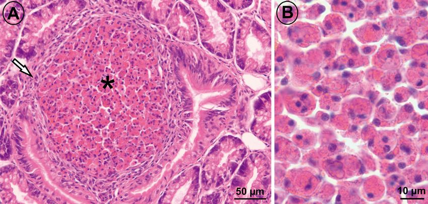
Fig. 1. In situ hybridisation (ISH) assay showing positive reaction (dark blue deposits). (A) Section through the digestive gland
*
of a mussel Mytilus sp. showing a granuloma ( ) with tiny dark blue deposits within them. (B) Higher magnification of the in
side of a granuloma showing dark blue deposits (arrows) indicative of positive ISH reaction within cells
Fig. 2. Micrographs of a histological section consecutive to that of the Fig. 1, stained with Harris’ haematoxylin and eosin. (A)
Large granuloma in the connective tissue of the digestive gland consisting of a mass of accumulated granulocytes ( ) sur-
rounded by layers of flattened, epithelioid cells (arrow). (B) Higher magnification of the inside of the granuloma showing
*
numerous granulocytes with phagocytosed cellsCharles et al.: Francisella halioticida in mussels in France 207
infiltration and diapedesis, were negative via ISH; 2016, Travers et al. 2016, Pépin et al. 2017, 2018,
furthermore, mussels with no significant histopatho- Charles et al. 2020). In fact, some of those authors
logy were also negative via ISH. Thus, infection with have even suggested a link between the presence of
F. halioticida appears to be associated with the host’s the granuloma lesions and mortality. In addition,
response and formation of granulomas (Fig. 2), which since 2014, mussels have not been the only molluscs
is common in vertebrates infected with other Francis- in France to be affected by unexplained mortalities;
ella species (Colquhoun & Duodu 2011). abalone (Haliotis tuberculata), cockles (Cerastoderma
edule), clams (Ruditapes philippinarum) and scallops
(Pecten maximus, Chlamys varia) have also been
4. DISCUSSION affected (François et al. 2015, Lupo et al. 2016, Osta
Amigo et al. 2018).
The occurrence of Francisella halioticida in French Considering the contagious and virulent nature of
areas affected by high mussel mortality was re- the Francisella spp. bacteria, and particularly F.
vealed, although it was not detected in 2 areas: St halioticida in scallops, the detection of this bacterial
Brieuc, which was not affected by abnormal mussel species in French mussels opens up a new path for
mortality, and Chausey’s Island, where mussels are further research into the cause of high mussel mor-
known to suffer from intense predation by ducks, talities that have been occurring in northern France
gulls and spider crabs, which were most likely con- for the past few years. A field approach combined
tributing to the mortalities (CRC Normandie-Mer du with laboratory challenges assessing the Koch’s clas-
Nord 2017 pers. comm. 2019). The findings from this sic criteria would make a contribution towards clari-
study support a hypothesis proposed by Charles et al. fying the role that F. halioticida plays in high levels of
(2020) concerning the possibility of missing an uncul- mussel mortality in northern France.
tivable bacterial pathogen with the classical medium
used for bacterial culture. This could also be the case
Data availability. The authors confirm that one part of the
of the network for the monitoring of mollusc patholo- data supporting the findings of this study is available within
gies (REPAMO), coordinated by the French institute the article and the other part is available on request from the
Ifremer, which monitors reports of mussel mortalities corresponding author.
and conducts bacteriology using traditional culture
Acknowledgements. The authors thank the Regional Shell-
media. Isolation and culture of F. halioticida requires
fish Committee of North Brittany for supporting sampling
a special culture medium (Modified Eugon Agar), in northern Brittany and the local mussel farmers for mak-
and it can take several weeks for the bacteria to grow ing this possible. The Regional Shellfish Committee of Nor-
(Kamaishi et al. 2010, Kawahara et al. 2018). The lim- mandy/North Sea (CRC Normandie/Mer du Nord) and the
Laboratory LABÉO financially supported this study. M.C.
ited sample size of this study prevents us from giving
received co-funding from the Normandy region and from
conclusive results; nevertheless, given that F. halioti- the laboratory LABÉO Frank Duncombe.
cida is known to be highly pathogenic in abalone and
Yesso scallops, its detection in mussels from Nor-
mandy and the northern Brittany areas affected by LITERATURE CITED
high mussel mortality highlights the need for further Allain G, Bernard I (2016) Les mortalités de moules en 2014
research concerning the association between mussel et 2015 vues par les professionnels. Compte-rendu de la
mortalities and infection with F. halioticida. There is phase 1: synthèse sur l’émergence, la propagation et
no geographical connection between the French l’installation des mortalités — Rapport technique. CRC
Bretagne Nord, Morlaix
mussel culture areas with the locations where F.
Béchemin C, Soletchnik P, Polsenaere P, Le Moine O and
halioicida was previously found, Canada and Japan, others (2014) Surmortalités de la moule bleue Mytilus
but the importation of M. yessoensis at the beginning edulis dans les Pertuis Charentais (mars 2014) — Rapport
of 1987 to a hatchery in Brittany for several aquacul- d’expertise. Ifremer. http://archimer.ifremer.fr/doc/00229/
ture trials (Buestel et al. 1988) could be considered as 34022/32387.pdf
Béchemin C, Soletchnik P, Polsenaere P, Le Moine O and
a potential hypothetical connection. It would be others (2015) Épisodes de mortalité massive de moules
interesting to retrospectively test for the presence of bleues observés en 2014 dans les Pertuis charentais. Bull
F. halioticida DNA in samples of past mortalities, Épid Santé Anim Alim 67:6−9
given that since 2015 several studies highlighted the Benabdelmouna A, Ledu C (2016) The mass mortality of
blue mussels (Mytilus spp.) from the Atlantic coast of
presence of numerous inflammatory granulomas in France is associated with heavy genomic abnormalities
French mussels affected by mortalities in the Atlantic as evidenced by flow cytometry. J Invertebr Pathol 138:
coasts without identifying any cause (Robert et al. 30−38208 Dis Aquat Org 140: 203–208, 2020 Benabdelmouna A, Saunier A, Ledu C, Travers MA, Morga B able cause of adductor muscle lesions in Yesso scallops (2018) Genomic abnormalities affecting mussels (Mytilus Patinopecten yessoensis cultured in southern Hokkaido, edulis-galloprovincialis) in France are related to ongoing Japan. Fish Pathol 53:78−85 neoplastic processes, evidenced by dual flow cytometry Kawahara M, Meyer GR, Lowe GJ, Kim E, Polinski MP, and cell monolayer analyses. J Invertebr Pathol 157:45−52 Yoshinaga T, Itoh N (2019) Parallel studies confirm Bernard I, Allain G (2017) Mortalités des moules en Bre- Francisella halioticida causes mortality in Yesso scallops tagne nord: bilan des connaissances — Rapport tech- Patinopecten yessoensis. Dis Aquat Org 135:127−134 nique. CRC Bretagne Nord, Morlaix Lupo C, Osta Amigo A, Fleury E, Robert S and others (2016) Bernard I, Charles M, Allain G, Burioli EAV and others Bilan 2015 du dispositif national de surveillance de la (2018) Bilan de l’observatoire des mortalités de moules santé des mollusques marins. Ifremer, La Tremblade. en Bretagne Nord pour la saison 2016−2017 et premiers https://archimer.ifremer.fr/doc/00324/43486/ éléments sur les organismes pathogènes présents — Meyer GR, Lowe GJ, Gilmore SR, Bower SM (2017) Disease Rapport technique. CRC Bretagne Nord, Morlaix and mortality among Yesso scallops Patinopecten yesso- Brevik ØJ, Ottem KF, Kamaishi T, Watanabe K, Nylund A ensis putatively caused by infection with Francisella halio- (2011) Francisella halioticida sp. nov., a pathogen of ticida. Dis Aquat Org 125:79−84 farmed giant abalone (Haliotis gigantea) in Japan. J Appl Normandie-Mer du Nord CRC (2017) Demande de déroga- Microbiol 111:1044−1056 tion pour la perturbation intentionnelle de goélands Buestel D, Comps M, Moriceau J, Paquotte P (1988) Pro- argentés sur l’archipel des îles Chausey. www.normandie. gramme de recherche pour la mise au point d’une méth- developpement-durable.gouv.fr/IMG/pdf/dossier_dde_ ode d’élevage des coquilles Saint-Jacques en Méditer- autorisation_chausey_2017.pdf ranée — Rapport d’avancement des travaux. Résultats Osta Amigo A, Robert S, Fleury E, Lupo C, Garcia C, Gea- obtenus en 1987. Ifremer, Sète iron P, Canier L (2018) Repamo 2016 — bulletins de la Canier L, Fleury E, Normand J, Garcia C, Geairon P (2018) surveillance janvier à juin et décembre 2016. Ifremer, La REPAMO 2017 — Bulletins de la surveillance — Janvier à Tremblade. https://archimer.ifremer.fr/doc/00480/59210/ Décembre 2017. Ifremer, La Tremblade. https://archimer. Pépin JF, Benabdelmouna A, Degremont L, Guesdon S and ifremer.fr/doc/00480/59184/ others (2017) Mortalités de moules bleues dans les Charles M (2019) Étude des organismes pathogènes, des secteurs mytilicoles charentais et vendéens: description conditions physiologiques et pathologiques impliqués et facteurs liés — MORBLEU. Ifremer. http://archimer. dans les mortalités anormales de moules (Mytilus sp.). ifremer.fr/doc/00391/50288/ PhD dissertation, University of Caen Normandy, Caen Pépin JF, Benabdelmouna A, Bierne N, Bouget JF and oth- Charles M, Bernard I, Villalba A, Oden E and others (2020) ers (2018) Mortalités de moules bleues dans les secteurs High mortality of mussels in northern Brittany — evalua- mytilicoles: description et facteurs liés — MORBLEU. tion of the involvement of pathogens, pathological condi- Ifremer, La Tremblade. https://archimer.ifremer.fr/doc/ tions and pollutants. J Invertebr Pathol 170:107308 00503/61503/ Colquhoun DJ, Duodu S (2011) Francisella infections in Robert S, Bouget JF, Gabellec R, Louis W and others (2016) farmed and wild aquatic organisms. Vet Res 42:47 Réseau national d’observation de la moule bleue Mytilus Combette A (2018) REPAMO 2018 — Bulletins de la surveil- edulis, Mytilobs, campagne 2015. Ifremer, La Tremblade. lance — Janvier à Décembre 2018. Ifremer, La Trem- https://www.researchgate.net/publication/306544440_ blade. https://archimer.ifremer.fr/doc/00473/58454/ Reseau_national_d%27observation_de_la_moule_bleue François C, Garcia C, Lupo C, Travers MA and others (2015) _Mytilus_edulis_MYTILOBS_Campagne_2015 Bilan 2014 du réseau Repamo — Réseau national de sur- Soletchnik P, Robert S (2016) Éléments de connaissance sur veillance de la santé des mollusques marins. Ifremer. la mortalité et la reproduction de la moule bleue (Mytilus http://archimer.ifremer.fr/doc/00256/36691/ edulis) sur la façade atlantique. Ifremer. https://ar- Kamaishi T, Miwa S, Goto E, Matsuyama T, Oseko N (2010) chimer.ifremer.fr/doc/00345/45634/45263.pdf Mass mortality of giant abalone Haliotis gigantea caused Travers MA, Pépin JF, Soletchnik P, Guesdon S, Le Moine O by a Francisella sp. bacterium. Dis Aquat Org 89:145−154 (2016) Mortalités de moules bleues dans les Pertuis Cha- Kawahara M, Kanamori M, Meyer G, Yoshinaga T, Itoh N rentais — MORBLEU. Ifremer. http://archimer.ifremer.fr/ (2018) Francisella halioticida, identified as the most prob- doc/00324/43539/ Editorial responsibility: Kimberly Reece, Submitted: March 12, 2020; Accepted: June 29, 2020 Gloucester Point, Virginia, USA Proofs received from author(s): August 6, 2020
You can also read