Geographical Distribution of Borrelia burgdorferi sensu lato in Ticks Collected from Wild Rodents in the Republic of Korea - MDPI
←
→
Page content transcription
If your browser does not render page correctly, please read the page content below
pathogens
Article
Geographical Distribution of Borrelia burgdorferi
sensu lato in Ticks Collected from Wild Rodents in
the Republic of Korea
Seong Yoon Kim , Tae-Kyu Kim, Tae Yun Kim and Hee Il Lee *
 Division of Vectors and Parasitic Diseases, Bureau of Infectious Disease Diagnosis Control, Korea Disease
 Control and Prevention Agency, 187 Osongsaengmyeong 2-ro, Cheongwon-gun, Cheongju-si 363-951,
 Chungcheongbuk-do 28159, Korea; gunbo0402@korea.kr (S.Y.K.); tkkim80@korea.kr (T.-K.K.);
 kty4588@korea.kr (T.Y.K.)
 * Correspondence: isak@korea.kr; Tel.: +82-43-719-8560; Fax: 82-43-719-8589

 Received: 21 September 2020; Accepted: 22 October 2020; Published: 22 October 2020

 Abstract: Lyme disease is a tick-borne zoonotic disease caused by Borrelia burgdorferi sensu lato
 (s. l.) via transmission cycles involving competent tick vectors and vertebrate reservoirs. Here,
 we determined the prevalence and distribution of Borrelia genospecies in 738 ticks of at least three
 species from wild rodents in nine regions of the Republic of Korea (ROK). Ticks were analyzed using
 nested PCR targeting partial flagellin B gene sequences, followed by sequence analysis. The prevalence
 of Borrelia infection was 33.6%, and the most common genospecies were B. afzelii (62.5%), B. valaisiana
 (31.9%), B. yangtzensis (2.4%), B. garinii (1.6%), and B. tanukii (1.6%). Borrelia afzelii was found in
 all regions except Jeju Island; this predominant genospecies was found in the northern and central
 sampling regions. Borrelia valaisiana, B. yangtzensis, and B. tanukii were found only in the southern
 regions with B. valaisiana being the most common, whereas B. yangtzensis and B. tanukii were only
 found on Jeju Island. Our study is the first to describe the nationwide prevalence of B. burgdorferi
 s. l. in ticks from wild rodents in the ROK. Continuous surveillance in ticks, animals, humans,
 and different regions is required to avoid disease distribution and possible transmission to humans in
 the ROK.

 Keywords: lyme disease; Borrelia burgdorferi sensu lato; tick; wild rodent

1. Introduction
      Lyme disease (LD) is the most prevalent tick-borne zoonotic disease and is common in all
temperate regions of the Northern Hemisphere, including Asia, Europe, America, Africa, and Oceania,
with more than 0.3 million clinical cases per year [1]. Borrelia burgdorferi sensu lato (s. l.) is the causative
agent of LD and comprises of at least 20 different named and unnamed genospecies [2]. Three of
these are the major causative agents of LD in humans and display different clinical manifestations and
geographical distributions. For instance, B. afzelii and B. garinii are known to cause dermatitis and
neuritis, respectively, in Eurasia, while B. burgdorferi sensu stricto (s. s) causes arthritis in the United
States and Western Europe [3–5].
      Borrelia burgdorferi sensu lato (s. l.) is mainly maintained via transmission cycles involving
competent tick vectors and vertebrate reservoirs [6]; therefore, ticks are used for epidemiological studies
of tick-borne pathogens [7]. Ixodes ricinus is typically found in Europe, while I. scapularis and I. pacificus
are found in North America, and I. persulcatus and I. nipponensis are found in Asia [4]. The larvae of hard
ticks are uninfected due to the lack of transovarial transmission; however, B. burgdorferi s. l. is acquired
by feeding on an infected animal reservoir. After molting to the next stage, the ticks can transmit

Pathogens 2020, 9, 866; doi:10.3390/pathogens9110866                            www.mdpi.com/journal/pathogens
Pathogens 2020, 9, 866                                                                                                      2 of 9

the pathogen to animals such as small mammals, which are common hosts for immature ticks [2].
Since blood-sucking vectors contain infected host blood and the pathogen itself, they are reliable
tools for demonstrating the existence of the pathogens in a specific area [7]. Moreover, micromammal
species have been widely investigated because they act as natural reservoirs in the Borrelia transmission
cycle [8].
     Borrelia burgdorferi sensu lato (s. l.) was first identified in the Republic of Korea (ROK) in Ixodes
ticks and the wild rodent Apodemus agrarius in 1993, with the first human case of LD reported in the
same year [9,10]. Lyme disease (LD) was designated as a national notifiable infectious disease in
2010, and two cases were identified in 2011. Since then, the number of cases has gradually increased;
23 cases have been reported in 2019, including 11 imported cases [11]. To date, various molecular
and serological studies have identified the pathogenic cause of LD in ticks and animals including
wild rodents, migratory birds, water deer, and dogs [12–20]. Borrelia afzelii and B. garinii were first
identified in I. persulcatus and wild rodents, respectively, from the Gangwon province. Moreover,
other genospecies of B. burgdorferi s. l., including B. tanukii, B. turdi, B. yangtzensis, B. bavariensis,
and B. valaisiana, have been identified in ticks and wild animals [9,16,18–22]. Regional information
regarding ticks, wild animals, and B. burgdorferi s. l. genospecies has been revealed sporadically in the
ROK; however, the main B. burgdorferi s. l. genospecies and its prevalence within tick distributions
remain unknown.
     In this study, we aimed to determine the prevalence and distribution of Borrelia genospecies in
ticks attached to wild rodents throughout ROK as part of a nationwide investigation.

2. Results

2.1. Tick Samples and Infection Rates
      First, we examined Borrelia infection in engorged ticks attached to 123 wild rodents collected
from nine regions. Apodemus agrarius (90.2%) was the predominant rodent species, followed by
Crocidura lasiura (8.9%) and Craseomys regulus (0.8%; data not shown). A total of 738 engorged ticks
were collected that included ticks from two genera and at least three species. Some Ixodes spp. cannot
be identified in their larval stage so we used the term “Ixodes spp.” to refer to these ticks (Table 1).
Ixodes nipponensis, I. angustus, and H. longicornis were collected from wild rodents and examined
individually. Overall, 33.6% (248 positive ticks among the 738 ticks examined) of the ticks tested
positive via PCR, including 56.5% of I. nipponensis, 30.7% of Ixodes spp., 11.1% of H. longicornis, and 3.1%
of I. angustus (Table 1).

                   Table 1. Prevalence of Borrelia genospecies in engorged ticks via nested PCR for flaB.

                                No. of                                             Borrelia Genospecies
      Species          Stage              Positive (%)
                                Ticks                    B. afzelii   B. garinii   B. valaisiana   B. yangtzensis   B. tanukii
     Ixodes spp.        larva     440    135    (30.7)      102           1             29                3             0
    I. nipponensis     nymph      193    109    (56.5)      53            3             50                2             1
                        larva     78      2     (2.6)        0            0              0                0             2
     I. angustus
                       nymph      18      1     (5.6)        0            0              0                0             1
   Haemaphysalis
                        larva      9      1     (11.1)       0            0              0                1             0
    longicornis
        Total                     738    248    (33.6)      155           4             79                6             4

2.2. Molecular Identification of B. burgdorferi s. l.
     A total of five B. burgdorferi s. l. genospecies were identified, of which the most common
genospecies were B. afzelii (62.5%), followed by B. valaisiana (31.9%), B. yangtzensis (2.4%), B. garinii
(1.6%), and B. tanukii (1.6%; Table 1). In the regional distribution, B. afzelii was found in all the inland
sampling regions except for Jeju Island, while Sejong and Uiseong located in the central inland regions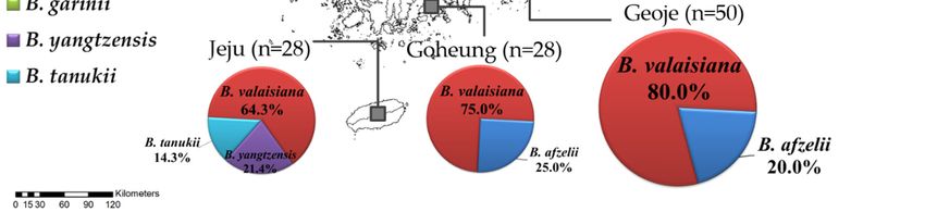
Pathogens 2020, 9, x FOR PEER REVIEW 2 of 9
regions
Pathogens 2020, 9,only
identified B. afzelii. Borrelia valaisiana was found in Geoje, Goheung, and Jeju
866 3 ofIsland,
9
which are located in the southern sampling regions, and displayed a higher prevalence than other
genospecies. Specifically, B. yangtzensis and B. tanukii were only found on Jeju Island (Figure 1).
only identified B. afzelii. Borrelia valaisiana was found in Geoje, Goheung, and Jeju Island, which are
located in the southern sampling regions, and displayed a higher prevalence than other genospecies.
Specifically, B. yangtzensis and B. tanukii were only found on Jeju Island (Figure 1).
Figure 1. A map
Figure 1. A of
maptheofBorrelia genospecies
the Borrelia detected
genospecies in theindifferent
detected sampling
the different regions
sampling of theof
regions ROK.
the ROK.
2.3. Phylogenetic Tree of B. burgdorferi s. l.
2.3. Phylogenetic Tree of B. burgdorferi s. l.
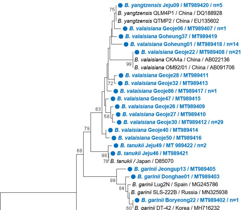
We selected 24 representative samples without duplicate sequences among the 248 ticks identified
We selected 24 representative samples without duplicate sequences among the 248 ticks
as positive for Borrelia genospecies and constructed a phylogenetic tree using the flagellin B (flaB)
identified as positive for Borrelia genospecies and constructed a phylogenetic tree using the flagellin
genes from several Borrelia genospecies sequences deposited in GenBank (Figure 2). The representative
B (flaB) genes from several Borrelia genospecies sequences deposited in GenBank (Figure 2). The
sequences of the five genospecies clustered with related reference sequences; however, B. yangtzensis
representative sequences of the five genospecies clustered with related reference sequences; however,
clustered together in a large clade closely related to B. valaisiana. No apparent clustering was observed
B. yangtzensis clustered together in a large clade closely related to B. valaisiana. No apparent clustering
based on the tick host or sampling region.
was observed based on the tick host or sampling region.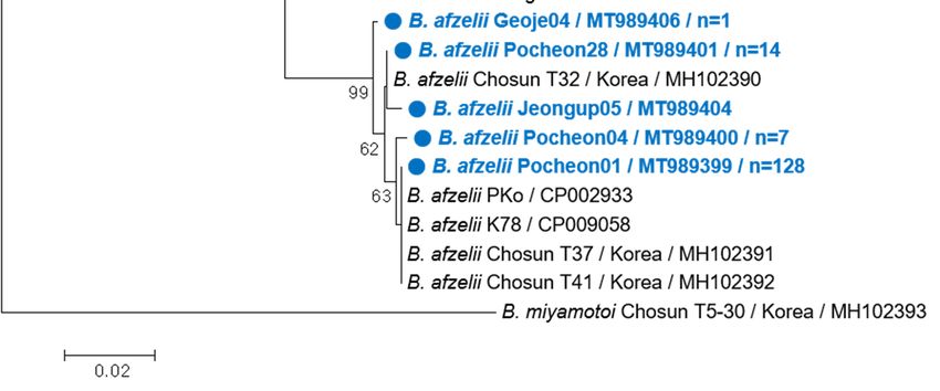
Pathogens 2020, 9, 866                                                                                                    4 of 9
 Pathogens 2020, 9, x FOR PEER REVIEW                                                                                      3 of 9

      Figure2.2. Phylogenetic
     Figure      Phylogenetic relationship       between Borrelia
                                 relationship between       Borrelia genospecies
                                                                      genospecies based
                                                                                    based on     partial flaB
                                                                                             on partial   flaBnucleotide
                                                                                                                nucleotide
     sequences
      sequences (303 nucleotide positions) constructed using the neighbor-joining method in
                 (303 nucleotide    positions)    constructed  using   the  neighbor-joining    method      in MEGA
                                                                                                               MEGA 5.2.
                                                                                                                       5.2.
     Node
      Node numbers
             numbers indicate
                       indicate the
                                  theproportion
                                       proportion of  ofbootstrap
                                                         bootstrapreplicates
                                                                     replicates (1000)
                                                                                 (1000)that
                                                                                         thatsupported
                                                                                               supported the thetopology
                                                                                                                 topology
     shown.   The cutoff
      shown. The   cutoff value
                          value for
                                  forthe
                                      theconsensus
                                           consensustree treewas
                                                              was  50%.
                                                                 50%.   TheThe  scale
                                                                             scale barbar   represents
                                                                                       represents    2% 2%    divergence.
                                                                                                         divergence.  The
     The sequences   identified  in this study    are indicated by  blue  circles. The number     of sequences
      sequences identified in this study are indicated by blue circles. The number of sequences (n) with an      (n) with
     an identical
      identical   genospecies
                genospecies   areare shown
                                   shown      if the
                                           if the     sequence
                                                   sequence     was
                                                              was     detected
                                                                   detected   inin more
                                                                                 more     than
                                                                                       than     one
                                                                                              one    case.
                                                                                                   case.

3. Discussion
 3. Discussion
       In this study, we performed the molecular detection and phylogenetic analysis of B. burgdorferi s. l.
        In this study, we performed the molecular detection and phylogenetic analysis of B. burgdorferi
in engorged ticks from wild rodents in nine regions of the ROK. Nested PCR revealed an overall Borrelia
 s. l. in engorged ticks from wild rodents in nine regions of the ROK. Nested PCR revealed an overall
infection rate of 33.6% among the 738 ticks analyzed, similar to the high prevalence rates found in ticks
 Borrelia infection rate of 33.6% among the 738 ticks analyzed, similar to the high prevalence rates
feeding on wild rodents in Malaysia (46.1%) and Taiwan (47.1%) [23,24]. However, previous studies
 found in ticks feeding on wild rodents in Malaysia (46.1%) and Taiwan (47.1%) [23,24]. However,
from the ROK have detected very low rates of B. burgdorferi s. l. in ticks feeding on wild animals
 previous studies from the ROK have detected very low rates of B. burgdorferi s. l. in ticks feeding on
such as dogs (0.2%, n = 562 ticks) [20], wild water deer (2.1%, n = 48 ticks) [19], migratory birds (3.7%,
 wild animals such as dogs (0.2%, n = 562 ticks) [20], wild water deer (2.1%, n = 48 ticks) [19], migratory
four positive pools of 108 tested pools among 212 ticks) [18], and wild rodents in northern Gyeonggi
 birds (3.7%, four positive pools of 108 tested pools among 212 ticks) [18], and wild rodents in northern
near the demilitarized zone (1.0%, n = 1618 ticks including 933 questing ticks) [25]. These differences in
 Gyeonggi near the demilitarized zone (1.0%, n = 1,618 ticks including 933 questing ticks) [25]. These
prevalence may be due to variations in the sampling methods for tick and host species, survey regions,
 differences in prevalence may be due to variations in the sampling methods for tick and host species,
the environment, and the seasonal timing of the survey.
 survey regions, the environment, and the seasonal timing of the survey.
        Sequence analysis, including phylogenetic analysis based on partial flaB sequences, confirmed
 that B. afzelii, B. valaisiana, B. garinii, B. yangtzensis, and B. tanukii, were detected in the ticks collected
Pathogens 2020, 9, 866 5 of 9
Sequence analysis, including phylogenetic analysis based on partial flaB sequences, confirmed that
B. afzelii, B. valaisiana, B. garinii, B. yangtzensis, and B. tanukii, were detected in the ticks collected in this
study and displayed isolated geographical distribution. For instance, B. afzelii and B. garinii were found
only inland, while B. afzelii was the main genospecies in the northern and central sampling regions.
Conversely, B. valaisiana, B. yangtzensis, and B. tanukii were only found in the southern sampling
regions, where B. valaisiana was the most common genospecies. Previous studies in the ROK have
identified B. afzelii and B. garinii in various tick species, animals, and patient sera throughout inland
areas [9,16,19–22,26]. In addition, other B. burgdorferi s. l. groups, such as B. valaisiana, have been
detected in I. nipponensis [27], whereas B. yangtzensis was reclassified via multilocus sequence typing in
A. agrarius from Haenam in the southernmost inland region [22]. Borrelia tanukii has been reported in
Ixodes tanuki from Japan [28]. In the ROK, a similar partial 16s rRNA sequence of B. tanukii has been
reported in ticks feeding on migratory birds on Hongdo Island [18]; however, it was unclear whether
the ticks were indigenous. Our study demonstrates for the first time that B. tanukii is indigenous to
ticks in the ROK. Many B. burgdorferi s. l. genospecies, including B. afzelii, B. garinii, B. burgdorferi s. s.,
B. bavariensis, and B. bissettii, are known to be pathogenic to humans, while B. lusitaniae, B. spielmanii,
and B. valaisiana are considered potentially pathogenic [3]. In this study, the identification of B. afzelii
and B. valaisiana in ticks that act as major Borrelia vectors indicates that LD transmission may be possible
via questing ticks in the ROK.
A previous study in which vegetation was swept and dragged for questing ticks identified
H. longicornis and H. flava as the dominant tick species, while I. nipponensis has been shown to be the
most frequently collected tick from small mammals [29,30]. Our study found that most of the ticks
collected from wild rodents were of the Ixodes genus, including Ixodes spp. (n = 440, 59.6%), I. nipponensis
(n = 193, 26.1%), and I. angustus (n = 96, 13.0%). Moreover, we found that Borrelia DNA was prevalent
in I. nipponensis, Ixodes spp., I. angustus, and H. longicornis; however, the prevalence varied between
species. Borrelia burgdorferi sensu lato (s. l.) was first identified from I. angustus in the ROK and has
since been found in several other tick species, such as I. granulatus, I. persulcatus, I. nipponensis, I. turdus,
and H. longicornis [9,18,19,27]. In experimental vector competence studies, I. angustus was found to be
a competent vector for transmitting B. burgdorferi s. s. to deer mice [31], while this study identified
Borrelia infection in 1/9 H. longicornis larvae. A previous study reported the first molecular detection of
B. afzelii in H. longicornis infesting wild Korean water deer (Hydropotes inermis) in the ROK [19]; however,
Sun et al. [32] showed that H. longicornis can carry but not transmit Borrelia as H. longicornis can only
maintain spirochetes for a shorter period than the digestion period of blood. Therefore, H. longicornis is
not the main Borrelia vector, despite being dominant in the ROK, indicating that Ixodes species may be
the main vector and carrier of Borrelia in the ROK. Because we used ticks harvested from wild rodents,
the Borrelia genospecies and infection rate in this study may have originated from the wild rodent itself.
Nevertheless, blood-sucking vectors are reliable tools for demonstrating the existence of pathogens in
a specific area [7]. Human biting cases by the Ixodes species was significantly lower (5.7%) than those
by other species in the ROK [33]. Considering the gradually increasing number of LD cases in the ROK
and the high Borrelia infection rate in ticks engorged with wild rodent blood in this study, monitoring
for Borrelia in non-engorged or questing ticks at different developmental stages should be carried out.
To our knowledge, this study is the first to describe the nationwide prevalence of B. burgdorferi s. l.
in engorged ticks from wild rodents in the ROK. The ticks were found to have a high infection rate
(33.6%) and a wide geographical distribution for the five detected genospecies of B. burgdorferi s. l.,
the causative agent of LD. Humans working in agricultural fields, visiting mountains and reservoirs
for recreational activities, or inhabiting residential areas may therefore be at risk of exposure to and
infection with Borrelia due to close contact with wild rodents and ticks. Thus, continuous surveillance
on various tick species, animals, humans, and different geographical regions is required to reduce
disease distribution and possible transmission to humans in the ROK.Pathogens 2020, 9, 866 6 of 9
4. Materials and Methods
4.1. Surveillance Localities and Periods
Ticks were collected from wild rodents during spring (March–May) and autumn
(September–November) in 2017 from nine regions in the ROK, namely Pocheon, Donghae, Sejong,
Boryeong, Uiseong, Jeongup, Geoje, Goheung, and Jeju Island (Figure 1). Rodent traps were installed
at several environmental sites, including cropped fields, reservoirs, waterways, and mountains.
The animal-handling protocol used in this study was reviewed and approved based on the guidelines
for ethical procedures and scientific care of the Institutional Animal Care and Use Committee of the
Korea Centers for Disease Control and Prevention (KCDC-089-17).
4.2. Collection of Wild Rodents and Ticks
At each site, 25 Sherman folding live traps (3 × 3.5 × 9 inches, BioQuip, Gardena, CA, USA) were
set up at five points at 3–5 m intervals with a peanut butter-spread biscuit. The rodent traps were
collected the next morning and transported to the laboratory. After euthanization using compressed
carbon dioxide, the rodents were suspended for 24 h over glass bowls filled with 70% EtOH to collect
ticks. The ticks were then recovered from the surface using a fine brush and stored in 70% EtOH at
−20 ◦ C for identification.
4.3. Identification of Ticks
The species and developmental stage of each tick were identified under a dissection microscope
using taxonomic identification keys according to Yamaguti et al. (1971) [34]. Individual ticks were
then placed in a 2 mL screw cap tube according to the host rodent, collection site, region, and season
and were stored at −20 ◦ C before DNA extraction.
4.4. Detection of Borrelia DNA in Ticks
Tick samples were homogenized in 450 µL of 1× phosphate-buffered saline (PBS) with 2.8 mm
ceramic (zirconium oxide) beads using a Precellys Evolution homogenizer (Bertin Technologies,
Bretonneux, France). The homogenates were centrifuged at 25,000× g for 10 min, and 400 µL of
the supernatant was used for DNA extraction using a MagMAX™ DNA Multi-Sample Ultra 2.0 Kit
(Applied Biosystems, Foster city, CA, USA) according to the manufacturer’s instructions. To detect
Borrelia DNA in tick samples, we targeted a partial flaB sequence, as described previously [34] (Table 2).
For the first PCR reaction, 5 µL of tick template DNA was added to AccuPower® HotStart PCR PreMix
(Bioneer, Daejeon, Korea) and incubated at 94 ◦ C for 5 min, followed by 35 cycles of 94 ◦ C for 30 s,
60 ◦ C for 45 s, and 72 ◦ C for 1 min, with a final amplification at 72 ◦ C for 10 min. Secondary nested PCR
was then performed using 1 µL of the primary PCR products under the following conditions: 94 ◦ C for
5 min, followed by 25 cycles of 94 ◦ C for 10 s, 48 ◦ C for 1 min, 72 ◦ C for 90 s, and a final extension
step at 72 ◦ C for 5 min. Positive and negative controls were included in each PCR set. Amplified PCR
products were visualized via 2.0% agarose gel electrophoresis after staining with Safe-Pinky DNA Gel
Staining Solution (10,000×) in water (GenDEPOT, Barker, TX, USA), yielding a predicted size of 347 bp.
To avoid cross-contamination, DNA extraction, amplification, and agarose gel electrophoresis were
performed in separate rooms.
Table 2. DNA sequences of primers used for nested PCR.
Targe Amplicon
Designation Sequence Reference
Specificity Length (bp)
BflaPAD 1st 50 -GATCARGCWCAAYATAACCAWATGCA-30
459
BflaPDU PCR 50 -AGATTCAAGTCTGTTTTGGAAAGC-30
Flagellin B [35]
BflaPBU 2nd 50 -GCTGAAGAGCTTGGAATGCAACC-30
347
BflaPCR PCR 50 -TGATCAGTTATCATTCTAATAGCA-30Pathogens 2020, 9, 866 7 of 9
4.5. Sequencing and Phylogenetic Analysis
All positive nested PCR products were sequenced in directions, using the PCR primers at Bioneer
Inc. (Daejeon, ROK). To identify Borrelia genospecies, the sequencing results were analyzed using the
BlastN program from the National Center for Biotechnology Information (NCBI, Bethesda, MD, USA).
The sequences obtained in this study were aligned using CLUSTALW and compared to published
sequences in GenBank (NIH, Montgomery, MD, USA). A phylogenetic tree was constructed based
on the DNA sequences of flaB from tick samples with a Borrelia infection using the neighbor-joining
method with the p-distance model in MEGA version 5.2. Bootstrap analysis was conducted using
1000 replicates to improve the confidence level of the phylogenetic tree. The GenBank accession
numbers of the genospecies sequences obtained in this study are presented in Figure 2.
Author Contributions: Formal Analysis, T.Y.K.; Investigation, S.Y.K. and T.-K.K.; Resources, T.-K.K.; Data Curation
and Writing—Original Draft Preparation, S.Y.K.; Conceptualization, Writing—Review & Editing, and Funding
Acquisition, H.I.L. All authors have read and agreed to the published version of the manuscript.
Funding: This study was financially supported by the Korea Centers for Disease Control and Prevention (Grant
No. 2016-NG55001-00).
Acknowledgments: We thank Su Yeon Kim from the Division of Pathogen Resource Management and the Division
of Bacterial Disease Research at the National Institute of Infectious Diseases for their technical expertise and help
with the positive control in this study. We also thank the fieldwork crew, Bong Gu Song, Hak Seon Lee, Gi Hun
Kim, Won Il Park, and Hyung Woo Lim, for their hard work and thoughtful contributions to the project. Lastly,
this paper is dedicated to the memory of our friend and colleague, Jong Yul Roh, who passed away in May 2020
after helping us initiate and organize this project.
Conflicts of Interest: The authors declare no conflict of interest.
References
1. Aucott, J.; Morrison, C.; Munoz, B.; Rowe, P.C.; Schwarzwalder, A.; West, S.K. Diagnostic challenges of early
Lyme disease: Lessons from a community case series. BMC Infect. Dis. 2009, 9, 79. [CrossRef] [PubMed]
2. Radolf, J.D.; Caimano, M.J.; Stevenson, B.; Hu, L.T. Of ticks, mice and men: Understanding the dual-host
lifestyle of Lyme disease spirochaetes. Nat. Rev. Microbiol. 2012, 10, 87–99. [CrossRef] [PubMed]
3. Rudenko, N.; Golovchenko, M.; Grubhoffer, L.; Oliver, J.H., Jr. Updates on Borrelia burgdorferi sensu lato
complex with respect to public health. Ticks Tick Borne Dis. 2011, 2, 123–128. [CrossRef] [PubMed]
4. Radolf, J.D.; Samuels, D.S. Borrelia: Molecular biology, host interaction and pathogenesis. Clin. Infect. Dis.
2011, 52, 965. [CrossRef]
5. Wang, G.; van Dam, A.P.; Schwartz, I.; Dankert, J. Molecular typing of Borrelia burgdorferi sensu lato:
Taxonomic, epidemiological, and clinical implications. Clin. Microbiol. Rev. 1999, 12, 633–653. [CrossRef]
6. Hengge, U.R.; Tannapfel, A.; Tyring, S.K.; Erbel, R.; Arendt, G.; Ruzicka, T. Lyme borreliosis. Lancet Infect. Dis.
2003, 3, 489–500. [CrossRef]
7. Sparagano, O.A.; Allsopp, M.T.; Mank, R.A.; Rijpkema, S.G.; Figueroa, J.V.; Jongejan, F. Molecular detection
of pathogen DNA in ticks (Acari: Ixodidae): A review. Exp. Appl. Acarol. 1999, 23, 929–960. [CrossRef]
8. Gern, L.; Humair, P.-F. Ecology of Borrelia burgdorferi sensu lato in Europe. In Lyme Borreliosis: Biology,
Epidemiology and Control, 1st ed.; Gray, J., Kahl, O., Lane, R.S., Stanek, G., Eds.; CABI: Wallingford, UK, 2002;
pp. 149–174.
9. Park, K.H.; Chang, W.H.; Schwan, T.G. Identification and characterization of Lyme disease spirochetes,
Borrelia burgdorferi sensu lato, isolated in Korea. J. Clin. Microbiol. 1993, 31, 1831–1837. [CrossRef]
10. Lee, M.G.; Chung, K.Y.; Choi, Y.S.; Cho, S.N. Lyme disease. Korean, J. Dermatol. 1993, 31, 601–605. [CrossRef]
11. Korea Centers for Disease Control and Prevention (KCDC). Diseases Web Statistics System. Available online:
http://www.cdc.go.kr/npt/biz/npp/ist/bass/bassAreaStatsMain.do (accessed on 15 July 2020).
12. Park, S.H.; Hwang, K.J.; Chu, H.; Park, M.Y. Serological detection of Lyme borreliosis agents in patients from
Korea, 2005-2009. Osong Public Health Res. Perspect. 2011, 2, 29–33. [CrossRef]
13. Moon, S.; Hong, Y.; Hwang, K.J.; Kim, S.; Eom, J.; Kwon, D.; Park, J.H.; Youn, S.K.; Sohn, A.
Epidemiological features and clinical manifestations of Lyme borreliosis in Korea during the period
2005–2012. Jpn. J. Infect. Dis. 2015, 68, 1–4. [CrossRef] [PubMed]Pathogens 2020, 9, 866 8 of 9
14. Bell, D.R.; Berghaus, R.D.; Patel, S.; Beavers, S.; Fernandez, I.; Sanchez, S. Seroprevalence of tick-borne
infections in military working dogs in the Republic of Korea. Vector Borne Zoonotic Dis. 2012, 12, 1023–1030.
[CrossRef] [PubMed]
15. Lee, S.H.; Yun, S.H.; Choi, E.; Park, Y.S.; Lee, S.E.; Cho, G.J.; Kwon, O.D.; Kwak, D. Serological Detection of
Borrelia burgdorferi among Horses in Korea. Korean J. Parasitol. 2016, 54, 97–101. [CrossRef] [PubMed]
16. Kee, S.; Hwang, K.-J.; Oh, H.-B.; Park, K.-S. Identification of Borrelia burgdorferi isolated in Korea using outer
surface protein A (OspA) serotyping system. Microbiol. Immunol. 1994, 38, 989–993. [CrossRef]
17. Choi, Y.J.; Han, S.H.; Park, J.M.; Lee, K.M.; Lee, E.M.; Lee, S.H.; Song, H.J.; Koh, Y.S.; Lee, K.W.; Jang, W.J.;
et al. First molecular detection of Borrelia afzelii in clinical samples in Korea. Microbiol. Immunol. 2007,
51, 1201–1207. [CrossRef]
18. Kang, J.G.; Kim, H.C.; Choi, C.Y.; Nam, H.Y.; Chae, H.Y.; Chong, S.T.; Klein, T.A.; Ko, S.; Chae, J.S. Molecular
detection of Anaplasma, Bartonella, and Borrelia species in ticks collected from migratory birds from Hong-do
Island, Republic of Korea. Vector Borne Zoonotic Dis. 2013, 13, 215–225. [CrossRef]
19. VanBik, D.; Lee, S.H.; Seo, M.G.; Jeon, B.R.; Goo, Y.K.; Park, S.J.; Rhee, M.H.; Kwon, O.D.; Kim, T.H.;
Geraldino, P.J.L.; et al. Borrelia species detected in ticks feeding on wild Korean water deer (Hydropotes inermis)
using molecular and genotypic analyses. J. Med. Entomol. 2017, 54, 1397–1402. [CrossRef]
20. Lee, S.H.; Goo, Y.K.; Geraldino, P.J.L.; Kwon, O.D.; Kwak, D. Molecular detection and characterization of
Borrelia garinii (Spirochaetales: Borreliaceae) in Ixodes nipponensis (Ixodida: Ixodidae) parasitizing a dog in
Korea. Pathogens 2019, 8, 289. [CrossRef]
21. Lee, S.H.; Jung, K.D.; Lee, J.H.; Kim, S.C.; Kim, J.H.; Jang, W.J.; Park, K.H. Characterization of Borrelia afzelii
isolated from Ixodes nipponensis and Apodemus agrarius in Chungju, Korea, by PCR-rFLP analyses of ospC
gene and rrf (5S)-rrl (23S) intergenic spacer. Microbiol. Immunol. 2002, 46, 677–683. [CrossRef]
22. Park, K.H.; Choi, Y.J.; Kim, J.; Park, H.J.; Song, D.; Jang, W.J. Reclassification of Borrelia spp. isolated in South
Korea using multilocus sequence typing. Jpn. J. Infect. Dis. 2018, 71, 350–353. [CrossRef]
23. Khoo, J.J.; Ishak, S.N.; Lim, F.S.; Mohd-Taib, F.S.; Khor, C.S.; Loong, S.K.; AbuBakar, S. Detection of a Borrelia
sp. from Ixodes granulatus ticks collected from rodents in Malaysia. J. Med. Entomol. 2018, 55, 1642–1647.
[CrossRef] [PubMed]
24. Chao, L.L.; Liu, L.L.; Shih, C.M. Prevalence and molecular identification of Borrelia spirochetes in Ixodes
granulatus ticks collected from Rattus losea on Kinmen Island of Taiwan. Parasit. Vectors 2012, 5, 167.
[CrossRef] [PubMed]
25. Chae, J.S.; Yu, D.H.; Shringi, S.; Klein, T.A.; Kim, H.C.; Chong, S.T.; Lee, I.Y.; Foley, J. Microbial pathogens
in ticks, rodents and a shrew in northern Gyeonggi-do near the DMZ, Korea. J. Vet. Sci. 2008, 9, 285–293.
[CrossRef] [PubMed]
26. Lim, S.; Irwin, P.J.; Lee, S.; Oh, M.; Ahn, K.; Myung, B.; Shin, S. Comparison of selected canine vector-borne
diseases between urban animal shelter and rural hunting dogs in Korea. Parasit. Vectors 2010, 3, 32. [CrossRef]
27. Masuzawa, T.; Fukui, T.; Miyake, M.; Oh, H.-B.; Cho, M.-K.; Chang, W.-H.; Imai, Y.; Yanagihara, Y.
Determination of members of a Borrelia afzelii-related group isolated from Ixodes nipponensis in Korea as
Borrelia valaisiana. Int. J. Syst. Bacteriol. 1999, 49, 1409–1415. [CrossRef]
28. Fukunaga, M.; Hamase, A.; Okada, K.; Nakao, M. Borrelia tanukii Sp. Nov. and Borrelia turdae Sp. Nov. found
from Ixodid ticks in Japan. Microbiol. Immunol. 1996, 40, 877–881. [CrossRef]
29. Kim, H.C.; Kim, J.H.; Jo, Y.S.; Chong, S.T.; Sames, W.J.; Klein, T.A.; Robbins, R.G. Records of Ixodes pomeranzevi
Serdyukova, 1941 (Acari: Ixodidae) from small mammals in northern Gyeonggi and Gangwon provinces,
Republic of Korea. Syst. Appl. Acarol. 2009, 14, 129–135. [CrossRef]
30. Kim, H.C.; Chong, S.T.; Sames, W.J.; Nunn, P.V.; Wolf, S.P.; Robbins, R.G.; Klein, T.A. Tick surveillance of
small mammals captured in Gyeonggi and Gangwon Provinces, Republic of Korea, 2004–2008. Syst. Appl.
Acarol. 2010, 15, 100–108. [CrossRef]
31. Peavey, C.A.; Lane, R.S.; Damrow, T. Vector competence of Ixodes angustus (Acari: Ixodidae) for
Borrelia burgdorferi sensu stricto. Exp. Appl. Acarol. 2000, 24, 77–84. [CrossRef]
32. Sun, Y.; Xu, R.M.; Guo, T.Y.; Zhang, P.H.; Cao, W.C. Incapability of Haemaphysalis longicornis and Dermacentor
nuttalli to acquire and trans-stadial transmit the Lyme spirochetes Borrelia garinii. Acta Parasitol. Med. Entomol.
Sin. 2003, 10, 174–180.Pathogens 2020, 9, 866 9 of 9
33. Yun, S.M.; Lee, W.G.; Ryou, J.; Yang, S.C.; Park, S.W.; Roh, J.Y.; Lee, Y.-J.; Park, C.; Han, M.G. Severe fever with
thrombocytopenia syndrome virus in ticks collected from humans, South Korea, 2013. Emerg. Infect. Dis.
2014, 20, 1358–1361. [CrossRef] [PubMed]
34. Yamaguti, N.; Tipton, V.J.; Keegan, H.L.; Toshioka, S. Ticks of Japan, Korea, and the Ryukyu islands. Brigham
Young Univ. Sci. Bull. Biol. Ser. 1971, 15, 1.
35. Sato, Y.; Konishi, T.; Hashimoto, Y.; Takahashi, H.; Nakaya, K.; Fukunaga, M.; Nakao, M. Rapid diagnosis of
lyme disease: Flagellin gene-based nested polymerase chain reaction for identification of causative Borrelia
species. Int. J. Infect. Dis. 1997, 2, 64–73. [CrossRef]
Publisher’s Note: MDPI stays neutral with regard to jurisdictional claims in published maps and institutional
affiliations.
© 2020 by the authors. Licensee MDPI, Basel, Switzerland. This article is an open access
article distributed under the terms and conditions of the Creative Commons Attribution
(CC BY) license (http://creativecommons.org/licenses/by/4.0/).You can also read