National Diagnostic Protocol - Synchytrium endobioticum The cause of potato wart
←
→
Page content transcription
If your browser does not render page correctly, please read the page content below
NDP 16 V2 - National Diagnostic Protocol for Synchytrium endobioticum

      National Diagnostic Protocol
                     Synchytrium endobioticum
                         The cause of potato wart

                                            NDP 16 V2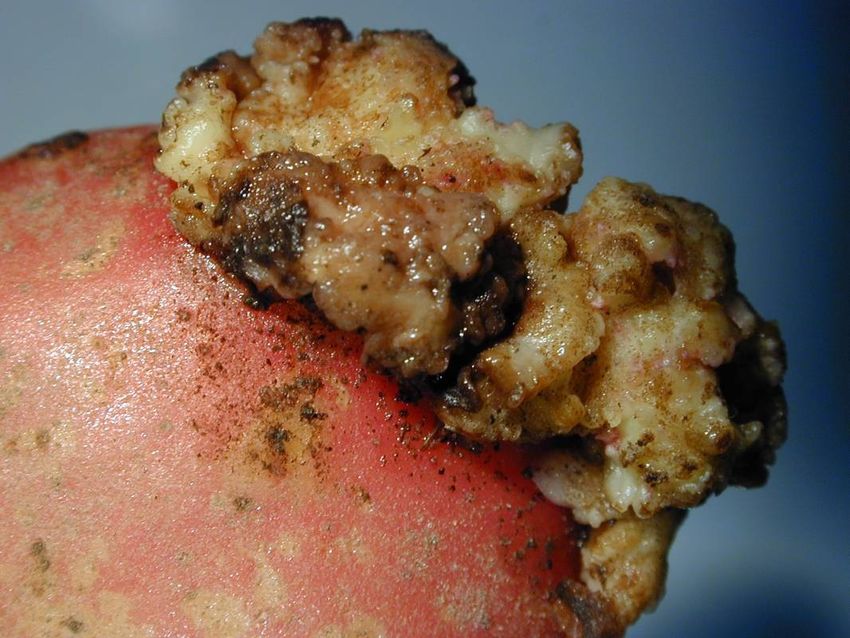
NDP 16 V2 - National Diagnostic Protocol for Synchytrium endobioticum

© Commonwealth of Australia

Ownership of intellectual property rights
Unless otherwise noted, copyright (and any other intellectual property rights, if any) in this publication is owned by
the Commonwealth of Australia (referred to as the Commonwealth).

Creative Commons licence
All material in this publication is licensed under a Creative Commons Attribution 3.0 Australia Licence, save for content
supplied by third parties, logos and the Commonwealth Coat of Arms.

Creative Commons Attribution 3.0 Australia Licence is a standard form licence agreement that allows you to copy,
distribute, transmit and adapt this publication provided you attribute the work. A summary of the licence terms is
available from http://creativecommons.org/licenses/by/3.0/au/deed.en. The full licence terms are available from
https://creativecommons.org/licenses/by/3.0/au/legalcode.

This publication (and any material sourced from it) should be attributed as: Subcommittee on Plant Health Diagnostics
(2017). National Diagnostic Protocol for Synchytrium endobioticum – NDP16 V2. (Eds. Subcommittee on Plant Health
Diagnostics). Authors Rowles-van Rijswijk, B, Cunnington, J and Luck, J. Reviewers Pascoe, I and Ho, W. ISBN 978-0-
9945113-8-6. CC BY 3.0.

Cataloguing data
Subcommittee on Plant Health Diagnostics (2017). National Diagnostic Protocol for Synchytrium endobioticum –
NDP16 V2. (Eds. Subcommittee on Plant Health Diagnostics). Authors Rowles-van Rijswijk, B, Cunnington, J and Luck, J.
Reviewers Pascoe, I and Ho, W. ISBN 978-0-9945113-8-6.

ISBN; 978-0-9945113-8-6

Internet
Report title is available at: http://plantbiosecuritydiagnostics.net.au/resource-hub/priority-pest-diagnostic-
resources/

Department of Agriculture and Water Resources

Street Address 1: 18 Marcus Clarke Street, Canberra City ACT 2601
Street Address 2: 7 London Circuit, Canberra City ACT 2601
Postal Address: GPO Box 858, Canberra City ACT 2601
Switchboard Phone: 02 6272 3933
Web: http://www.agriculture.gov.au

Inquiries regarding the licence and any use of this document should be sent to: copyright@agriculture.gov.au.

The Australian Government acting through the Department of Agriculture and Water Resources has exercised due care
and skill in the preparation and compilation of the information and data in this publication. Notwithstanding, the
Department of Agriculture and Water Resources, its employees and advisers disclaim all liability, including liability for
negligence, for any loss, damage, injury, expense or cost incurred by any person as a result of accessing, using or
relying upon any of the information or data in this publication to the maximum extent permitted by law.

   ii                                                                     Subcommittee on Plant Health Diagnostics

NDP 16 V2 - National Diagnostic Protocol for Synchytrium endobioticum

Purpose
National Diagnostic Protocols (NDPs) are diagnostic protocols for the unambiguous taxonomic
identification of plant pests. NDPs:

    •    are a verified information resource for plant health diagnosticians
    •    are consistent with ISPM No. 27 – Diagnostic Protocols for Regulated Pests
    •    provide a nationally consistent approach to the identification of plant pests enabling
         transparency when comparing diagnostic results between laboratories; and,
    •    are endorsed by regulatory jurisdictions for use (either within their own facilities or when
         commissioning from others) in a pest incursion.
Where an International Plant Protection Convention (IPPC) diagnostic protocol exists it should be
used in preference to NDPs although NDPs may contain additional information to aid diagnosis. IPPC
protocols are available on the IPPC website:

https://www.ippc.int/core-activities/standards-setting/ispms

Process
NDPs are facilitated and endorsed by the Subcommittee on Plant Health Diagnostics (SPHD). SPHD
reports to Plant Health Committee and is Australia’s peak technical and policy forum for plant health
diagnostics.

NDPs are developed and endorsed according to Reference Standards developed and maintained by
SPHD. Current Reference Standards are available at
http://plantbiosecuritydiagnostics.net.au/sphd/sphd-reference-standards/

NDPs are living documents. They are updated every 5 years or before this time if required (i.e. when
new techniques become available).

Document status
This version of the National Diagnostic Protocol (NDP) for Synchytrium endobioticum is current as at
the date contained in the version control box below.

                 PEST STATUS                              Not present in Australia
                 PROTOCOL NUMBER                          NDP 16
                 VERSION NUMBER                           V2
                 PROTOCOL STATUS                          Endorsed
                 ISSUE DATE                               2017
                 REVIEW DATE                              2021
                 ISSUED BY                                SPHD
The most current version of this document is available from the SPHD website:
http://plantbiosecuritydiagnostics.net.au/resource-hub/priority-pest-diagnostic-resources/

Further information
Inquiries regarding technical matters relating to this project should be sent to:
sphds@agriculture.gov.au

   iii                                                                  Subcommittee on Plant Health Diagnostics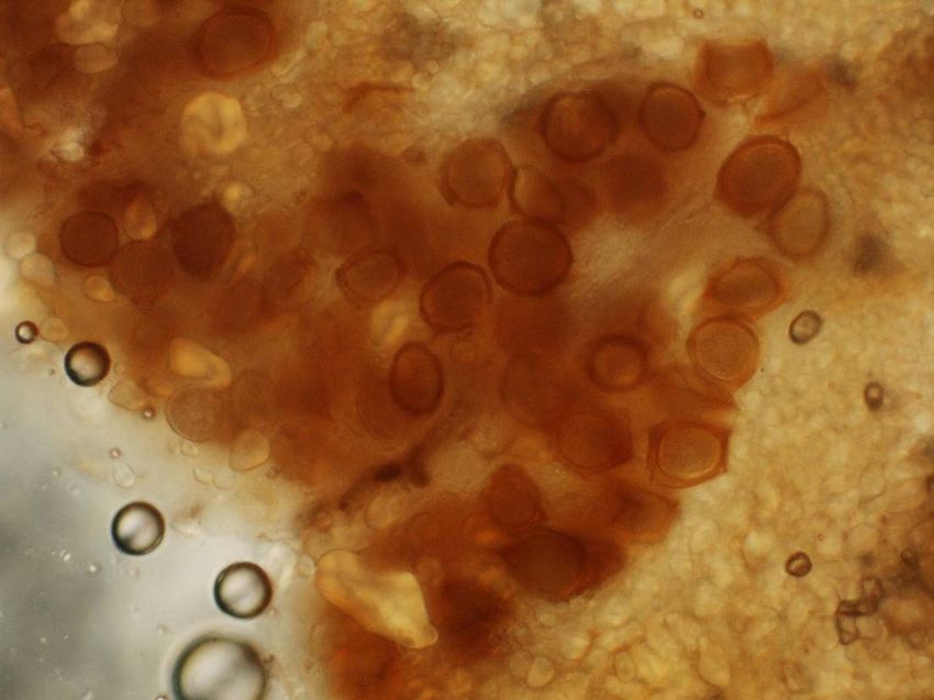
NDP 16 V2 - National Diagnostic Protocol for Synchytrium endobioticum

Contents
1    INTRODUCTION........................................................................................................................................ 2
2    TAXONOMIC INFORMATION ................................................................................................................ 3
3    DETECTION ................................................................................................................................................ 4
     3.1         Symptoms......................................................................................................................................................4
4    IDENTIFICATION...................................................................................................................................... 7
     4.1         Microscopic examination for sporangia ...........................................................................................7
     4.2         Morphology ..................................................................................................................................................7
5    CONTACTS FOR FURTHER INFORMATION.................................................................................... 14
6    ACKNOWLEDGEMENTS........................................................................................................................ 15
7    REFERENCES ............................................................................................................................................ 16
     7.1         Related Websites ..................................................................................................................................... 17
8    APPENDICES ............................................................................................................................................ 18
     8.1         Life Cycle..................................................................................................................................................... 18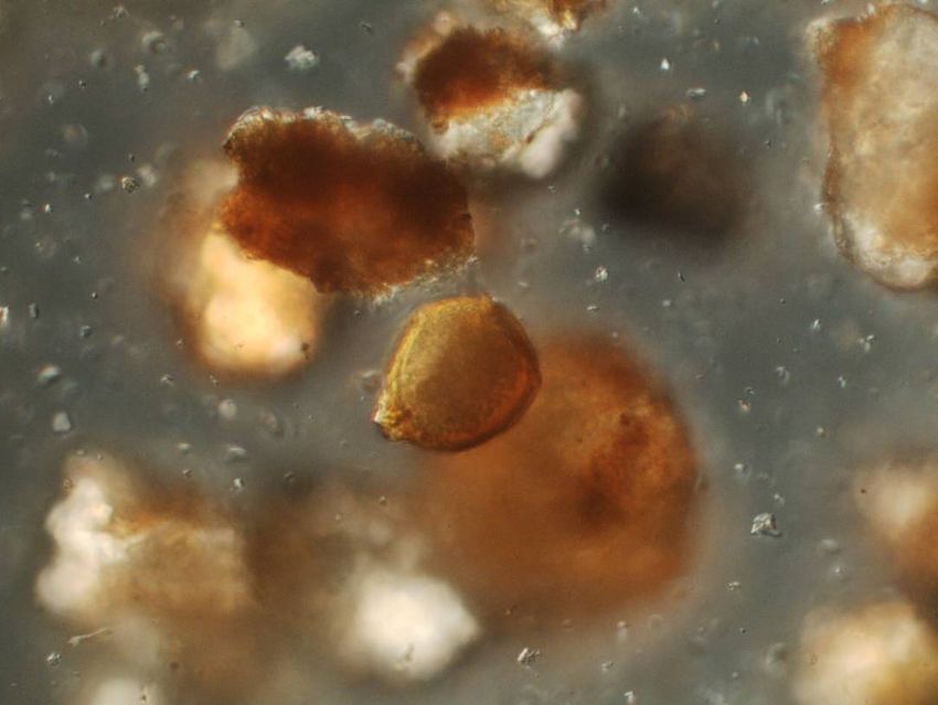
NDP 16 V2 - National Diagnostic Protocol for Synchytrium endobioticum

1            INTRODUCTION
Potato wart disease is caused by the soil-borne fungus Synchytrium endobioticum (Schilb.) Percival
(Chytridiomycetes: Chytridiales). The principal symptom is a warty, cauliflower-like gall which
develops on tubers and tuber initials. The main cultivated host affected by potato wart is potato
(Solanum tuberosum L.), although other wild Solanum species are known to be infected in Mexico. In
addition, a number of Solanaceous crops (including tomato) and weeds have been artificially
inoculated. Experimentally, this fungus can also infect species within the genera Nicotiana, Physalis,
Capsicastrum, Datura, Duboisia, Hyoscyamus, Lycium, Nicandra and Schizanthus (Hampson 1993;
DEFRA 2011).

Potato wart can be destructive under favourable conditions and entire crops can be lost as a result of
heavily infected potato tubers being virtually entirely converted to warts. The fungus targets
meristematic tissue and infects all below ground parts of the plant except root tissue. In tubers,
infection initially occurs in eye tissue and gradually spreads causing massive hypertrophy and
hyperplasia, frequently involving the entire tuber. The tumour is composed of simple, large
parenchyma cells crowded with starch grains and organised into a malformed and compressed foliar
branch system. In near-immune potato cultivars, the warts remain superficial and scab-like, while in
resistant potato cultivars the zoospore dies soon after invasion by necrotic abortion (hypersensitive
reaction) of the infected tissue (Hampson and Proudfoot 1974; Pratt 1976; Hooker 1990). Very long
periods of survival of the winter sporangia prevent replanting of infested fields for up to 30 years or
longer (Pratt 1976).

This diagnostic manual was developed based on available published methods, and in consultation with
international experts. It describes potato wart symptoms and outlines a microscopic procedure to
confirm the presence of S. endobioticum spores in potato tissue.

    2                                                                   Subcommittee on Plant Health Diagnostics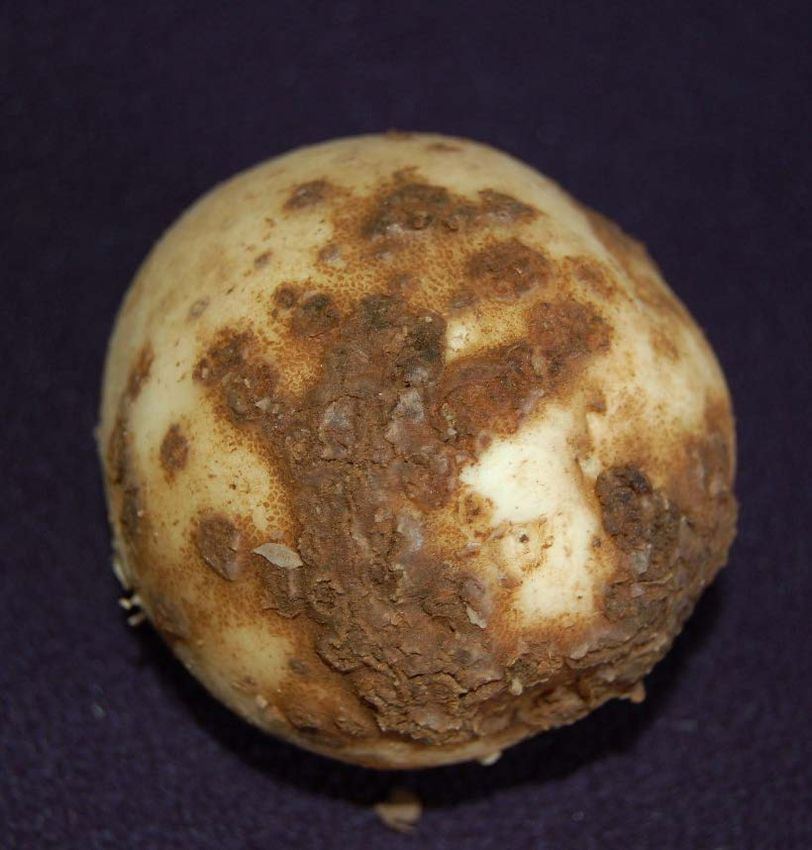
NDP 16 V2 - National Diagnostic Protocol for Synchytrium endobioticum

2            TAXONOMIC INFORMATION
Kingdom:            Fungi

Phylum:             Chytridiomycota

Class:              Chytridiomycetes

Order:              Chytridiales

Family:             Synchytriaceae

Genus:              Synchytrium

Species:            Synchytrium endobioticum (Schilb.) Pervical

Synonyms:           Chrysophlyctis endobioticum Schilb. (variant spelling C. endobiotica), Synchytrium
solani Massee

Common name: potato wart

    3                                                                   Subcommittee on Plant Health Diagnostics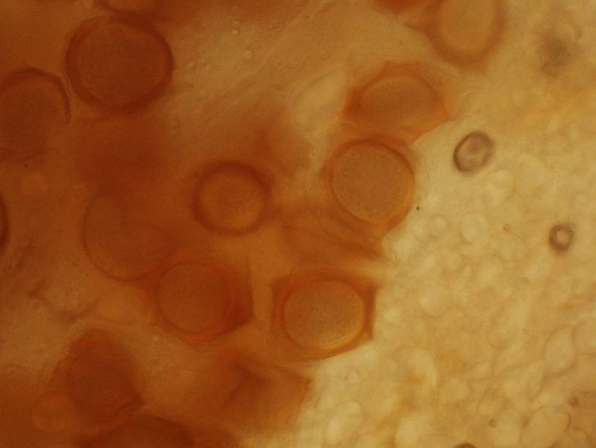
NDP 16 V2 - National Diagnostic Protocol for Synchytrium endobioticum
3 DETECTION
The detection of Synchytrium endobioticum relies on symptomatology and the microscopic detection
and identification of fungal spores. At the time of writing there were no rapid molecular diagnostic
techniques available for the identification of this pathogen. Molecular protocols specific for the
detection of potato wart have since been developed, and will need to be included once adequately
validated. However the current microscopic techniques are sufficient for identification.
S. endobioticum can be detected in soil using a wet-sieving technique (Pratt 1976; EPPO/OEPP 2003),
which separates S. endobioticum resting sporangia from soil particles of similar size by chloroform
flotation and centrifugation. Spores are then examined microscopically. For detection from soil, a
tomato bioassay (Hampson and Haard 1980; Hampson 1981) can be used to detect viable spores.
Stachewicz (1999) provides a method for soil sampling specifically for the detection of potato wart.
Methods for soil detection are not included in this protocol.
3.1 Symptoms
Potato plants infected by S. endobioticum do not usually show symptoms in above ground parts,
although occasionally there may be a reduction in vigour, and rarely, development of small greenish-
yellow warty growths at the stem base. Normally, however, all symptoms are confined to below
ground parts of the plant and no evidence of infection is seen until potato tubers are harvested
(Hampson 1993; DEFRA 2011).
The fungus causes symptoms of infection on all underground potato parts except true roots, with buds
on stems, stolons and tubers the main centres of infection (MPI 2009).
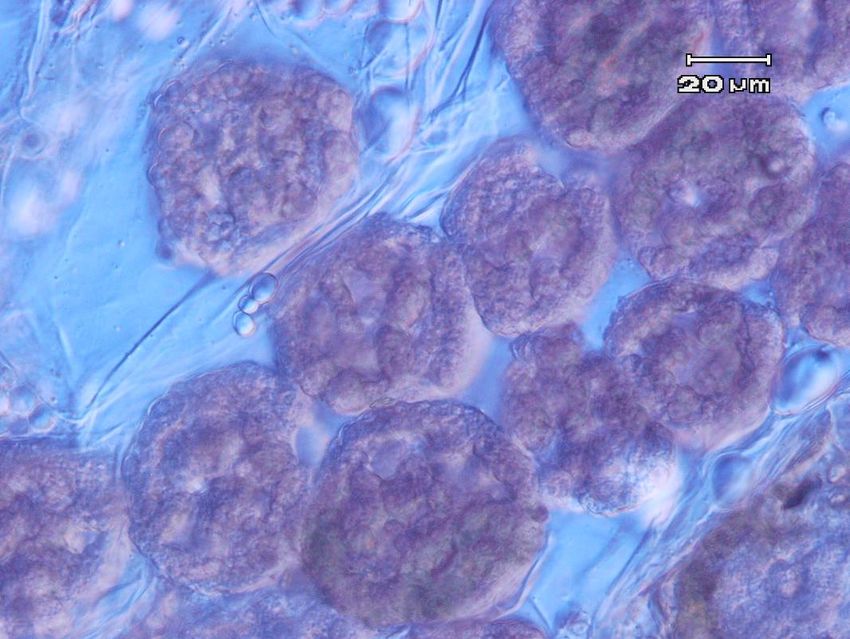
The most distinguishing symptom of potato wart disease is the gall, a characteristic warty, cauliflower-
like swelling that may be as small as a pin or as large as a fist, which forms on developing tubers
(Figure 1). The gall is soft and pulpy in texture, with a surface that is rough and corrugated, giving it
the warty appearance. The galls when formed underground or in storage, are the same colour as the
potato skin, however, if exposed to light, they will turn green. As the warts age they darken and decay
(Hampson 1993; DEFRA 2011)
Tubers may bear more than one warty outgrowth. If infected early, potato tubers can become so
distorted and spongy that they are almost unrecognisable, with entire tubers replaced by warts. In
some cases, a severe infestation of the tip of the stolon where the tuber is normally formed, will cause
a gall to develop instead of a tuber, subsequently destroying the potato crop by preventing tuber
production (Hampson 1993; MPI 2009; DEFRA 2011).
Some symptoms of powdery scab (Figure 2) and bud proliferation (Figure 3) can be mistaken for wart
disease (DEFRA 2011). However powdery scab spore balls are very different in appearance from
winter sporangia of S. endobioticum and microscopic examination quickly reveals the spongy
appearance of many spore balls made up of many small cysts. Some smut symptoms can also be
confused (Figure 4) however this disease is also not in Australia.
4 Subcommittee on Plant Health Diagnostics
NDP 16 V2 - National Diagnostic Protocol for Synchytrium endobioticum

                                                      B

                                           A

                                                                               C

  A

Figure 1. Warty galls on underground potato plant parts caused by Synchytrium endobioticum
infection.

Sources. A: © Plant Health & Environment Laboratory, Ministry for Primary Industries, New Zealand.
B: © Peter Wilkins, AsureQuality, New Zealand. C: © Department for Environment, Food and Rural
Affairs, UK.

   5                                                                    Subcommittee on Plant Health Diagnostics
NDP 16 V2 - National Diagnostic Protocol for Synchytrium endobioticum

Figure 2. Powdery scab (© SARDI)                                   Figure 3. Sprout proliferation on tuber
                                                                   (© SARDI)

Figure 4. Potato tuber in Bolivia showing symptoms of infection with potato smut (Thecaphora
solani). © William M. Brown Jr., Bugwood.org

   6                                                                    Subcommittee on Plant Health Diagnostics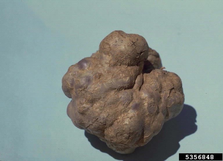
NDP 16 V2 - National Diagnostic Protocol for Synchytrium endobioticum

4            IDENTIFICATION
4.1          Microscopic examination for sporangia
Potato tubers can be examined directly by mounting a small portion of tissue from a warty growth in
lactic acid on a microscope slide.

Equipment required
        •   Scalpel blades
        •   Dissecting microscope
        •   Compound microscope (interference contrast optics preferred)
        •   Glass microscope slides and coverslips
        •   Small flame such as a cigarette lighter of spirit lamp (optional)
        •   Lactic acid (C3H6O3) ~98%

Method
        1. Using a scalpel blade make two incisions into the warty outgrowth, as thin as possible (less
           than 1 mm thick) and 3 mm long
        2. Remove the thin section of potato tissue and place on a glass microscope slide
        3. Add a droplet of lactic acid large enough to wet the entire section of potato tissue
        4. Place a coverslip over the potato tissue
        5. Examine the prepared slide with a compound microscope using at least the X40 objective.
           If sections are too thick or if it is difficult to visualise the sporangium morphology
           adequately, prepare another section, place in lactic acid and tease out with a pair of fine
           needles under the dissecting microscope, before adding a coverslip
        6. Optional: heat the microscope slide with the hand-held flame to remove any bubbles in the
           lactic acid

4.2          Morphology
    1. Compare structures seen with those illustrated in Section 4.2.1
    2. Winter sporangia (survival structure - see Appendix) are thick-walled and golden brown and
       embedded in the wart tissue, each sporangium filling its host cell almost completely. The thick
       outer wall is furrowed, irregularly thickened and bears prominent ridges which give the
       otherwise subglobose sporangium an angular appearance in median view. Winter sporangia
       measure 25 – 75µm (mean 50µm) in diameter
    3. The prominent ridges on the outer wall are diagnostic for S. endobioticum and serve to
       distinguish winter sporangia of this fungus from any other structures or organisms that might
       be encountered in potato warts or soil
    4. Summer sporangia (see Appendix) are of similar size to winter sporangia but are transparent
       and thin-walled and are unlikely to be found in mature warts. They lack the characteristic
       ridged wall of winter sporangia and their morphology is not diagnostic for potato wart.
    5. Other species of Synchytrium may be encountered in potato soils but are unlikely to be found in
       potato wart tissue. These other species lack the characteristic ridges on the winter sporangia.
    6. Powdery scab (caused by Spongospora subterranea) causes irregular scabby outgrowths on

    7                                                                   Subcommittee on Plant Health Diagnostics
NDP 16 V2 - National Diagnostic Protocol for Synchytrium endobioticum
potato tubers, but microscopic examination of scab contents reveals ovoid, irregular or
elongate, spongy spore balls composed of multiple, closely single spores (Fig. 15).
7. Potato smut (caused by Thecaphora solani) also causes warty swellings on potato tubers, but
the wart tissue contains black spores when examined microscopically (Fig 16). Potato smut is
absent from Australia.
4.2.1 Images to aid identification of Synchytrium endobioticum
sporangia
Figure 5. Live S. endobioticum sporangium: (L) resting and (R) with contents rounding off (Pratt 1976)
Figure 6. Dead S. endobioticum sporangium: (L) showing plasmolysis of contents and (R) without
contents (Pratt 1976)
8 Subcommittee on Plant Health DiagnosticsNDP 16 V2 - National Diagnostic Protocol for Synchytrium endobioticum Figure 7. Various stages of Synchytrium endobioticum sporangia, as published in Hampson et al. (1994). 1. Resting spores of S. endobioticum in potato gall tissue (bar = 25µm). The tissue was stained with Fleming’s triple stain: although some shrinkage took place, the spore walls conform to the surrounding cell walls. 2-5 Germinating resting spores of S. endobioticum (bar = 20µm). 2. Early stage with content in process of flowing into the extruded vesicle. 3. Similar stage, showing attachments to wall (arrow). 4. Mid stage. The connection to the mesospore wall are clear (arrows). 5. End stage. The filled vesicle darkens with time. 9 Subcommittee on Plant Health Diagnostics
NDP 16 V2 - National Diagnostic Protocol for Synchytrium endobioticum

Figure 8. (L) Group of S. endobioticum sporangia isolated from decayed warty tissue. (R) Two S.
endobioticum resting sporangia (Hedworth Foulkes 1910)

Figure 9. Synchytrium endobioticum resting sporangia with ruptured cell walls (Hedworth Foulkes
1910)

Figure 10. Starch grains in potato tissue, at 100, 400 and 1000 times magnification (left to right)
(Source: http://botit.botany.wisc.edu/images/130/Plant_Cell/starch_grains)

   10                                                                   Subcommittee on Plant Health Diagnostics
NDP 16 V2 - National Diagnostic Protocol for Synchytrium endobioticum

Figure 11. S. endobioticum resting spores at 20X (© IDCR-PHEL Ministry for Primary Industries, New
Zealand).

Figure 12. S. endobioticum resting spores at 40X (© IDCR-PHEL Ministry for Primary Industries, New
Zealand).

   11                                                                   Subcommittee on Plant Health Diagnostics
NDP 16 V2 - National Diagnostic Protocol for Synchytrium endobioticum

Figure 13. S. endobioticum individual resting spores (© IDCR-PHEL Ministry for Primary Industries,
New Zealand)

Figure 14. A single S. endobioticum resting spore in sol particles at 40X (© IDCR-PHEL Ministry for
Primary Industries, New Zealand)

   12                                                                   Subcommittee on Plant Health Diagnostics
NDP 16 V2 - National Diagnostic Protocol for Synchytrium endobioticum

Figure 15. Spongospora subterranean spore balls at 40X (© IDCR-PHEL Ministry for Primary
Industries, New Zealand)

Figure 16. Thecaphora solani Spore balls from potato tuber, 400X. - BPI 179335. Chalkley, D.
Systematic Mycology and Microbiology Laboratory, ARS, USDA.

   13                                                                   Subcommittee on Plant Health Diagnostics
NDP 16 V2 - National Diagnostic Protocol for Synchytrium endobioticum

5            CONTACTS FOR FURTHER
             INFORMATION
AUSTRALIA                                               AUSTRALIA

James Cunnington                                        Dolf De Boer

DAWR                                                    Victorian Department of Primary Industries

james.cunnington@agriculture.gov.au                     dolf.deboer@ecodev.vic.gov.au

                                                        THE NETHERLANDS

                                                        Peter Bonants

                                                        Plant Research International B.V.

                                                        Business Unit Biointeractions & Plant Health

                                                        PO BOX 16

                                                        6700 AA Wageningen

                                                        The Netherlands

                                                        Phone: 31 317 476213

                                                        peter.bonants@wur.nl

…..

      14                                                                Subcommittee on Plant Health Diagnostics
NDP 16 V2 - National Diagnostic Protocol for Synchytrium endobioticum
6 ACKNOWLEDGEMENTS
This diagnostic manual was originally compiled by:
Bonny Rowles-van Rijswijk, Dr James Cunnington, Dr Joanne Luck
Department of Primary Industries, Knoxfield
Private Mail Bag 15
Ferntree Gully Delivery Centre
Victoria 3156, Australia
Reviewed and edited by Ian Pascoe, Consulting Mycologist, pascoeig@bigpond.net.au
Procedures verified by Ministry for Primary Industries (previously MAF Biosecurity New Zealand).
…….
15 Subcommittee on Plant Health DiagnosticsNDP 16 V2 - National Diagnostic Protocol for Synchytrium endobioticum
7 REFERENCES
DEFRA (2011) Department for Environment, Food and Rural Affairs
https://planthealthportal.defra.gov.uk/assets/factsheets/pwd.pdf [accessed 20 Feb 2017]
EPPO/OEPP (2003) Synchytrium endobioticum: soil tests and descheduling of previously infested plots.
EPPO Standards - Phytosanitary procedures PM 3/59 (2),
http://archives.eppo.int/EPPOStandards/PM3_PROCEDURES/pm3-059-2-en.pdf
Hampson MC (1979) Potato wart disease in Newfoundland. Canadian Agriculture 24:20-23.
Hampson MC (1981) Infection of additional hosts of Synchytrium endobioticum, the causal agent of
potato wart disease: 3. Tomato as an assay tool in potato wart disease. Canadian Plant Disease
Survey 61:15-18.
Hampson MC (1993) History, biology and control of potato wart disease in Canada. Canadian Journal
of Plant Pathology 15:223-244.
Hampson MC and Haard NF (1980) Pathogenesis of Synchytrium endobioticum: 1. Infection responses
in potato and tomato. Canadian Journal of Plant Pathology 2:143-147.
Hampson MC and Proudfoot KG (1974) Potato wart disease, its introduction to North America,
distribution and control problems in Newfoundland. FAO Plant Protection Bulletin 22:53-64.
Hampson MC, Yang AF and Bal AK (1994) Ultrastructure of Synchytrium endobioticum resting spores
and enhancement of germination using snails. Mycologia 86:733-740.
Hedworth Foulkes P. (1910) Wart diseases of potatoes. Synchytrium endobioticum, Percival. Chronicle
Office, Shrewsbury, UK.
Hooker WJ (ed.) (1990) Compendium of Potato Diseases. American Phytopathological Society,
Michigan, USA, pg. 36
Matskiv TI, Melnik PA and Golik IV (1998) Definition and distribution of aggressive pathotypes of
Synchytrium endobioticum in Ukraine. Bulletin OEPP/EPPO 28:539-542.
MPI (2009) Potato Wart. Source: http://www.biosecurity.govt.nz/pests/potato-wart). Accessed
September 2016.
Pratt MA (1976) A wet-sieving and flotation technique for the detection of resting sporangia of
Synchytrium endobioticum in soil. Annals of Applied Biology 86:21-29.
Stachewicz H (1999) Phytosanitary procedures; Synchytrium endobioticum: soil tests and descheduling
of previously infested plots. Bulletin OEPP/EPPO Bulletin 29:225-231.
Walker JC (1983) CMI Descriptions of Pathogenic Fungi and Bacteria; No. 755, Synchytrium
endobioticum. Commonwealth Mycological Institute, Surrey, UK.
Weiss F (1925) The conditions of infection in Potato wart. American Journal of Botany 12:413-443.
16 Subcommittee on Plant Health DiagnosticsNDP 16 V2 - National Diagnostic Protocol for Synchytrium endobioticum
7.1 Related Websites
Australasia
http://www.biosecurity.govt.nz/pests/potato-wart
North America
http://www.inspection.gc.ca/english/plaveg/pestrava/synend/tech/synende.shtml
http://massnrc.org/pests/pestFAQsheets/potatowart.html
https://www.aphis.usda.gov/aphis/ourfocus/planthealth/plant-pest-and-disease-programs/pests-
and-diseases/SA_Nematode/SA_Potato/CT_Potatowart
Useful references:
EPPO/OEPP (2004) EPPO Standards - Diagnostic protocols for regulated pests PM 7/28, Synchytrium
endobioticum. Bulletin OEPP/EPPO Bulletin 34:213-218
Glynne MD (1924) Infection experiments with wart disease on potatoes. Synchytrium endobioticum
(Schilb.) Perc. Annals of Applied Biology 12:34-60.
Hampson MC and Thompson PR (1977) A quantitative method to examine large numbers of soil
samples for Synchytrium endobioticum, the cause of potato wart disease. Plant Soil 46:659-664.
Hampson MC, Wood SL and Coombes JW (1996) Detection of resting spores of Synchytrium
endobioticum in soil from vehicles at Port-aux-Basques, Newfoundland. Canadian Journal of Plant
Pathology 18:59-63.
Kole AP (1965) Resting-spore germination in Synchytrium endobioticum. Het kiemen van de rustspore
by Synchytrium endobioticum. Netherlands Journal of Plant Pathology 71:72-78.
..
17 Subcommittee on Plant Health DiagnosticsNDP 16 V2 - National Diagnostic Protocol for Synchytrium endobioticum
8 APPENDICES
8.1 Life Cycle
Synchytrium endobioticum belongs to the fungal phylum Chytridiomycota, a large, primitive group of
fungi regarded as basal to the true fungi. The fungus is an obligate parasite that depends entirely on its
host, Solanum tuberosum for development and reproduction. Its simple, coenocytic thalli do not
develop hyphae but develop into sporangia containing large numbers of flagellate zoospores.
As S. endobioticum does not produce hyphae, infection is initiated by spherical haploid zoospores 1.5-
3.0 µm in diameter, which penetrate epidermal cells of meristematic tissues of growing points, buds,
stolon tips, or young leaf primordia of the potato tuber and lower stem. The invaded and surrounding
cells enlarge, and rapid cell division following infection from either zygotes or haploid zoospores
causes an increase in meristematic tissue, providing additional infection courts. This can occur
relatively quickly, as zoospores encyst and penetrate epidermal cells of susceptible tissue
approximately 2 hours after formation. The zoospores swell into prosori, and then develop into sori.
The prosori are oval, aseptate, smooth, thick walled, light golden brown, 40-50 µm in diameter, and
usually lie at the bottom of the infected plant cell. There may be up to 4 prosori per infected plant cell.
Contents escape the prosorus through the prosorus outer wall to form an ovoid, flattened or spherical
haploid sorus of sporangia with each sorus containing 1-9 hyaline, thin-walled summer sporangia
which quickly release zoospores to initiate new infection sites (Hampson and Proudfoot 1974; Hooker
1990; Walker 1983).
Winter sporangia (resting spores or meiosporangia) are the characteristic survival and dispersal
spores of the fungus and are released from decaying warts (Fig 2). Under normal environmental
conditions, the hyperplasic tissue quickly senesces and the disease passes through the “black wart” or
“black scab” stage. The sub-epidermal tumour tissue is packed with winter sporangia and as the wart
decays, sporangia are released into the surrounding soil. They are golden brown, spheroidal,
measuring 35-80 µm in diameter. The thick sporangium wall is prominently ridged, and this
morphology is diagnostic for Synchytrium endobioticum. These winter sporangia can survive between
potato crop rotations for very long periods of time and can remain viable for at least 30 years.
Techniques have been developed to test soils for presence of winter sporangia and to determine
numbers of viable sporangia in old infested soils (Pratt 1976; EPPO/OEPP 2003).
After release from the decaying wart tissue, sporangia germinate to release zoospores into free water
(Hampson and Proudfoot 1974; Hampson 1993). “Germination of both resting [winter] and soral
[summer] sporangia occurs in water, and there is an indispensable minimum of water for the
distribution of the motile cells. If the soil-moisture content does not at any time reach saturation,
germination is prevented, but if it is constantly near saturation, infection is repressed, probably
through the reaction on the host. The most favourable condition is periodic flooding, followed by
drainage and aeration. Infection may occur, if the temperature is favourable, in soil that is wet at
insufficient intervals to afford a normal crop” (Weiss 1925). After studying disease peaks, Hampson
(1979) found that the fungus could control its own germination rate, to some extent, with dormancy
varying from a few months to thirty years or more. This long period of infectiveness creates one of the
major problems in controlling potato wart disease (Hampson and Proudfoot 1974). The second major
problem in controlling potato wart disease is that by natural mutation, adaptation and hybridisation,
18 Subcommittee on Plant Health DiagnosticsNDP 16 V2 - National Diagnostic Protocol for Synchytrium endobioticum the fungus readily changes its pathogenic properties to develop races favoured by the local environmental conditions (Hooker 1990; Matskiv et al. 1998). …… 19 Subcommittee on Plant Health Diagnostics
You can also read