Next-Generation Sequencing of Cerebrospinal Fluid for the Diagnosis of VZV-Associated Rhombencephalitis
←
→
Page content transcription
If your browser does not render page correctly, please read the page content below
J. Integr. Neurosci. 2023; 22(2): 36
                                                                                                               https://doi.org/10.31083/j.jin2202036

Original Research
Next-Generation Sequencing of Cerebrospinal Fluid for the Diagnosis
of VZV-Associated Rhombencephalitis
Jingzhe Han1,† , Zhihua Si1,† , Na Wei1 , Duanhua Cao1 , Ye Ji2 , Zhilei Kang3 , Jianguo Zhu1, *
1 Department  of Neurology, Hengshui People’s Hospital, 050000 Hengshui, Hebei, China
2 Department  of Neurological Function Examination, Hengshui People’s Hospital, 050000 Hengshui, Hebei, China
3 Department of MRI, Hengshui People’s Hospital, 050000 Hengshui, Hebei, China

*Correspondence: hyzhjg@126.com (Jianguo Zhu)
† These authors contributed equally.

 Academic Editor: Gernot Riedel
  Submitted: 11 August 2022 Revised: 3 September 2022 Accepted: 8 September 2022 Published: 16 February 2023

Abstract
Background: Rhombencephalitis (RE) is a general term for a group of inflammatory diseases of the rhombencephalon caused by dif-
ferent etiologies. Patients of RE caused by the varicella-zoster virus (VZV) are sporadic in medical practice. The VZV-RE is easily
misdiagnosed and causes a poor prognosis for patients. Methods: In this study, we analyzed the clinical symptoms and imaging features
of five patients with VZV-RE diagnosed by the next-generation sequencing (NGS) technique of cerebrospinal fluid. Magnetic resonance
imaging (MRI) examination was used to characterize the imaging of the patients. The McNemar test was used to analyze the cerebrospinal
fluid testing (CSF) values and MRI test of the 5 patients. Results: We finally used NGS technology to confirm the diagnosis in 5 patients
with VZV-RE. MRI revealed T2/FLAIR high signal lesions in the patients’ medulla oblongata, pons, and cerebellum. All patients had
early signs of cranial nerve palsy; some had herpes or pain in the corresponding cranial nerve distribution areas. The patients develop
headaches, fever, nausea, vomiting, and other signs and symptoms of brainstem cerebellar involvement. McNemar’s test showed no
statistical difference between multi-mode MRI and CSF values for diagnosing VZV-RE (p = 0.513). Conclusions: This study showed
that patients with herpes in the skin and mucous membranes at the distribution area of the cranial nerves and with the underlying disease
were prone to RE. We suggest that the NGS analysis should be considered and selected based on the level of parameters, such as MRI
lesion characteristics.

Keywords: varicella-zoster virus; rhombencephalitis; cerebrospinal fluid; next-generation sequencing; magnetic resonance imaging

 1. Introduction                                                                ever, both the isolation and identification of VZV are very
                                                                                time-consuming, and the detection rate of the virus is usu-
     Rhombencephalitis (RE) is a rare rhombencephalon
                                                                                ally low. At the same time, PCR is limited by the nucleic
inflammatory syndrome involving the brain stem and cere-
                                                                                acid fragments bound by its primers. Therefore, both of
bellum, which is caused by numerous etiologies [1]. The
                                                                                these assays are limited in clinical application.
rhombencephalon consists of metencephalon and myelen-
cephalon. The metencephalon includes the pons, cere-                                  Next-generation sequencing (NGS) technology is
bellum, and pontine part of the fourth ventricle. The                           characterized by unrestricted targeting primers, fast detec-
myelencephalon consists of the medulla oblongata and the                        tion speed, and a high detection rate, which can quickly and
medulla oblongata of the fourth ventricle. The causes of RE                     efficiently diagnose infectious diseases of the central ner-
include infectious, autoimmune, and paraneoplastic syn-                         vous system. Since it was first reported in 2014, NGS has
dromes. The most common infectious agents are Listeria,                         been progressively promoted and applied in detecting cere-
enterovirus 71, and herpes viruses, while RE caused by the                      brospinal fluid pathogens [3–5]. There are many causes
varicella-zoster virus (VZV) is very rare [2].                                  of RE, while VZV-RE is relatively rare clinically. Clin-
      VZV belongs to a double-stranded DNA herpes virus.                        icians are prone to delay the diagnosis and treatment of
When a primary infection occurs in a patient, VZV often                         the disease, which can easily lead to a poor patient prog-
lurks in the patient’s cranial nerves, dorsal nerve roots, and                  nosis. Therefore, this study intends to analyze the clinical
autonomic ganglia. When cell-mediated immunity is re-                           and imaging characteristics of five patients with VZV-RE
duced in the elderly and immunosuppressed patients, VZV                         diagnosed by cerebrospinal fluid NGS techniques to im-
can become activated and secondary to infectious disease in                     prove clinicians’ knowledge of the disease; Provide readers
the central nervous system. Activation of VZV is an infec-                      with an understanding of rhombencephalitis and, in partic-
tious disease secondary to the central nervous system. Virus                    ular, to increase their awareness of the clinical and imag-
isolation combined with polymerase chain reaction (PCR)                         ing features of VZV-associated rhombencephalitis; Enrich
detection is the “gold standard” for diagnosing VZV. How-                       the imaging features of this infectious disease of the cen-

                Copyright: © 2023 The Author(s). Published by IMR Press.
                This is an open access article under the CC BY 4.0 license.
Publisher’s Note: IMR Press stays neutral with regard to jurisdictional claims in published maps and institutional affiliations.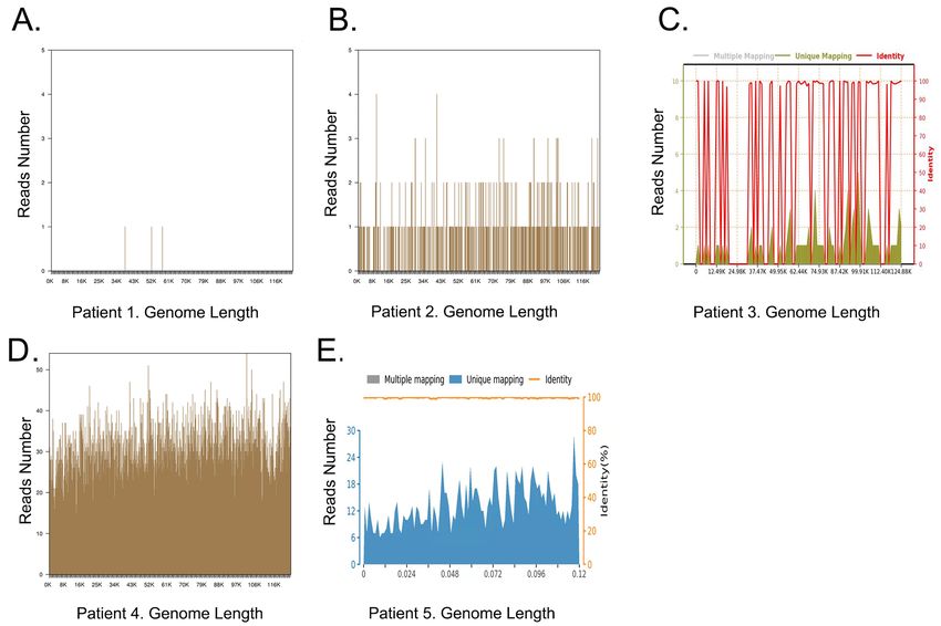
tral nervous system and to select the appropriate pathogen       constructed through end-repair, poly(A)-tailing, adapter
screening modality promptly for early diagnosis, treatment,      ligation, and PCR amplification. Roller amplification tech-
and improved prognosis of the VZV-associated rhomben-            nology was used to increase single-stranded circular DNA
cephalitis patients.                                             by 2–3 sets to obtain DNA nanospheres. (4) Sequencing:
                                                                 The DNA nanospheres were loaded on the sequencing chip,
2. Materials and Methods                                         and the BGISEQ-50 sequencing platform (Institute of Med-
2.1 Patients                                                     ical Laboratory, Tianjin BGI Technology Co., Ltd.Tianjin,
                                                                 China) was used to sequence the DNA nanospheres.
      A total of 5 patients (4 males and one female) with
VZV-RE were hospitalized and diagnosed in the Depart-                  After the data analysis and quality control sequencing
ment of Neurology of our hospital from January 2019 to           data were put on the machine, the reads with low quality,
January 2021. The median age of the patients was 63.8            low complexity, and sequencing length 1 is defined as positive.
clinical features were consistent with the symptoms and
signs of RE (cranial nerve palsy, cerebellar ataxia, long        2.2.2 Antibodies Detection
pyramidal signs, disturbance of consciousness, etc.); (2) Pa-
tients with lumbar puncture cerebrospinal fluid examina-               Indirect immunofluorescence assay (IIF) was used for
tion: white blood cell (WBC) count >8 × 10⁶ /L; (3) Imag-        autoimmune encephalitis antibodies. The Cell-based as-
ing examination was consistent with the imaging features of      say (CBA) has high diagnostic specificity and sensitivity
RE (T2/FLAIR hyperintense lesions were mainly located in         for RE. The initial dilution titers of cerebrospinal fluid
the pons, medulla oblongata, cerebellum and other parts);        testing (CSF) and serum were 1:1 and 1:10, respectively
(4) NGS of cerebrospinal fluid detected human VZV se-            (Supplementary Fig. 1).
quences; (5) Clinical and laboratory tests have ruled out ev-
                                                                 2.3 Statistical Methods
idence of other microbial infection; (6) Patients who signed
informed consent.                                                      Means ± standard deviations were used to represent
      Exclusion criteria of patients were: (1) Non-infectious    continuous data that conformed to a normal distribution;
encephalitis, such as autoimmune encephalitis (AE), para-        medians (25%, 75%) were used for continuous data that
neoplastic marginal encephalitis (PLE), neuromyelitis op-        did not conform to a normal distribution. The chi-square
tica spectrum diseases (NMOSDs), immune rheumatic dis-           test was used for statistical analysis of the count data. The
eases involving the central nervous system; (2) Recent 4-        McNemar test was used to analyze the CSF values and MRI
week history of vaccination; (3) Patients who had con-           test results for the 5 patients. The patients were divided into
traindications to lumbar puncture or inability to obtain cere-   three groups according to the number of leukocytes in the
brospinal fluid.                                                 CSF: the mild group (500 × 109 /L).
2.2 Methods                                                      The imaging diagnosis was based on the MRI’s number of
2.2.1 NGS of Cerebrospinal Fluid                                 brainstem lesions. It was divided into three groups: the mild
                                                                 group (No brainstem lesions), the moderate group (Brain-
       NGS of Cerebrospinal fluid: (1) Specimen collec-
                                                                 stem lesions), and the severe group (Including lesions out-
tion: 1–2 mL cerebrospinal fluid was obtained by lum-
                                                                 side the brainstem).
bar puncture in patients, dripped into test tubes and stored
in a refrigerator at –80 °C for 30 min and used for NGS
assays. (2) Sample extraction and quality control: The
                                                                 3. Results
DNA of cerebrospinal fluid samples was extracted using a         3.1 Clinical Findings
micro-sample genomic DNA extraction kit (DP316, TIAN-                  We used the NGS of CSF to identify the DNA se-
GEN BIOTECH, Beijing, China), and fragmented into                quence of VZV. The patients’ clinical features with VZV-
200–300 bp fragments by DNA cutting ultrasonic disrup-           RE were summarized in Table 1 and Fig. 1. The patients’
tor(Bioruptor Pico, Diagenode, Belgium). After quality           lumbar puncture pressure values, cerebrospinal fluid pro-
control of the pieces’ size by a 2100 Bioanalyzer, the quality   tein concentration, glucose, chloride, total blood choles-
control DNA library’s concentration was detected by quan-        terol, triglyceride, the WBC and lymphocyte counts, and
titative PCR. (3) Library construction: DNA libraries were       the number of sequences (NGS) are summarized in Ta-

2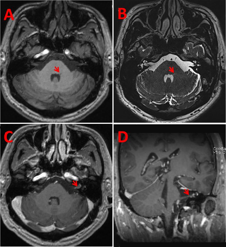
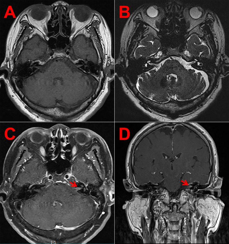
Fig. 1. Nucleotide Position along Human alphaherpesvirus 3 (Varicella-zoster virus) Genome. (A) Patient 1. (B) Patient 2. (C)
Patient 3. (D) Patient 4. (E) Patient 5.

ble 2. The oligoclonal bands (OCB), aquaporin 4 (AQP4),
myelin oligodendrocyte glycoprotein (MOG) antibodies,
anti-GQ1b antibodies, and autoimmune encephalitis anti-
bodies in all of the patients were negative. All five patients
ruled out the diagnosis of HIV, syphilis, and hepatitis B in-
fection. The patient’s autoantibody spectrum and immune
abnormalities were both normal.

3.2 Case 1
     A 66-year-old man presented with cerchnus and dys-
phagia three days pre-hospitalization. This patient showed
dysarthria and slowly pharyngeal reflex. The brain MRI
showed low T1, high T2 and DWI signals in the medulla
oblongata’s left dorsal lateral position (Fig. 2A–C). Seven
days later, the patient showed peripheral facial paralysis and
herpes of the external auditory canal. Brain MRI reexami-
nation revealed high DWI signals in the left cerebellar pe-
duncles (Fig. 2D). This patient was finally diagnosed with
VZV-RE and treated with acyclovir and immunoglobulins.
After 12 weeks of follow-up, the patient still had left-sided
                                                                 Fig. 2. MRI results of patient 1. The MRI showed low
peripheral facial palsy symptoms.
                                                                 T1-MPRAGE, high T2 (3D-CISS), and DWI signals in the left
3.3 Case 2                                                       medulla (A–C). Reexamination of brain MRI revealed high DWI
      A 41-year-old male patient developed facial distortion     signals in the left brachium pontis (D).
and dizziness 7 days before admission. The patient had
many mottled blisters on the left auricle, and peripheral fa-    of the internal auditory canal (Fig. 3C,D). The patient was
cial palsy occurred on the left side. MRI testing results        treated with acyclovir and immunoglobulins. Six months
revealed low T1 and high T2 signals in the left cerebellar       after discharge, the patient’s hearing loss in the left ear had
medius of the patient’s (Fig. 3A,B). The MRI showed the          not improved.
enhanced vestibulocochlear and facial nerve at the bottom

                                                                                                                              3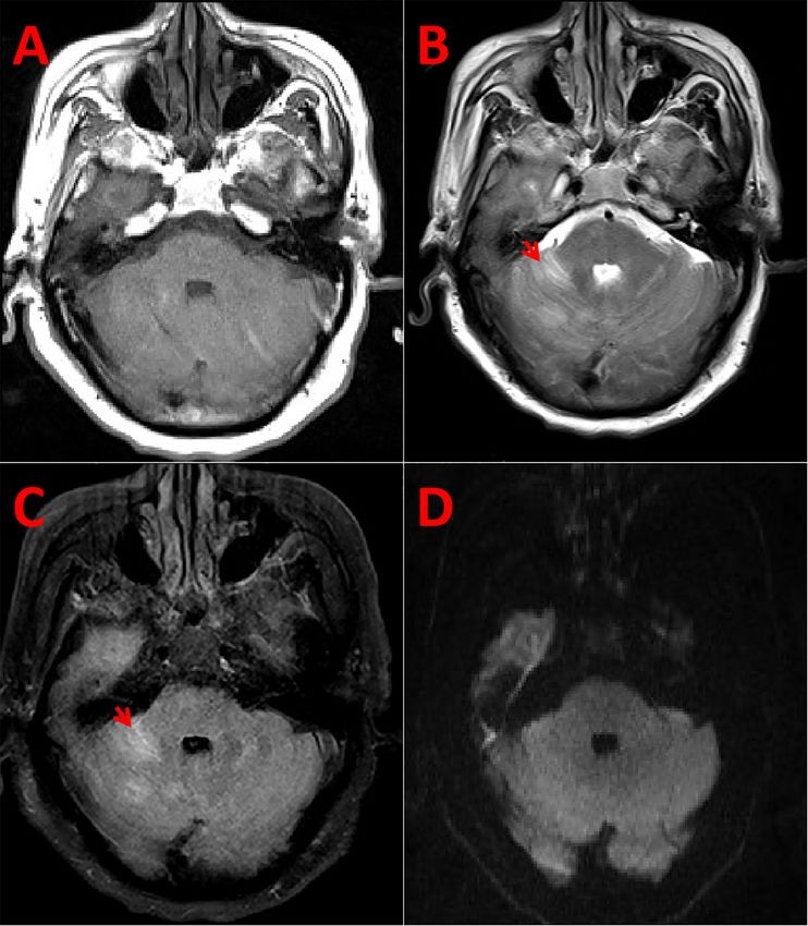
4

                                                                              Table 1. Clinical features of the five patients with VZV-RE.
    Case NO. Age Cranial nerve          Underlying          Location of     Fever Headache Epilepsy Meningeal          Antibody testing    Neuroimaging features                        Treatment        Prognosis
                                         disease              herpes                                irritation
    1         66 VII, VIII, IX, X Hypertension                Auricle       None   None      None      None        OCB, AQP4, MOG       High T2 signals, FLAIR and DWI signals in Acyclovir + im- Peripheral
                                                                                                                  antibodies anti-GQ1b the left medulla and left brachium pontis. munoglobulins facioplegia
                                                                                                                  antibody, AE antibody
                                                                                                                      are all negative
    2         41      VII, VIII            NO                 Auricle       None    None     None      None                 idem           MRI of the brain after admission revealed high Acyclovir + im- Hearing loss
                                                                                                                                           T2, FLAIR, and DWI signals in the left pedun- munoglobulins
                                                                                                                                           culus cerebellaris medius. The gadolinium-
                                                                                                                                           enhanced MRI bottom showed that the facial
                                                                                                                                           nerve and vestibulocochlear nerve at the inter-
                                                                                                                                           nal auditory canal was enhanced.
    3         65      VII, VIII     Hypertension   External                 Yes     Yes      None      Yes                  idem           Head MRI of the brainstem showed no signifi- Acyclovir        Peripheral
                                    and diabetes auditory canal                                                                            cant abnormalities. Contrast-enhanced MRI of                  facioplegia
                                                                                                                                           the internal auditory canal revealed abnormal
                                                                                                                                           enhancement of the auditory nerve on the left
                                                                                                                                           internal auditory canal’s inner surface.
    4         72        VII         Hypertension             External       Yes     Yes       Yes      Yes                  idem           Head MRI revealed high T2 and FLAIR sig- Acyclovir + im- Death
                                                           auditory canal                                                                  nals in the brainstem and cerebellum.    munoglobulins
    5         75        VII          Hypertension    External     Yes     Yes      None      Yes                idem            Head MRI showed T2/Flair hyperintensity in Acyclovir + im- Peripheral
                                     and diabetes auditory canal                                                                the back of the left pons.                 munoglobulins facioplegia
    VZV, Varicella-Zoster Virus; RE, Rhombencephalitis; DWI, Diffusion-Weighted Imaging; FLAIR, Fluid attenuated inversion recovery; T2, T2 weighted image; MRI, Magnetic Resonance Imaging; OCB,
    Oligoclonal Bands; AQP4, Aquaporin Protein 4; MOG, Myelin Oligodendrocyte Glycoprotein; AE, Autoimmune Encephalitis.

                                                                            Table 2. Laboratory features of the five patients with VZV-RE.
                                                                                                                 CSF
    NO.
          Pressure (mmH2 O)       WBC   (×106   cells/L)     LYM (%) (60–70)          Protein (g/L)     Glucose (mmol/L)         Chloride (mmol/L)     NGS (number of       Total cholesterol       Triglycerides
               (80–180)                  (
Fig. 4. MRI results of patient 3. MRI showed T1-MPRAGE
Fig. 3. MRI results of patient 2. MRI showed low T1-MPRAGE
                                                                     and high T2 (3D-CISS) signals in the brainstem (A,B). Head
and high T2 (3D-CISS) in the left brachium pontis (A,B). The fa-
                                                                     MRI enhancement showed that the central canal’s facial nerve and
cial nerve and vestibular nerve in the central canal showed an en-
                                                                     vestibular nerve had an enhanced signal (C,D, red arrow).
hanced sign in head MRI (red arrow), and the brain stem showed
no abnormal enhanced signal (C,D).
                                                                     brainstem and cerebellum (Fig. 5A–D). Cytologic analysis
                                                                     revealed abnormal cerebrospinal fluid cytology. The sig-
                                                                     nificant cellular abnormalities were manifested by lympho-
3.4 Case 3
                                                                     cyte reaction, activated monocytes, 2% plasma cells, 2%
      A 65-year-old woman was admitted due to a deviation            erythrophage, and the whole field of erythrocytes. The pa-
of the mouth for seven days and headache accompanied by              tient was diagnosed with VZV-RE and meningoencephalitis
dizziness with fever for one day. Fast-phase nystagmus to-           and received acyclovir and immunoglobulin combined with
ward the right level was seen in both eyes of the patient.           ventilator-assisted therapy.
The patient showed herpes in the left side of the external au-
ditory canal, peripheral facial paralysis, and periodic limb         3.6 Case 5
movement disorder. Contrast-enhanced MRI of the internal                   The patient was a 75-year-old man admitted due to
auditory canal revealed abnormal enhancement of the audi-            deviated mouth for seven days and fever accompanied by
tory nerve on the left internal auditory canal (Fig. 4D). Al-        psycho-behavioural abnormalities for three days. The pa-
though there was no abnormal signal on MRI (Fig. 4A–C),              tient presented drowsiness, irritability, suspected herpes in
VZV-RE could still be diagnosed with brainstem involve-              the left auricle, and peripheral facial palsy on the left side.
ment symptoms and signs. The patient was treated with                The patient’s neck resistance was positive. The patient’s
acyclovir. Two months after discharge, the patient’s dizzi-          head MRI showed a high density of Flair in the left cerebral
ness improved, and he could walk independently but still             bridge (Fig. 6). The patient was diagnosed with VZV-RE.
had left peripheral facial paralysis.                                After combined treatment with acyclovir and immunoglob-
                                                                     ulin, the patient’s consciousness became clear, psychiatric
3.5 Case 4                                                           symptoms disappeared. Three-month follow-ups showed
      A 72-year-old man was admitted due to a headache               that the patient regained the ability to live normally but still
for five days and convulsions accompanied by unconscious-            had peripheral facial palsy symptoms.
ness for one day. The patient developed a coma with a GCS
of 4 (E1V1M2), a pupil diameter of 2.0 mm, and a slow light          3.7 McNemar Test for Diagnostic Methods
reflex was detected. He presented with a herpes scab in                   The McNemar’s test was used to analyze the CSF val-
the left external auditory canal, left peripheral facial paral-      ues and MRI test of the 5 patients (Table 3). In the present
ysis, and uncooperative muscle strength of the extremities.          study, there were no diagnostic differences in any of the
The patient had a positive response to the neck resistance           three indices (p = 0.400).
test. MRI of the brain revealed high FLAIR signals in the

                                                                                                                                   5
Table 3. McNemar’s test was used to analyze the results of
CSF values and MRI test.
No-severe Severe Total p
No-severe 3 1 4 0.400
Group
Severe 0 1 1
Total 3 2 5
can be involved. Patients with this disease may present
with clinical features such as long pyramidal tract signs
(corticospinal tract, spinothalamic tract, posterior column),
symptoms of cerebellar involvement (hemiataxia, vertigo,
cerebellar dysarthria), disturbance of consciousness, and
respiratory failure [1]. Some patients may also be ac-
companied by the symptoms such as headache, fever, and
meningismus. MRI has a significant diagnostic value
for RE patients. The main imaging manifestations are
T2/FLAIR high signal lesions in the brain, medulla oblon-
gata, upper cervical spine and cerebellum. Some patients
may have only clinical signs and symptoms but lack typical
MRI imaging [2]. In this study, all patients met the clinical
manifestations of RE, and T2/FLAIR hyperintense lesions
Fig. 5. MRI results of patient 4. The MRI of the head showed in the medulla oblongata, pons, cerebellum, and other parts
high T2/Flair signals (B and C, red arrow) and normal T1/DWI were observed on head MRI. Although there was no clear
signals in brachium pontis (A and D). A, T1; B,T2; C, Flair; D abnormal signal in the brainstem, ataxia on the left side was
DWI.
observed, suggesting that the brainstem or cerebellum was
also affected. These MRI changes in the patient were con-
sistent with the diagnostic imaging features of RE.
There are numerous causes of RE, focusing on eti-
ology and differential diagnosis [1,2]. A range of infec-
tious and non-infectious diseases can cause the develop-
ment of RE. In this study, the results of oligoclonal bands
(OCB), AQP4, MOG antibodies, anti-GQ1b antibodies, au-
toimmune encephalitis antibodies and autoantibody tests in
the patients’ cerebrospinal fluid were not found to be ab-
normal. These test results largely exclude the possibility
of RE triggered by immune-related diseases. In this study,
all patients underwent lumbar puncture, and the results sug-
gested an “inflammatory” response (elevated cerebrospinal
fluid leukocyte count), and the diagnosis of infectious RE
was largely confirmed. Due to the wide variety of infec-
tious agents causing RE, conventional diagnostic methods
are more challenging to reach a final diagnosis. In contrast,
NGS technology can obtain information on pathogenic mi-
croorganisms directly from the patient’s diseased brain tis-
sue and cerebrospinal fluid, which will significantly shorten
Fig. 6. MRI results of patient 5. MRI showed high Flair signals
the diagnostic testing period for RE patients. In one study,
(red arrow) in the left medulla.
Guan et al. [4] used NGS to test the cerebrospinal fluid of
four patients suspected of having viral encephalitis. They
4. Discussion detected two cases of herpes simplex virus type 1 (HSV1),
one case of herpes simplex virus type 2 (HSV2), and one pa-
RE is encephalitis involving the brainstem and/or tient with VZV with genome-wide coverage of 12%–98%,
cerebellum. Cranial nerve palsy is the disease’s most com- respectively [4]. Their results indicate the significant value
mon symptom, with a median incidence of about 75% of NGS for diagnosing pathogenic CNS infections. In the
(67%–86%), and the VII, VI, IX, X, and V cranial nerves present study, NGS was performed on the cerebrospinal
6fluid in all patients. Our results showed that the patients de- palsy caused by VZV infection. It has been found that cy-
veloped VZV CNS infection. It further clarifies the diagno- totoxic edema can be triggered in the early stages of nerve
sis of VZV-RE and excludes RE caused by other pathogens. cell damage caused by VZV infection, with DWI showing
In addition, the differential diagnosis needs to consider the a high signal [9]. However, the brightness of the DWI sig-
possibility of paraneoplastic syndrome-associated RE and nal was significantly lower in patients with acute cerebral
brainstem lymphoma. In this study, no lymphoma-like en- infarction, and the signal duration was more transient. In
hancement was seen in the brainstem lesions, and the pa- this study, although some patients exhibited high DWI sig-
tient’s follow-up and disease progression did not support nal intensities, these signals were significantly lower than
the lymphoma diagnosis. Based on the patient’s history those of patients with acute cerebral infarction. Instead,
and clinical auxiliary examination, the RE diagnosis can these high-intensity areas coincided with the vascular distri-
be ruled out as caused by progressive multifocal leukoen- bution areas but overlapped with the routes of cranial nerves
cephalopathy and radiation encephalopathy. VZV is neu- into the cranial brain. Therefore, we believe it is more likely
rophilic; when it infects patients, it often lurks in cranial that the VZV virus directly invades the brainstem.
nerves, dorsal spinal nerve root, and autonomic ganglion. In this study, although all patients received active an-
In the elderly or immunosuppressed, the virus can reactivate tiviral and immunosuppressive therapy, short-term follow-
and extend the nerve retrogradely into the skull resulting in up showed poor prognosis, indicating that once damage oc-
Encephalitis, meningitis, myelitis, and acute cerebrovascu- curred in the brain stem and parenchyma, the patient’s re-
lar diseases. In this study, the patients were all older, and covery period would be significantly prolonged. Simulta-
most had underlying diseases such as hypertension and di- neously, NGS technology found that the number of VZV
abetes, activating factors for VZV. copy sequences was closely related to patients’ prognoses.
A high copy number of genes tested for VZV in RE pa-
This study showed that patients with early cranial
tients indicates a high number of viruses in the brainstem
nerve palsy or without herpes or pain in the correspond-
and severe brain tissue destruction, often indicating a poor
ing distribution area, developed signs and symptoms such
prognosis.
as headache, fever, nausea, vomiting, and involvement of
the brainstem and cerebellum a few days later. These clini- We used the McNemar test to compare the CSF value
cal features may be essential indicators for the latent activa- and MRI of the five patients. The results showed no statis-
tion of VZV, leading to the development of VZV-RE. Early tically significant difference in diagnostic efficacy between
damage to cranial nerves suggested the entry pathway of the two methods (p > 0.05). We considered that the results
VZV. For example, patient 1 had retrograde entry through that were derived might be due to the lower clinical inci-
the glossopharyngeal vagus and facial nerves, and other pa- dence of the VZV-RE’s, which leads to a relatively small
tients had retrograde entry mainly through the facial and number of cases that could be included in this study. This
vestibular nerves. This study also predicted that in patients deficiency has limited the conclusions of the study to some
with cutaneous and mucosal herpes in the cranial nerves extent. Therefore, studies on VZV-RE still need to expand
distribution area, VZV is prone to retrograde into the skull the sample size further, analyze clinical and imaging fea-
and cause the development of RE, which deserves the at- tures, achieve early diagnosis and treatment, and improve
tention of clinicians. Limited case reports have shown that patient prognosis. Moreover, clinicians must test patients
VZV-RHS can be accompanied by damage to the patient’s with RE for NGS as soon as possible to avoid unnecessary
brainstem, which is consistent with what was reported in treatment delays.
this study [6–8]. In the present study, we also found that
early VZV-RE brainstem injury sites were mainly related 5. Conclusions
to cranial nerve connection brainstem sites and connecting The clinical occurrence of VZV-RE is relatively rare.
nuclei, such as the suspected nucleus dorsolateral medulla However, in patients with clinical and imaging features
oblongata and the facial nucleus at the base of the fourth consistent with RE’s diagnosis, the relationship between the
ventricle. We suggest that the location of the injury may distribution of the involved cranial nerve and the brainstem
be related to the neurophilic properties of VZV, retrograde lesion should be focused on. We suggest that the NGS anal-
infection along the nerve, and the early nerve myelin’s in- ysis should be considered and selected based on the param-
hibitory effect on the virus’s spread. Therefore, these fea- eters such as MRI lesion characteristics and the presence or
tures can serve as imaging manifestations that distinguish absence of enhancement in the cranial nerve pool segment.
VZV infection from other infectious diseases causing RE. The possibility of RE-VZV should be focused on if the site
T2 imaging (3D-CISS) showed significant signal enhance- of early brainstem injury in RE patients is mainly related
ment of the facial and vestibular nerves in the medial audi- to the brainstem site of the involved cranial nerve connec-
tory canal. T2 and its enhancement allow early detection of tion and its associated nuclei. Cutaneous herpes in the cra-
the site and extent of injury in RE patients and allow assess- nial nerve distribution of RE patients can be highly sugges-
ment of the patient’s prognosis. Therefore, this diagnostic tive of VZV-RE and should be promptly tested for VZV-
method is also very effective in patients with cranial nerve DNA or cerebrospinal fluid mNGS. Although this study
7showed no statistical difference between MRI and CSF in Funding
diagnosing VZV-RE, the smaller sample size may limit the This work was supported by the Projects in Science
present results. This study provides insight into the imag- and Technique Plans of Hengshui City (grant number:
ing features of VZV-associated rhombencephalitis and the 2019014078Z).
pathogenic model of VZV retrograde cranial nerve entry
causing rhombencephalitis. This study shows that looking Conflict of Interest
for early VZV infection evidence can lead to early diagnosis
The authors declare no conflict of interest.
and treatment of RE patients and improve their prognosis.
Supplementary Material
Abbreviations
Supplementary material associated with this article
RE, Rhombencephalitis; VZV, varicella-zoster virus;
can be found, in the online version, at https://doi.org/10.
NGS, next-generation sequencing; MRI, magnetic reso-
31083/j.jin2202036.
nance imaging; PCR,polymerase chain reaction; AE, au-
toimmune encephalitis; PLE, paraneoplastic marginal en- References
cephalitis; NMOSDs, neuromyelitis optica spectrum dis- [1] Jubelt B, Mihai C, Li TM, Veerapaneni P. Rhombencephali-
eases; IIF, Indirect immunofluorescence assay; CBA, Cell- tis/brainstem encephalitis. Current Neurology and Neuroscience
based assay; CSF, cerebrospinal fluid testing; OCB, oligo- Reports. 2011; 11: 543–552.
clonal bands; AQP4, aquaporin 4; MOG, myelin oligoden- [2] Campos LG, Trindade RA, Faistauer Â, Pérez JA, Vedolin
LM, Duarte JÁ. Rhombencephalitis: pictorial essay. Radiologia
drocyte glycoprotein; DWI, diffusion-weighted imaging;
Brasileira. 2016; 49: 329–336.
FLAIR, fluid attenuated inversion recovery; HSV1, sim- [3] Wilson MR, Naccache SN, Samayoa E, Biagtan M, Bashir H, Yu
plex virus type 1; HSV2, herpes simplex virus type 2; CNS, G, et al. Actionable Diagnosis of Neuroleptospirosis by next-
Central Nervous System; RHS, Ramsay-Hunt syndrome; Generation Sequencing. New England Journal of Medicine.
3D-CISS, 3D-Constructive interference in the steady state. 2014; 370: 2408–2417.
[4] Guan H, Shen A, Lv X, Yang X, Ren H, Zhao Y, et al. Detec-
tion of virus in CSF from the cases with meningoencephalitis
Author Contributions by next-generation sequencing. Journal of NeuroVirology. 2016;
JH, ZS and JZ conceived and designed the experi- 22: 240–245.
ments; DC, YJ and ZK analyzed the data; YJ and ZK con- [5] Yao M, Zhou J, Zhu Y, Zhang Y, Lv X, Sun R, et al. Detec-
tion of Listeria monocytogenes in CSF from Three Patients with
tributed reagents and materials; JH and NW wrote the pa-
Meningoencephalitis by Next-Generation Sequencing. Journal
per. of Clinical Neurology. 2016; 12: 446–451.
[6] Ryu J, Park KA, Oh SY, Kim SJ, Cho K, Min JH, et al. Periop-
Ethics Approval and Consent to Participate tic neuritis related with varicella-zoster virus infection preceding
sixth cranial nerve palsy and progressive outer retinal necrosis in
This study was obtained with the informed consent of
an immunocompetent patient. Journal of the Neurological Sci-
all participants. And this study was approved by the Ethics ences. 2017; 373: 155–156.
Committee of Hengshui People’s Hospital, code 2019-1- [7] Androudi S. Varicella zoster Virus. Acta Ophthalmologica.
024. 2011; 89.
[8] Sauerbrei A. Diagnosis, antiviral therapy, and prophylaxis of
varicella-zoster virus infections. European Journal of Clinical
Acknowledgment
Microbiology and Infectious Diseases. 2016; 35: 723–734.
We thank anonymous reviewers for excellent criticism [9] Douglas JE, Buch VP, Mamourian AC. Varicella zoster-induced
of the article. magnetic resonance imaging abnormalities of the trigeminal nu-
cleus. Journal of the Neurological Sciences. 2015; 359: 57–58.
8You can also read